Endoscopic Treatment of Chronic Subdural Hematoma Combined with Inner Subdural Hygroma
- Affiliations
-
- 1Department of Neurosurgery, Daegu Catholic University College of Medicine, Daegu, Korea
- KMID: 2545346
- DOI: http://doi.org/10.3340/jkns.2023.0064
Abstract
Objective
: A chronic subdural hematoma (CSDH) is a collection of bloody fluid located in the subdural space and encapsulated by neo-membranes. An inner subdural hygroma (ISH) is observed between the inner membrane of a CSDH and the brain surface. We present six cases of CSDH combined with ISH treated via endoscopy.
Methods
: Between 2011 and 2022, among the 107 patients diagnosed with CSDH in our institute, six patients were identified as presenting with CSDH combined with ISH and were included in this study. Preoperative computerized tomography (CT) and magnetic resonance imaging (MRI) were performed simultaneously, and endoscopic surgery for aspiration of the hematoma was performed in all cases of CSDH combined with ISH.
Results
: The mean age of patients was 71 years (range, 66 to 79). The patients were all male. In two cases, the ISH was not identified on CT, but was clearly seen on MRI in all patients. The inner membrane of the CSDH was tense and bulging after draining of the CSDH in endoscopic view due to the high pressure of the ISH. After fenestration of the inner membrane of the CSDH and aspiration of the ISH, the membrane was sunken down due to the decreasing pressure of the ISH. There was one recurrence in post-operative 2-month follow up. The symptoms improved in all patients after surgery, and there were no surgery-related complications.
Conclusion
: CSDH combined with ISH can be diagnosed on imaging, and endoscopic surgery facilitates safe and effective treatment.
Keyword
Figure
-

Fig. 1. A : Preoperative computerized tomography showing the chronic subdural hematoma (white arrow) and inner subdural hygroma (open arrow). B : The chronic subdural hematoma (white arrowhead) of trabecular type and homogeneous inner subdural hygroma (open arrowhead) on T2-weighted image. C : The inner membrane (curved arrow) between the chronic subdural hematoma and inner subdural hygroma is showed as the dark signal on the T2-weighted image.
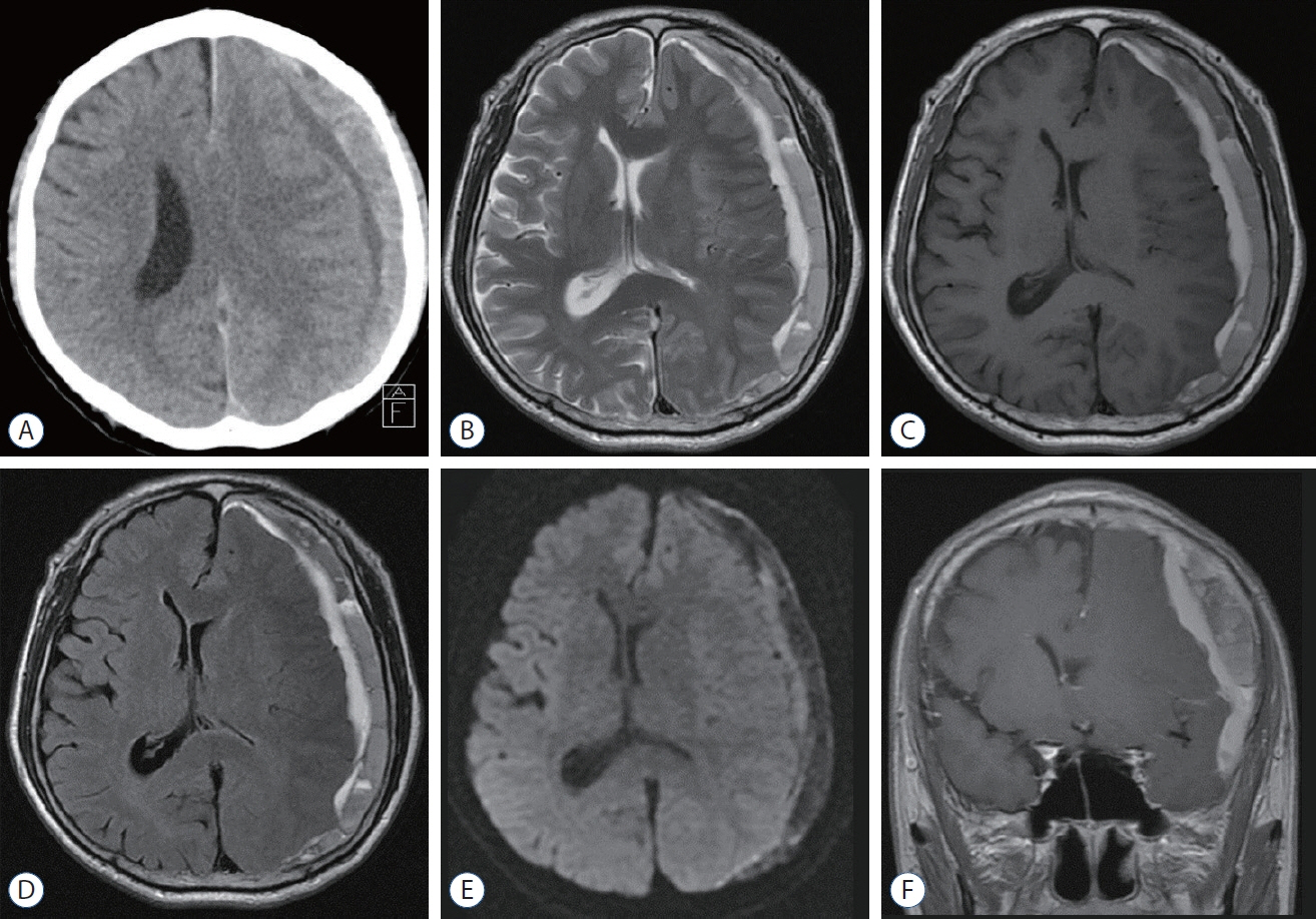
Fig. 2. A : Preoperative computerized tomography showing the chronic subdural hematoma and inner subdural hygroma on the left side. B-F : Preoperative magnetic resonance sequences showing the chronic subdural hematoma and inner subdural hygroma; T2 weighted image (B), T1- weighted image (C), flair (D), diffusion-weighted imaging (E), and coronal T1 weighted image (F).
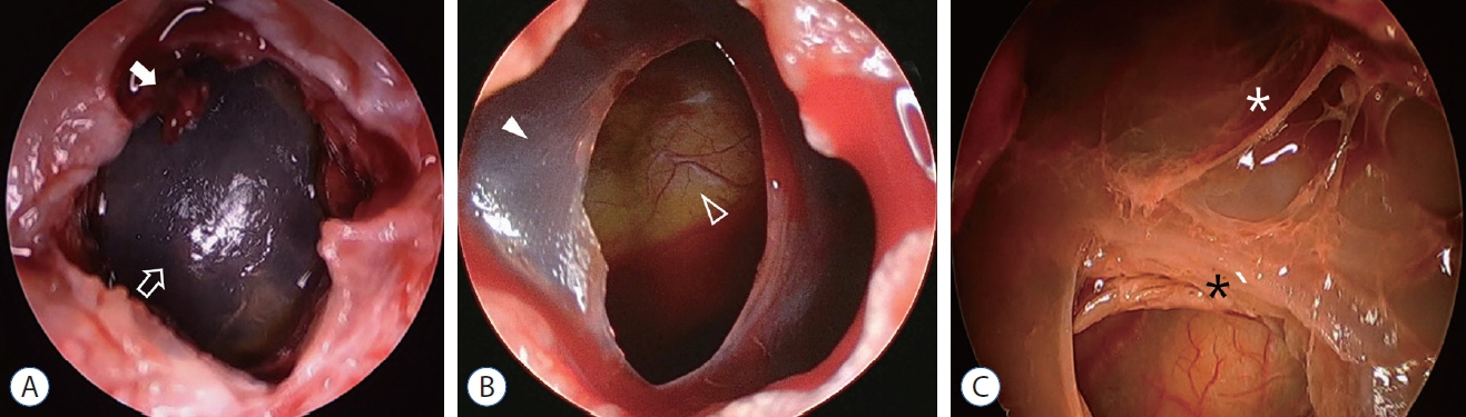
Fig. 3. A : Endoscopic view after opening of dura and outer membrane of chronic subdural hematoma. The remnant of the outer membrane (white arrow) and the inner membrane of chronic subdural hematoma (open arrow). The inner membrane is very tense and translucent but not clear. B : After fenestration of the inner membrane and draining of the inner subdural hygroma, the inner membrane (white arrowhead) is sunken down and the brain surface (open arrowhead) was showed clearly. C : Endoscopic view of a recurrent case. The inner membrane is very thick (black asterisk). The fibrous septa (white asterisk) of trabecular type are revealed in the chronic subdural hematoma space.
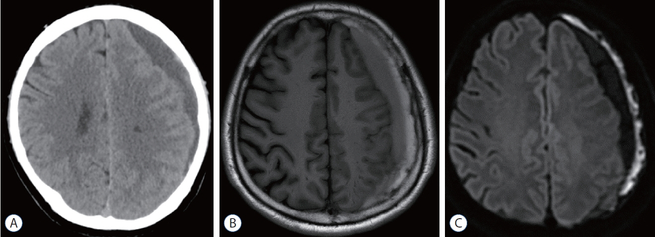
Fig. 4. Preoperative images of illustrative case 1. A : Inner subdural hygroma is not clearly seen on computerized tomography scan. B and C : The chronic subdural hematoma and inner subdural hygroma with compressed left hemisphere is showed on T1-weighted image (B) and diffusion-weighted imaging (C).
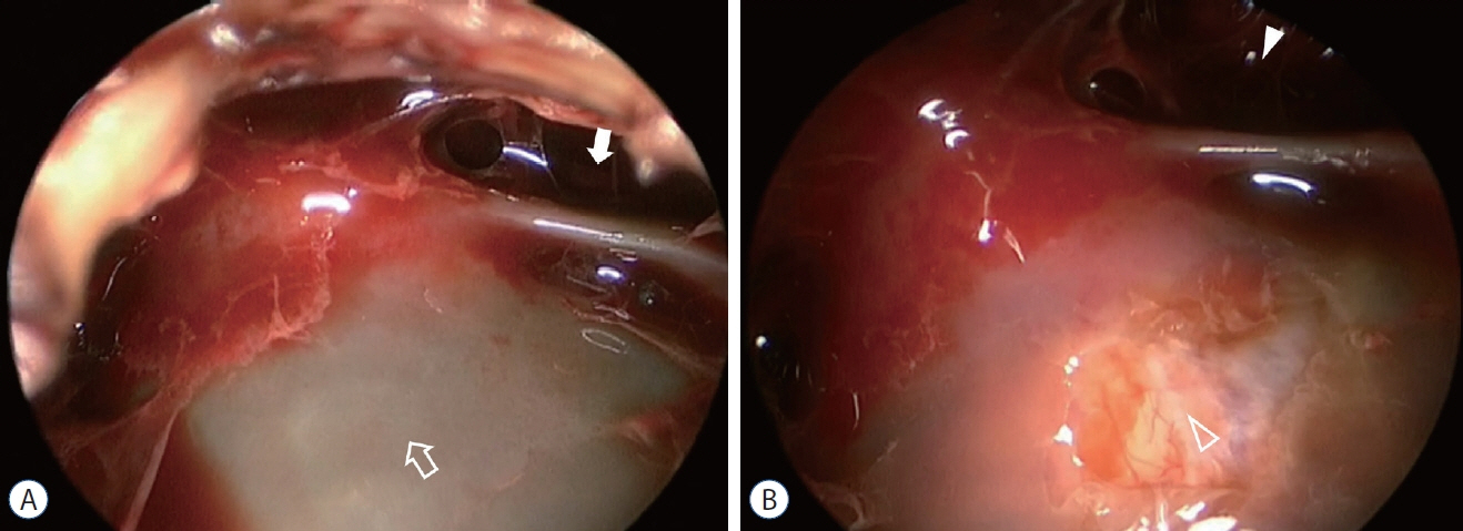
Fig. 5. Endoscopic view of illustrative case 1. A : The bulging inner membrane of the chronic subdural hematoma (open arrow) and narrowed chronic subdural hematoma space (white arrow). B : After fenestration of the inner membrane, the chronic subdural hematoma space is widened (white arrowhead) and the brain surface (open arrowhead) is showing via the opening of the inner membrane.
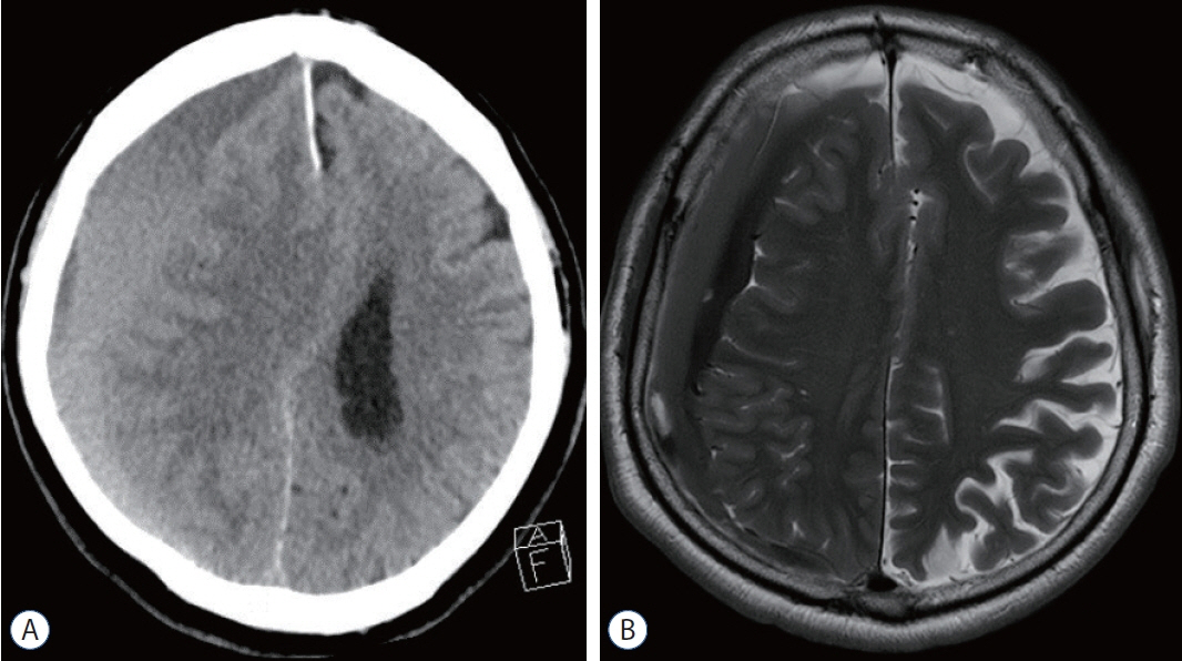
Fig. 6. Preoperative image of illustrative case 2. A : The separated type of chronic subdural hematoma on the right-side hemisphere is demonstrated on computerized tomography, however, inner subdural hygroma is not identified. B : The inner subdural hygroma is revealed well on magnetic resonance imaging.
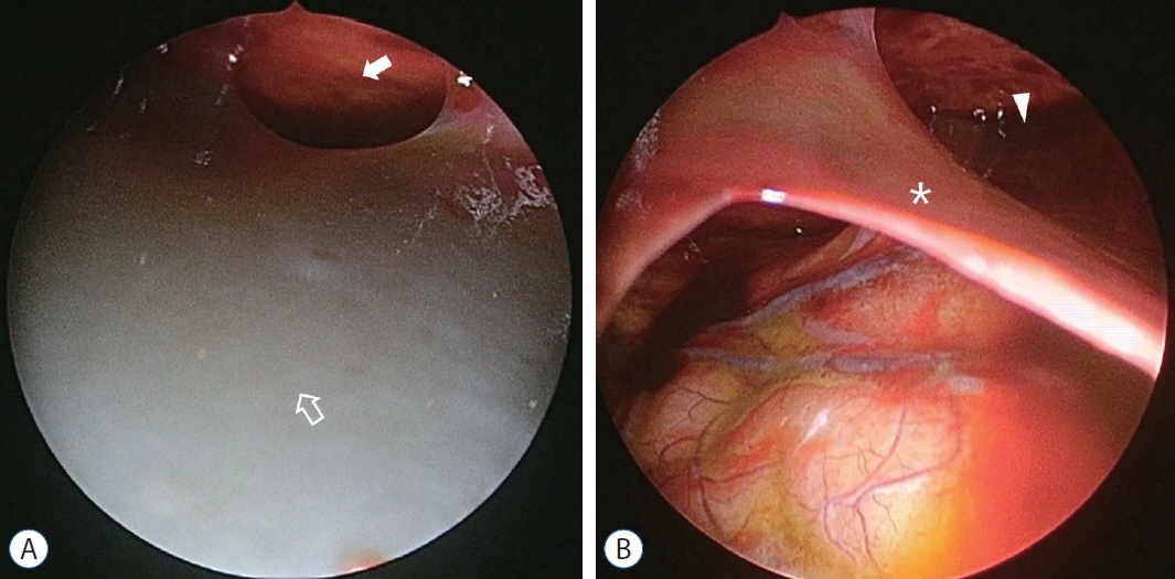
Fig. 7. Endoscopic view of illustrative case 2. A : The inner membrane is tense and gray (open arrow), and the chronic subdural hematoma space is narrowed (white arrow). B : After incision of the inner membrane (white asterisk), the inner membrane is sunken down, and the spaces of chronic subdural hematoma and inner subdural hygroma is observed widely. The remnant chronic subdural hematoma (white arrowhead) is also observed.
Reference
-
References
1. Almenawer SA, Farrokhyar F, Hong C, Alhazzani W, Manoranjan B, Yarascavitch B, et al. Chronic subdural hematoma management: a systematic review and meta-analysis of 34,829 patients. Ann Surg. 259:449–457. 2014.2. Berhouma M, Jacquesson T, Jouanneau E. The minimally invasive endoscopic management of septated chronic subdural hematomas: surgical technique. Acta Neurochir (Wien). 156:2359–2362. 2014.3. Cai Q, Guo Q, Zhang F, Sun D, Zhang W, Ji B, et al. Evacuation of chronic and subacute subdural hematoma via transcranial neuroendoscopic approach. Neuropsychiatr Dis Treat. 15:385–390. 2019.4. Chan DY, Chan DT, Sun TF, Ng SC, Wong GK, Poon WS. The use of atorvastatin for chronic subdural haematoma: a retrospective cohort comparison study. Br J Neurosurg. 31:72–77. 2017.5. Deng J, Wang F, Wang H, Zhao M, Chen G, Shangguan H, et al. Efficacy of neuroendoscopic treatment for septated chronic subdural hematoma. Front Neurol. 12:765109. 2021.6. Du B, Xu J, Hu J, Zhong X, Liang J, Lei P, et al. A clinical study of the intra-neuroendoscopic technique for the treatment of subacute-chronic and chronic septal subdural hematoma. Front Neurol. 10:1408. 2020.7. Edlmann E, Giorgi-Coll S, Whitfield PC, Carpenter KLH, Hutchinson PJ. Pathophysiology of chronic subdural haematoma: inflammation, angiogenesis and implications for pharmacotherapy. J Neuroinflammation. 14:108. 2017.8. Fan G, Ding J, Wang H, Wang Y, Liu Y, Wang C, et al. Risk factors for the development of chronic subdural hematoma in patients with subdural hygroma. Br J Neurosurg. 35:1–6. 2021.9. Goto H, Ishikawa O, Nomura M, Tanaka K, Nomura S, Maeda K. Magnetic resonance imaging findings predict the recurrence of chronic subdural hematoma. Neurol Med Chir (Tokyo). 55:173–178. 2015.10. Guo S, Gao W, Cheng W, Liang C, Wu A. Endoscope-assisted surgery vs. burr-hole craniostomy for the treatment of chronic subdural hematoma: a systemic review and meta-analysis. Front Neurol. 11:540911. 2020.11. Hohenstein A, Erber R, Schilling L, Weigel R. Increased mRNA expression of VEGF within the hematoma and imbalance of angiopoietin-1 and -2 mRNA within the neomembranes of chronic subdural hematoma. J Neurotrauma. 22:518–528. 2005.12. Ichimura S, Takahara K, Nakaya M, Yoshida K, Fujii K. Neuroendoscopic technique for recurrent chronic subdural hematoma with small craniotomy. Turk Neurosurg. 30:701–706. 2020.13. Kageyama H, Toyooka T, Tsuzuki N, Oka K. Nonsurgical treatment of chronic subdural hematoma with tranexamic acid. J Neurosurg. 119:332–337. 2013.14. Kawasaki T, Kurosaki Y, Fukuda H, Kinosada M, Ishibashi R, Handa A, et al. Flexible endoscopically assisted evacuation of acute and subacute subdural hematoma through a small craniotomy: preliminary results. Acta Neurochir (Wien). 160:241–248. 2018.15. Kwon SM, Lee MH, Seo Y, Kim YI, Oh HJ, Kim KH, et al. A radiological assessment of chronic subdural hematomas. Korean J Neurotrauma. 18:12–21. 2022.16. Lee KS. How to treat chronic subdural hematoma? Past and now. J Korean Neurosurg Soc. 62:144–152. 2019.17. Lee SH, Choi JI, Lim DJ, Ha SK, Kim SD, Kim SH. The potential of diffusion-weighted magnetic resonance imaging for predicting the outcomes of chronic subdural hematomas. J Korean Neurosurg Soc. 61:97–104. 2018.18. Lin GX, Chen CM, Kim JS, Song KS. The transformation of intracranial subdural hygroma to chronic subdural hematoma following endoscopic spinal surgery: a case report. J Neurol Surg A Cent Eur Neurosurg. 83:502–506. 2022.19. Mobbs R, Khong P. Endoscopic-assisted evacuation of subdural collections. J Clin Neurosci. 16:701–704. 2009.20. Murakami H, Hirose Y, Sagoh M, Shimizu K, Kojima M, Gotoh K, et al. Why do chronic subdural hematomas continue to grow slowly and not coagulate? Role of thrombomodulin in the mechanism. J Neurosurg. 96:877–884. 2002.21. Nakaguchi H, Tanishima T, Yoshimasu N. Factors in the natural history of chronic subdural hematomas that influence their postoperative recurrence. J Neurosurg. 95:256–262. 2001.22. Nakaguchi H, Yoshimasu N, Tanishima T. Relationship between the natural history of chronic subdural hematoma and enhancement of the inner membrane on post-contrast CT scan. No Shinkei Geka. 31:157–164. 2003.23. Pripp AH, Stanišić M. The correlation between pro- and anti-inflammatory cytokines in chronic subdural hematoma patients assessed with factor analysis. PLoS One. 9:e90149. 2014.24. Sato S, Suzuki J. Ultrastructural observations of the capsule of chronic subdural hematoma in various clinical stages. J Neurosurg. 43:569–578. 1975.25. Sherrod BA, Baker C, Gamboa N, McNally S, Grandhi R. Preoperative MRI characteristics predict chronic subdural haematoma postoperative recurrence: a meta-analysis. Br J Neurosurg. 35:527–531. 2021.26. Shimizu S, Mochizuki T, Osawa S, Kumabe T. Intraoperative ultrasonography during drainage for chronic subdural hematomas: a technique to release isolated deep-seated hematomas--technical note. Neurol Med Chir (Tokyo). 55:761–765. 2015.27. Singh H, Patir R, Vaishya S, Miglani R, Gupta A, Kaur A. Endoscopic evacuation of septated chronic subdural hemorrhage - technical considerations, results, and outcome. Surg Neurol Int. 13:8. 2022.28. Sun TF, Boet R, Poon WS. Non-surgical primary treatment of chronic subdural haematoma: preliminary results of using dexamethasone. Br J Neurosurg. 19:327–333. 2005.29. Takahashi S, Yazaki T, Nitori N, Kano T, Yoshida K, Kawase T. Neuroendoscope-assisted removal of an organized chronic subdural hematoma in a patient on bevacizumab therapy--case report. Neurol Med Chir (Tokyo). 51:515–518. 2011.30. Tsutsumi K, Maeda K, Iijima A, Usui M, Okada Y, Kirino T. The relationship of preoperative magnetic resonance imaging findings and closed system drainage in the recurrence of chronic subdural hematoma. J Neurosurg. 87:870–875. 1997.31. Weir B, Gordon P. Factors affecting coagulation: fibrinolysis in chronic subdural fluid collections. J Neurosurg. 58:242–245. 1983.32. Yan K, Gao H, Zhou X, Wu W, Xu W, Xu Y, et al. A retrospective analysis of postoperative recurrence of septated chronic subdural haematoma: endoscopic surgery versus burr hole craniotomy. Neurol Res. 39:803–812. 2017.33. Yang W, Huang J. Chronic subdural hematoma: epidemiology and natural history. Neurosurg Clin N Am. 28:205–210. 2017.
- Full Text Links
-
- Actions
-
Cited
- CITED
-
- Close
- Share
-
- Similar articles
-
- Chronic Subdural Hematoma Superimposed on Posttraumatic Subdural Hygroma: A Report of Three Cases
- Inteacerebral and Brain Stem Hemorrhage Following Evacuation of Chronic Subdural Hematoma and Hygroma
- Chronic Subdural Hematoma Secondary to Traumatic Subdural Hygroma
- Evolution of Chronic Subdural Hematoma based on Brain CT findings and Appropriate Treatment Methods
- Clinical Analysis of Chronic Subdural Hematoma Originated from Traumatic Subdural Hygroma
