Pattern of lip retraction according to the presence of lip incompetence in patients with Class II malocclusion
- Affiliations
-
- 1Department of Orthodontics, Institute of Craniofacial Deformity, College of Dentistry, Yonsei University, Seoul, Korea
- KMID: 2545043
- DOI: http://doi.org/10.4041/kjod22.260
Abstract
Objective
The aim of this retrospective study was to compare changes in hard tissue and soft tissue after the four first premolars were extracted with anterior teeth retraction according to the presence or absence of lip incompetence.
Methods
Patients who underwent the four first premolars were extracted with anterior teeth retraction were divided into competent (n = 20) and incompetent lip (n = 20) groups. Cephalometric measurements for hard tissue and soft tissue changes were performed pre-treatment and post-treatment.
Results
In the competent group, the upper and lower lips retreated by 2.88 mm and 4.28 mm, respectively, and in the incompetent group by 4.13 mm and 5.57 mm, respectively; the differences between the two groups were significant (p < 0.05). A strong positive correlation between retraction of the upper lip and upper incisors was observed in both groups (p < 0.05), whereas a correlation between retraction of the lower lip and lower incisors was only found in the incompetent group. A simple linear regression analysis showed that the pattern of lip retraction following the retraction of the anterior teeth was more predictable in the incompetent group than in the competent group.
Conclusions
These findings suggest that the initial evaluation of lip incompetence in patients with skeletal Class II is essential for the accurate prediction of the soft tissue changes following retraction of the anterior teeth in premolar extraction treatment. Therefore, sufficient explanation should be provided during patient consultations.
Keyword
Figure
-

Figure 1 Flow diagram of patient selection. ANB, A point- nasion-B point.
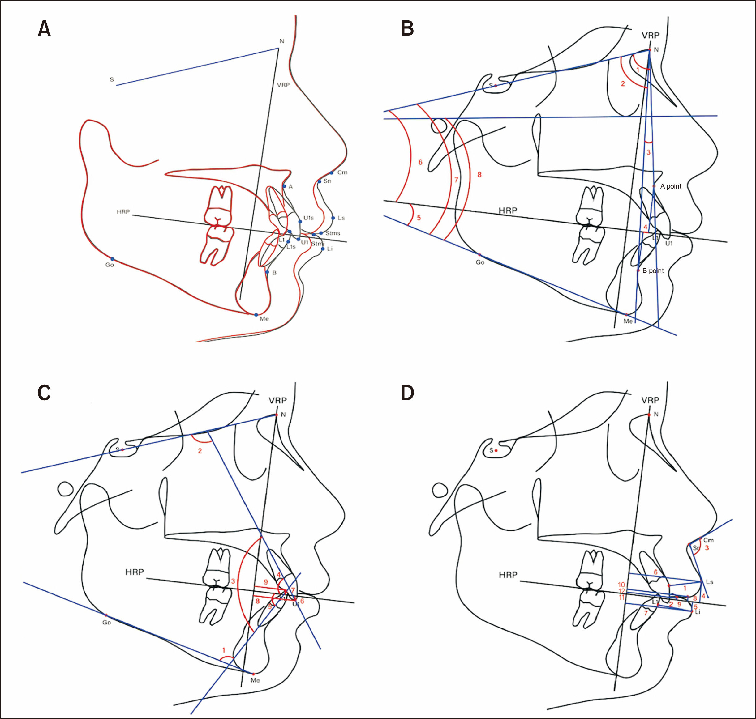
Figure 2 Cephalometric landmarks and reference planes. A, Reference planes for treatment changes. B, Skeletal measurements. 1, SNA (deg); 2, SNB (deg); 3, ANB (deg) 4, Wits appraisal, AO-BO (mm); 5, Occlusal plane to GoMe (deg); 6, Occlusal plane to SN (deg); 7, SN-GoMe (deg); 8, FMA (deg). C, 1, IMPA (deg); 2, U1 to SN (deg); 3, Interincisal angle (deg); 4, U1 to HRP (deg); 5, L1 to HRP (deg); 6, U1 to HRP (mm); 7, L1 to HRP (mm); 8, U1 to VRP (mm); 9, L1 to VRP (mm). D, 1, Upper lip thickness; 2, Lower lip thickness; 3, Nasolabial angle (deg); 4, Ls to HRP (mm); 5, Li to HRP (mm); 6, Ls to VRP (mm); 7, Li to VRP (mm); 8, Stms to HRP (mm); 9, Stmi to HRP (mm); 10, Stms to VRP (mm); 11, Stmi to VRP (mm); 12, Stms to Stmi (mm). S, sella; N, nasion; A, point A; B, point B; Go, gonion; Me, menton; U1s, upper central incisor surface; L1s, lower central incisor surface; U1, upper central incisor edge; L1, lower central incisor edge; Sn, subnasale; Cm, columella; Ls, labrale superioris; Li, labrale inferioris; Stms, stomion superius; Stmi, stomion inferius; HRP, horizontal reference plane; VRP, vertical reference plane; SNA, sella-nasion-A point; SNB, sella-nasion-B point; ANB, A point-nasion-B point; FMA, Frankfort-mandibular plane angle; IMPA, incisor-mandibular plane angle.

Figure 3 Scatter plot of the hard tissue versus soft tissue changes in both group and comparison of treatment changes between the two groups. A, U1 to VRP versus Ls to VRP in both groups. B, L1 to VRP versus Li to VRP in both groups. C, Lip retraction pattern in the competent group (vertical arrows indicate mutual lip pressure that may resist posterior displacement of the lips according to incisor retraction, indicated by horizontal arrows). D, Lip retraction pattern in the incompetent group (curved arrows represent free lip retraction without resistance). U1, upper central incisor edge; VRP, vertical reference plane; Ls, labrale superioris; L1, lower central incisor edge; Li, labrale inferioris.
Reference
-
1. Kang CS, Kim KH, Choy KC. 2000; The vertical changes of the lip and perioral soft tissue resulting from incisor retraction. Korean J Orthod. 30:185–96. https://e-kjo.org/journal/view.html?uid=897&vmd=Full.2. Caplan MJ, Shivapuja PK. 1997; The effect of premolar extractions on the soft-tissue profile in adult African American females. Angle Orthod. 67:129–36. https://pubmed.ncbi.nlm.nih.gov/9107377/. Erratum in: Angle Orthod 1997;67:240. DOI: 10.1043/0003-3219(1997)067<0129:TEOPEO>2.3.CO;2. PMID: 9107377.3. Son BH, Park YC. 1984; A roentgenocephalometric study of teeth and profile changes in orthodontically treated patient with four bicuspid extractions. J Korean Dent Assoc. 22:429–38. https://pubmed.ncbi.nlm.nih.gov/6590710/.4. Hershey HG. 1972; Incisor tooth retraction and subsequent profile change in postadolescent female patients. Am J Orthod. 61:45–54. https://doi.org/10.1016/0002-9416(72)90175-3. DOI: 10.1016/0002-9416(72)90175-3. PMID: 4500186.5. Talass MF, Talass L, Baker RC. 1987; Soft-tissue profile changes resulting from retraction of maxillary incisors. Am J Orthod Dentofacial Orthop. 91:385–94. https://doi.org/10.1016/0889-5406(87)90391-x. DOI: 10.1016/0889-5406(87)90391-X. PMID: 3472457.6. Leonardi R, Annunziata A, Licciardello V, Barbato E. 2010; Soft tissue changes following the extraction of premolars in nongrowing patients with bimaxillary protrusion. A systematic review. Angle Orthod. 80:211–6. https://pubmed.ncbi.nlm.nih.gov/19852663/. DOI: 10.2319/010709-16.1. PMID: 19852663. PMCID: PMC8978740.7. Burstone CJ. 1967; Lip posture and its significance in treatment planning. Am J Orthod. 53:262–84. https://doi.org/10.1016/0002-9416(67)90022-x. DOI: 10.1016/0002-9416(67)90022-X. PMID: 5227460.8. Hassan AH, Turkistani AA, Hassan MH. 2014; Skeletal and dental characteristics of subjects with incompetent lips. Saudi Med J. 35:849–54. https://pubmed.ncbi.nlm.nih.gov/25129185/. PMID: 25129185.9. Legan HL, Burstone CJ. 1980; Soft tissue cephalometric analysis for orthognathic surgery. J Oral Surg. 38:744–51. https://pubmed.ncbi.nlm.nih.gov/6932485/. PMID: 6932485.10. Trisnawaty N, Ioi H, Kitahara T, Suzuki A, Takahashi I. 2013; Effects of extraction of four premolars on vermilion height and lip area in patients with bimaxillary protrusion. Eur J Orthod. 35:521–8. https://doi.org/10.1093/ejo/cjs035. DOI: 10.1093/ejo/cjs035. PMID: 22573908.11. Lee KJ, Park YC, Hwang CJ, Kim YJ, Choi TH, Yoo HM, et al. 2011; Displacement pattern of the maxillary arch depending on miniscrew position in sliding mechanics. Am J Orthod Dentofacial Orthop. 140:224–32. https://doi.org/10.1016/j.ajodo.2010.05.020. DOI: 10.1016/j.ajodo.2010.05.020. PMID: 21803260.12. Aniruddh YV, Ravi K, Edeinton A. 2016; Comparative evaluation of soft tissue changes in Class I borderline patients treated with extraction and nonextraction modalities. Dental Press J Orthod. 21:50–9. https://doi.org/10.1590/2177-6709.21.4.050-059.oar. DOI: 10.1590/2177-6709.21.4.050-059.oar. PMID: 27653264. PMCID: PMC5029316.13. Vig PS, Cohen AM. 1979; Vertical growth of the lips: a serial cephalometric study. Am J Orthod. 75:405–15. https://doi.org/10.1016/0002-9416(79)90162-3. DOI: 10.1016/0002-9416(79)90162-3. PMID: 285616.14. Goel S, Bansal M, Kalra A. 2004; A preliminary assessment of cephalometric orthodontic superimposition. Eur J Orthod. 26:217–22. https://doi.org/10.1093/ejo/26.2.217. DOI: 10.1093/ejo/26.2.217. PMID: 15130046.15. Björk A, Skieller V. 1983; Normal and abnormal growth of the mandible. A synthesis of longitudinal cephalometric implant studies over a period of 25 years. Eur J Orthod. 5:1–46. https://doi.org/10.1093/ejo/5.1.1. DOI: 10.1093/ejo/5.1.1. PMID: 6572593.16. Huja SS, Grubaugh EL, Rummel AM, Fields HW, Beck FM. 2009; Comparison of hand-traced and computer-based cephalometric superimpositions. Angle Orthod. 79:428–35. https://doi.org/10.2319/052708-283.1. DOI: 10.2319/052708-283.1. PMID: 19413396.17. Kim TK, Ryu YK. 1994; Lip profile changes after orthodontic tooth movement in female adult with bimaxillary protrusion. Korean J Orthod. 24:135–48. https://e-kjo.org/journal/view.html?uid=1306&vmd=Full.18. Kim K, Choi SH, Choi EH, Choi YJ, Hwang CJ, Cha JY. 2017; Unpredictability of soft tissue changes after camouflage treatment of Class II division 1 malocclusion with maximum anterior retraction using miniscrews. Angle Orthod. 87:230–8. https://doi.org/10.2319/042516-332.1. DOI: 10.2319/042516-332.1. PMID: 27768390. PMCID: PMC8384362.19. Hayashida H, Ioi H, Nakata S, Takahashi I, Counts AL. 2011; Effects of retraction of anterior teeth and initial soft tissue variables on lip changes in Japanese adults. Eur J Orthod. 33:419–26. https://doi.org/10.1093/ejo/cjq095. DOI: 10.1093/ejo/cjq095. PMID: 20966067.20. Bloom LA. 1961; Perioral profile changes in orthodontic treatment. Am J Orthod Dentofacial Orthop. 47:371–9. https://doi.org/10.1016/0002-9416(61)90177-4. DOI: 10.1016/0002-9416(61)90177-4.
- Full Text Links
-
- Actions
-
Cited
- CITED
-
- Close
- Share
-
- Similar articles
-
- Evaluation of nasolabial angle in adult patients with skeletal Class III malocclusion
- A cephalometric study on the soft-tissue profile changes following the incisor retraction
- A cephalometric study of soft tissue profile changes associated with orthodontic treatment
- Correlations between orbicularis oris and mentalis muscle activity and craniofacial morphology in normal occlusion and Class III malocclusion
- Correlations between muscle activities of orbicularis oris, mentalis, buccinator and suprahyoid and craniofacial morphology in Class II division 1 malocclusion with incompetent lips and normal occlusion
