J Korean Med Sci.
2023 Aug;38(31):e253. 10.3346/jkms.2023.38.e253.
The Present and Future of Artificial Intelligence-Based Medical Image in Diabetes Mellitus: Focus on Analytical Methods and Limitations of Clinical Use
- Affiliations
-
- 1Department of Medical Informatics, College of Medicine, The Catholic University of Korea, Seoul, Korea
- 2Division of Endocrinology and Metabolism, Department of Internal Medicine, Seoul St. Mary’s Hospital, College of Medicine, The Catholic University of Korea, Seoul, Korea
- KMID: 2544962
- DOI: http://doi.org/10.3346/jkms.2023.38.e253
Abstract
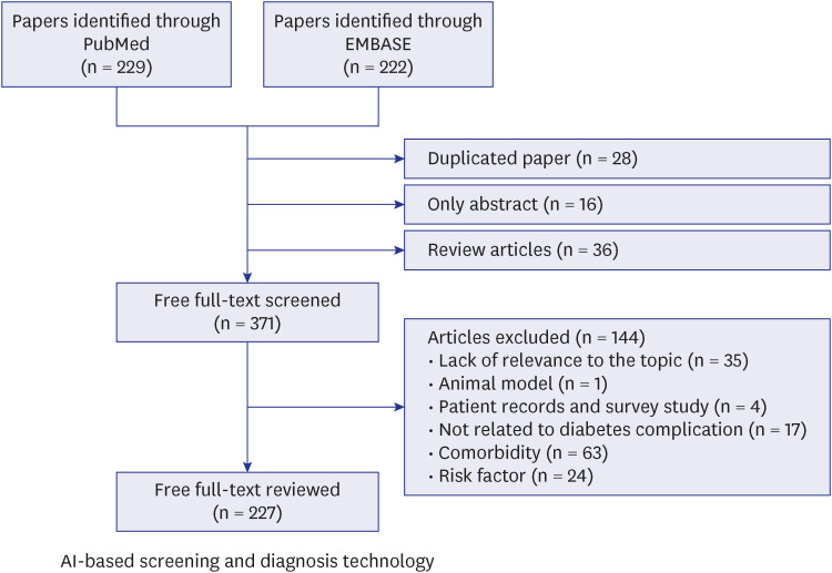
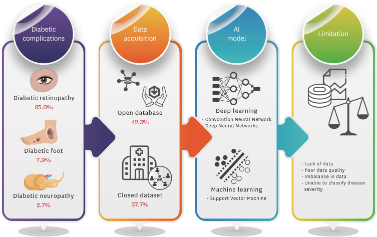
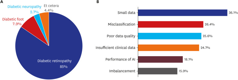
- Artificial intelligence (AI)-based diagnostic technology using medical images can be used to increase examination accessibility and support clinical decision-making for screening and diagnosis. To determine a machine learning algorithm for diabetes complications, a literature review of studies using medical image-based AI technology was conducted using the National Library of Medicine PubMed, and the Excerpta Medica databases. Lists of studies using diabetes diagnostic images and AI as keywords were combined. In total, 227 appropriate studies were selected. Diabetic retinopathy studies using the AI model were the most frequent (85.0%, 193/227 cases), followed by diabetic foot (7.9%, 18/227 cases) and diabetic neuropathy (2.7%, 6/227 cases). The studies used open datasets (42.3%, 96/227 cases) or directly constructed data from fundoscopy or optical coherence tomography (57.7%, 131/227 cases). Major limitations in AI-based detection of diabetes complications using medical images were the lack of datasets (36.1%, 82/227 cases) and severity misclassification (26.4%, 60/227 cases). Although it remains difficult to use and fully trust AI-based imaging analysis technology clinically, it reduces clinicians’ time and labor, and the expectations from its decision-support roles are high. Various data collection and synthesis data technology developments according to the disease severity are required to solve data imbalance.
Keyword
Figure
Reference
-
1. Park JH, Ha KH, Kim BY, Lee JH, Kim DJ. Trends in cardiovascular complications and mortality among patients with diabetes in South Korea. Diabetes Metab J. 2021; 45(1):120–124. PMID: 33290647.2. Kim J, Yoon SJ, Jo MW. Estimating the disease burden of Korean type 2 diabetes mellitus patients considering its complications. PLoS One. 2021; 16(2):e0246635. PMID: 33556138.3. Chawla A, Chawla R, Jaggi S. Microvasular and macrovascular complications in diabetes mellitus: distinct or continuum? Indian J Endocrinol Metab. 2016; 20(4):546–551. PMID: 27366724.4. Kim H, Jung DY, Lee SH, Cho JH, Yim HW, Kim HS. Long-term risk of cardiovascular disease among type 2 diabetes patients according to average and visit-to-visit variations of HbA1c levels during the first 3 years of diabetes diagnosis. J Korean Med Sci. 2023; 38(4):e24. PMID: 36718561.5. Crasto W, Patel V, Davies MJ, Khunti K. Prevention of microvascular complications of diabetes. Endocrinol Metab Clin North Am. 2021; 50(3):431–455. PMID: 34399955.6. Bowling FL, Rashid ST, Boulton AJ. Preventing and treating foot complications associated with diabetes mellitus. Nat Rev Endocrinol. 2015; 11(10):606–616. PMID: 26284447.7. Kwan CC, Fawzi AA. Imaging and biomarkers in diabetic macular edema and diabetic rRetinopathy. Curr Diab Rep. 2019; 19(10):95. PMID: 31473838.8. Polak JF, Szklo M, Kronmal RA, Burke GL, Shea S, Zavodni AE, et al. The value of carotid artery plaque and intima-media thickness for incident cardiovascular disease: the multi-ethnic study of atherosclerosis. J Am Heart Assoc. 2013; 2(2):e000087. PMID: 23568342.9. Itri JN, Tappouni RR, McEachern RO, Pesch AJ, Patel SH. Fundamentals of diagnostic error in imaging. Radiographics. 2018; 38(6):1845–1865. PMID: 30303801.10. Pesapane F, Codari M, Sardanelli F. Artificial intelligence in medical imaging: threat or opportunity? Radiologists again at the forefront of innovation in medicine. Eur Radiol Exp. 2018; 2(1):35. PMID: 30353365.11. Nomura A, Noguchi M, Kometani M, Furukawa K, Yoneda T. Artificial intelligence in current diabetes management and prediction. Curr Diab Rep. 2021; 21(12):61. PMID: 34902070.12. Mintz Y, Brodie R. Introduction to artificial intelligence in medicine. Minim Invasive Ther Allied Technol. 2019; 28(2):73–81. PMID: 30810430.13. Mahadevaiah G, Rv P, Bermejo I, Jaffray D, Dekker A, Wee L. Artificial intelligence-based clinical decision support in modern medical physics: Selection, acceptance, commissioning, and quality assurance. Med Phys. 2020; 47(5):e228–e235. PMID: 32418341.14. Shin J, Kim J, Lee C, Yoon JY, Kim S, Song S, et al. Development of various diabetes prediction models using machine learning techniques. Diabetes Metab J. 2022; 46(4):650–657. PMID: 35272434.15. Lee S, Jeon S, Kim HS. A study on methodologies of drug repositioning using biomedical big data: a focus on diabetes mellitus. Endocrinol Metab (Seoul). 2022; 37(2):195–207. PMID: 35413782.16. Chaki J, Ganesh ST, Cidham SK, Theertan SA. Machine learning and artificial intelligence based diabetes mellitus detection and self-management: a systematic review. J King Saud Univ Comput Inf Sci. 2022; 34(6 Pt B):3204–3225.17. Anaya-Isaza A, Mera-Jiménez L, Zequera-Diaz M. An overview of deep learning in medical imaging. Inform Med Unlocked. 2021; 26:100723.18. Liu R, Li Q, Xu F, Wang S, He J, Cao Y, et al. Application of artificial intelligence-based dual-modality analysis combining fundus photography and optical coherence tomography in diabetic retinopathy screening in a community hospital. Biomed Eng Online. 2022; 21(1):47. PMID: 35859144.19. Malerbi FK, Andrade RE, Morales PH, Stuchi JA, Lencione D, de Paulo JV, et al. Diabetic retinopathy screening using artificial intelligence and handheld smartphone-based retinal camera. J Diabetes Sci Technol. 2022; 16(3):716–723. PMID: 33435711.20. Gao Q, Amason J, Cousins S, Pajic M, Hadziahmetovic M. Automated identification of referable retinal pathology in teleophthalmology setting. Transl Vis Sci Technol. 2021; 10(6):30.21. Lu L, Ren P, Lu Q, Zhou E, Yu W, Huang J, et al. Analyzing fundus images to detect diabetic retinopathy (DR) using deep learning system in the Yangtze River delta region of China. Ann Transl Med. 2021; 9(3):226. PMID: 33708853.22. Tang F, Luenam P, Ran AR, Quadeer AA, Raman R, Sen P, et al. Detection of diabetic retinopathy from ultra-widefield scanning laser ophthalmoscope images: a multicenter deep learning analysis. Ophthalmol Retina. 2021; 5(11):1097–1106. PMID: 33540169.23. Lo YC, Lin KH, Bair H, Sheu WH, Chang CS, Shen YC, et al. Epiretinal membrane detection at the ophthalmologist level using deep learning of optical coherence tomography. Sci Rep. 2020; 10(1):8424. PMID: 32439844.24. Tang MC, Teoh SS, Ibrahim H, Embong Z. Neovascularization detection and localization in fundus images using deep learning. Sensors (Basel). 2021; 21(16):5327. PMID: 34450766.25. Wu J, Hu R, Xiao Z, Chen J, Liu J. Vision transformer-based recognition of diabetic retinopathy grade. Med Phys. 2021; 48(12):7850–7863. PMID: 34693536.26. Toğaçar M, Ergen B, Tümen V. Use of dominant activations obtained by processing OCT images with the CNNs and slime mold method in retinal disease detection. Biocybern Biomed Eng. 2022; 42(2):646–666.27. Wang TY, Chen YH, Chen JT, Liu JT, Wu PY, Chang SY, et al. Diabetic macular edema detection using end-to-end deep fusion model and anatomical landmark visualization on an edge computing device. Front Med (Lausanne). 2022; 9:851644. PMID: 35445051.28. Tang F, Wang X, Ran AR, Chan CK, Ho M, Yip W, et al. A multitask deep-learning system to classify diabetic macular edema for different optical coherence tomography devices: a multicenter analysis. Diabetes Care. 2021; 44(9):2078–2088. PMID: 34315698.29. Guo Y, Hormel TT, Xiong H, Wang J, Hwang TS, Jia Y. Automated segmentation of retinal fluid volumes from structural and angiographic optical coherence tomography using deep learning. Transl Vis Sci Technol. 2020; 9(2):54. PMID: 33110708.30. Arcadu F, Benmansour F, Maunz A, Michon J, Haskova Z, McClintock D, et al. Deep learning predicts OCT measures of diabetic macular thickening from color fundus photographs. Invest Ophthalmol Vis Sci. 2019; 60(4):852–857. PMID: 30821810.31. Khandakar A, Chowdhury ME, Reaz MB, Ali SH, Kiranyaz S, Rahman T, et al. A novel machine learning approach for severity classification of diabetic foot complications using thermogram images. Sensors (Basel). 2022; 22(11):4249. PMID: 35684870.32. Goyal M, Reeves ND, Rajbhandari S, Ahmad N, Wang C, Yap MH. Recognition of ischaemia and infection in diabetic foot ulcers: Dataset and techniques. Comput Biol Med. 2020; 117:103616. PMID: 32072964.33. Goyal M, Reeves ND, Rajbhandari S, Yap MH. Robust methods for real-time diabetic foot ulcer detection and localization on mobile devices. IEEE J Biomed Health Inform. 2019; 23(4):1730–1741. PMID: 30188841.34. Yogapriya J, Chandran V, Sumithra MG, Elakkiya B, Shamila Ebenezer A, Suresh Gnana Dhas C. Automated detection of infection in diabetic foot ulcer images using convolutional neural network. J Healthc Eng. 2022; 2022:2349849. PMID: 35432819.35. Dremin V, Marcinkevics Z, Zherebtsov E, Popov A, Grabovskis A, Kronberga H, et al. Skin complications of diabetes mellitus revealed by polarized hyperspectral imaging and machine learning. IEEE Trans Med Imaging. 2021; 40(4):1207–1216. PMID: 33406038.36. Wright DE, Mukherjee S, Patra A, Khasawneh H, Korfiatis P, Suman G, et al. Radiomics-based machine learning (ML) classifier for detection of type 2 diabetes on standard-of-care abdomen CTs: a proof-of-concept study. Abdom Radiol (NY). 2022; 47(11):3806–3816. PMID: 36085379.37. Tallam H, Elton DC, Lee S, Wakim P, Pickhardt PJ, Summers RM. Fully Automated abdominal CT biomarkers for type 2 diabetes using deep learning. Radiology. 2022; 304(1):85–95. PMID: 35380492.38. Shen D, Wu G, Suk HI. Deep learning in medical image analysis. Annu Rev Biomed Eng. 2017; 19(1):221–248. PMID: 28301734.39. Yamashita R, Nishio M, Do RK, Togashi K. Convolutional neural networks: an overview and application in radiology. Insights Imaging. 2018; 9(4):611–629. PMID: 29934920.40. LeCun Y, Bengio Y, Hinton G. Deep learning. Nature. 2015; 521(7553):436–444. PMID: 26017442.41. Anwar SM, Majid M, Qayyum A, Awais M, Alnowami M, Khan MK. Medical image analysis using convolutional neural networks: a review. J Med Syst. 2018; 42(11):226. PMID: 30298337.42. Aggarwal A, Mittal M, Battineni G. Generative adversarial network: an overview of theory and applications. IJIM Data Insights. 2021; 1(1):100004.43. Stahlschmidt SR, Ulfenborg B, Synnergren J. Multimodal deep learning for biomedical data fusion: a review. Brief Bioinform. 2022; 23(2):bbab569. PMID: 35089332.44. Zang P, Hormel TT, Wang X, Tsuboi K, Huang D, Hwang TS, et al. A diabetic retinopathy classification framework based on deep-learning analysis of OCT angiography. Transl Vis Sci Technol. 2022; 11(7):10.45. Kim HS, Kim DJ, Yoon KH. Medical big data is not yet available: why we need realism rather than exaggeration. Endocrinol Metab (Seoul). 2019; 34(4):349–354. PMID: 31884734.46. Dai L, Wu L, Li H, Cai C, Wu Q, Kong H, et al. A deep learning system for detecting diabetic retinopathy across the disease spectrum. Nat Commun. 2021; 12(1):3242. PMID: 34050158.47. Jagan Mohan N, Murugan R, Goel T, Mirjalili S, Roy P. A novel four-step feature selection technique for diabetic retinopathy grading. Phys Eng Sci Med. 2021; 44(4):1351–1366. PMID: 34748191.48. Park JI, Lee HY, Kim H, Lee J, Shinn J, Kim HS. Lack of acceptance of digital healthcare in the medical market: addressing old problems raised by various clinical professionals and developing possible solutions. J Korean Med Sci. 2021; 36(37):e253. PMID: 34581521.49. Chollet F. Xception: deep learning with depthwise separable convolutions. In : Proceedings of the 2017 IEEE Conference on Computer Vision and Pattern Recognition (CVPR); 2017 Jul 21-26; Honolulu, HI, USA. Piscataway, NJ, USA: IEEE;2017. p. 1251–1258.50. Iqbal T, Ali H. Generative adversarial network for medical images (MI-GAN). J Med Syst. 2018; 42(11):231. PMID: 30315368.51. Goodfellow I, Pouget-Abadie J, Mirza M, Xu B, Warde-Farley D, Ozair S, et al. Generative adversarial networks. Commun ACM. 2020; 63(11):139–144.52. Zhu JY, Park T, Isola P, Efros AA. Unpaired image-to-image translation using cycle-consistent adversarial networks. In : Proceedings of the 2017 IEEE International Conference on Computer Vision (ICCV); 2017 Oct 22-29; Venice, Italy. Piscataway, NJ, USA: IEEE;2017. p. 2223–2232.53. AbdelMaksoud E, Barakat S, Elmogy M. A computer-aided diagnosis system for detecting various diabetic retinopathy grades based on a hybrid deep learning technique. Med Biol Eng Comput. 2022; 60(7):2015–2038. PMID: 35545738.54. Zhang K, Liu X, Xu J, Yuan J, Cai W, Chen T, et al. Deep-learning models for the detection and incidence prediction of chronic kidney disease and type 2 diabetes from retinal fundus images. Nat Biomed Eng. 2021; 5(6):533–545. PMID: 34131321.55. Kim HS. Decision-making in artificial intelligence: is it always correct? J Korean Med Sci. 2020; 35(1):e1. PMID: 31898430.56. Tsuneki M. Deep learning models in medical image analysis. J Oral Biosci. 2022; 64(3):312–320. PMID: 35306172.57. Suganyadevi S, Seethalakshmi V, Balasamy K. A review on deep learning in medical image analysis. Int J Multimed Inf Retr. 2022; 11(1):19–38. PMID: 34513553.
- Full Text Links
- Actions
-
Cited
- CITED
-
- Close
- Share
- Similar articles
-
- Artificial Intelligence in Pathology
- Artificial Intelligence in Medicine: Beginner's Guide
- Application of artificial intelligence for diagnosis of early gastric cancer based on magnifying endoscopy with narrow-band imaging
- Prognostication of Hepatocellular Carcinoma Using Artificial Intelligence
- Machine Learning Application in Diabetes and Endocrine Disorders


