Study of frontal and ethmoid sinus of sinonasal complex along with olfactory fossa: anatomical considerations for endoscopic sinus surgery
- Affiliations
-
- 1Department of Anatomy, All India Institute of Medical Sciences, Bhopal, India
- KMID: 2544078
- DOI: http://doi.org/10.5115/acb.22.230
Abstract
- The Functional endoscopic sinus surgery through transnasal approach is a common modality of treatment for disorders of the nasal cavity, paranasal air sinuses as well as cranial cavity. The olfactory fossa (OF) is located along the superior aspect of cribriform plate which varies in shape and depth. This variable measurement of the depth of OF is mostly responsible for greater risk of intracranial infiltration during endoscopic procedures in and around the nasal cavity. The morphology of frontal and ethmoid sinus (ES) vary from simple to complex. This cadaveric study is planned to improve the ability of the otolaryngologist, radiologist to understand the possible morphological variations and plan steps of less invasive “precision surgery” to have a safe and complication free procedures. A total of 37 human head regions were included in the study. For classification of OF, Modified Kero’s classification was used. The size, shape and cells of frontal and ES were noted. We found, type II (60.8%) OF was more common followed by type I (29.7%) than type III (9.5%). The shape of frontal sinus was comma shaped (55.4%) followed by oval (18.9%) than irregular (16.2%). Most common two cells type of ES was seen in 50.0% of both anterior and posterior ES. Out of 74 ES, 8.1% of Onodi cells and 14.9% of agger nasi cells were seen.
Keyword
Figure
-
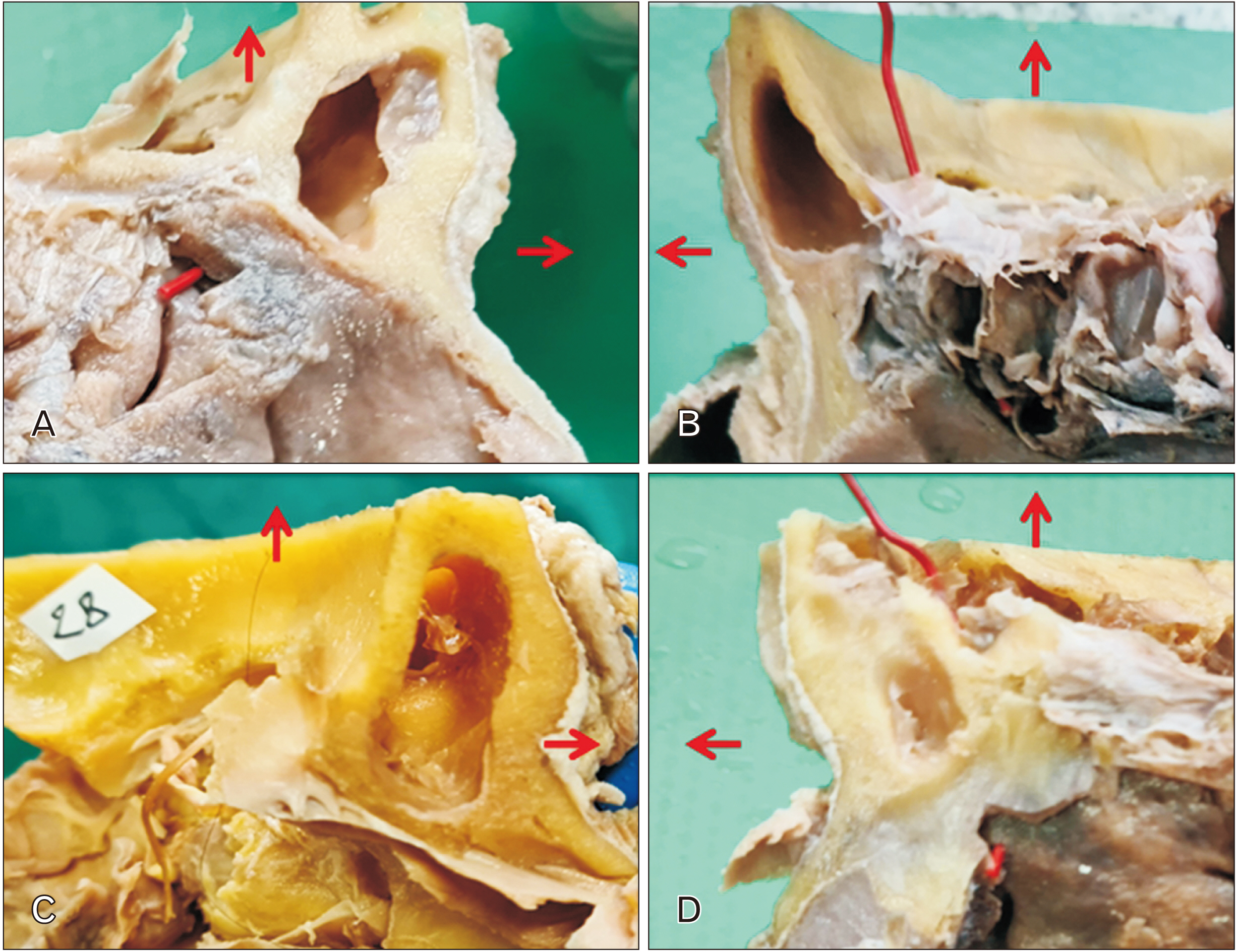
Fig. 1 Showing different shapes of the frontal sinus. (A) Irregular. (B) Comma shaped. (C) Oval. (D) Hypoplasia of frontal sinus. Horizontal arrows are directed towards the anterior aspect and upward arrows indicate a superior aspect of a specimen.

Fig. 2 Showing frontal sinus (A) Eccentric septa inside the frontal sinus. (B) Aplasia of frontal sinus on right side marked by 2. (C) Irregular frontal sins with lateral expansion in roof of orbit marked by a star. Arrow in the figure is directed to indicate anterior aspect of the specimen.
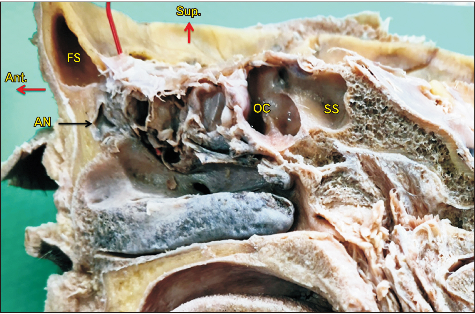
Fig. 3 Sagittal section of head for paranasal sinus. AN, agger nasi cell; FS, frontal sinus; OC, onodi cell; SS, sphenoid sinus; Ant., anterior; Sup., superior. Aspect of specimen are indicated by red arrows.
Reference
-
References
1. Rontal M, Rontal E. 1991; Studying whole-mounted sections of the paranasal sinuses to understand the complications of endoscopic sinus surgery. Laryngoscope. 101(4 Pt 1):361–6. DOI: 10.1002/lary.1991.101.4.361. PMID: 1895850.
Article2. Mosher HP. 1929; LXXII. Symposium on the ethmoid: the surgical anatomy of the ethmoidal labyrinth. Ann Otol Rhinol Laryngol. 38:869–901. DOI: 10.1177/000348942903800401.3. Zinreich S, Kuhn F, London NR, Kennedy D, Solaiyappan M, Hosemann W. 2020; 3D CT stereoscopic imaging: an improved anatomical understanding of the anterior ethmoid sinus and frontal sinus drainage pathway. Rhinol Online. 3:202–20. DOI: 10.4193/RHINOL/20.061. PMID: 59d94f39ac46421f827f2eb6afca5692.
Article4. Almushayti ZA, Almutairi AN, Almushayti MA, Alzeadi HS, Alfadhel EA, AlSamani AN. 2022; Evaluation of the Keros classification of olfactory fossa by CT scan in Qassim region. Cureus. 14:e22378. DOI: 10.7759/cureus.22378. PMID: 35321069. PMCID: PMC8935634.
Article5. Vaid S, Vaid N. 2015; Normal anatomy and anatomic variants of the paranasal sinuses on computed tomography. Neuroimaging Clin N Am. 25:527–48. DOI: 10.1016/j.nic.2015.07.002. PMID: 26476378.
Article6. Keros P. 1962; On the practical value of differences in the level of the lamina cribrosa of the ethmoid. Z Laryngol Rhinol Otol. 41:809–13. German. PMID: 14032071.7. V AM, Santosh B. 2017; A study of clinical significance of the depth of olfactory fossa in patients undergoing endoscopic sinus surgery. Indian J Otolaryngol Head Neck Surg. 69:514–22. DOI: 10.1007/s12070-017-1229-8. PMID: 29238684. PMCID: PMC5714917.
Article8. Moore KL, Dalley AF, Agur AMR. Moore KL, Dalley AF, Agur AMR, editors. 2013. Head: paranasal sinuses. Clinically Oriented Anatomy. 7th ed. Wolters Kluwer/Lippincott Williams & Wilkins Health;p. 960–4.9. Standring S. 2016. Gray's anatomy: the anatomical basis of clinical practice. 41st ed. Churchill Livingstone;p. 696–9.10. Elwany S, Medanni A, Eid M, Aly A, El-Daly A, Ammar SR. 2010; Radiological observations on the olfactory fossa and ethmoid roof. J Laryngol Otol. 124:1251–6. DOI: 10.1017/S0022215110001313. PMID: 20529392.
Article11. Kaplanoglu H, Kaplanoglu V, Dilli A, Toprak U, Hekimoğlu B. 2013; An analysis of the anatomic variations of the paranasal sinuses and ethmoid roof using computed tomography. Eurasian J Med. 45:115–25. DOI: 10.5152/eajm.2013.23. PMID: 25610263. PMCID: PMC4261488.
Article12. Sahin C, Yilmaz YF, Titiz A, Ozcan M, Ozlugedik S, Unal A. 2007; Analysis of ethmoid roof and cranial base in Turkish population. KBB BBC Dergisi. 15:1–6.13. Solares CA, Lee WT, Batra PS, Citardi MJ. 2008; Lateral lamella of the cribriform plate: software-enabled computed tomographic analysis and its clinical relevance in skull base surgery. Arch Otolaryngol Head Neck Surg. 134:285–9. DOI: 10.1001/archotol.134.3.285. PMID: 18347254.14. Souza SA, Souza MMA, Idagawa M, Wolosker AMB, Ajzen SA. 2008; Computed tomography assessment of the ethmoid roof: a relevant region at risk in endoscopic sinus surgery. Radiol Bras. 41:143–7. DOI: 10.1590/S0100-39842008000300003.15. Bista M, Maharjan M, Kafle P, Shrestha S, KC T. 2010; Computed tomographic assessment of lateral lamella of cribriform plate and comparison of depth of olfactory fossa. JNMA J Nepal Med Assoc. 49:92–5. DOI: 10.31729/jnma.104. PMID: 21485590. PMID: da02be8ff1d0454293c429f923eb63b4.16. Başak S, Karaman CZ, Akdilli A, Mutlu C, Odabaşi O, Erpek G. 1998; Evaluation of some important anatomical variations and dangerous areas of the paranasal sinuses by CT for safer endonasal surgery. Rhinology. 36:162–7. PMID: 9923058.17. Yüksel Aslier NG, Karabay N, Zeybek G, Keskinoğlu P, Kiray A, Sütay S, Ecevit MC. 2016; The classification of frontal sinus pneumatization patterns by CT-based volumetry. Surg Radiol Anat. 38:923–30. DOI: 10.1007/s00276-016-1644-7. PMID: 26884400.
Article18. Nikolova S, Toneva D, Georgiev I, Lazarov N. 2018; Digital radiomorphometric analysis of the frontal sinus and assessment of the relation between persistent metopic suture and frontal sinus development. Am J Phys Anthropol. 165:492–506. DOI: 10.1002/ajpa.23375. PMID: 29266191.
Article19. Çakur B, Sumbullu MA, Durna NB. 2011; Aplasia and agenesis of the frontal sinus in Turkish individuals: a retrospective study using dental volumetric tomography. Int J Med Sci. 8:278–82. DOI: 10.7150/ijms.8.278. PMID: 21537381. PMCID: PMC3085142.
Article20. Krogman WM. 1962. The human skeleton in forensic medicine. Thomas;DOI: 10.2307/278093.21. Gulisano M, Pacini P, Orlandini GE. 1978; Frontal sinus dimensions in relation to the cranial index: anatomo-radiologic findings. Boll Soc Ital Biol Sper. 54:66–9. Italian.22. Özdemir A, Arslan S. 2018; Incidence of agger nasi and frontal cells and their relation to frontal sinusitis in a Turkish population: a CT study. Anatomy. 12:71–5. DOI: 10.2399/ana.18.050.
Article23. Kayalioglu G, Oyar O, Govsa F. 2000; Nasal cavity and paranasal sinus bony variations: a computed tomographic study. Rhinology. 38:108–13. PMID: 11072655.24. Bradley DT, Kountakis SE. 2004; The role of agger nasi air cells in patients requiring revision endoscopic frontal sinus surgery. Otolaryngol Head Neck Surg. 131:525–7. DOI: 10.1016/j.otohns.2004.03.038. PMID: 15467630.
Article25. Driben JS, Bolger WE, Robles HA, Cable B, Zinreich SJ. 1998; The reliability of computerized tomographic detection of the Onodi (Sphenoethmoid) cell. Am J Rhinol. 12:105–11. DOI: 10.2500/105065898781390325. PMID: 9578928.
Article26. Weinberger DG, Anand VK, Al-Rawi M, Cheng HJ, Messina AV. 1996; Surgical anatomy and variations of the Onodi cell. Am J Rhinol. 10:365–72. DOI: 10.2500/105065896781794851.
Article27. Arslan H, Aydinlioğlu A, Bozkurt M, Egeli E. 1999; Anatomic variations of the paranasal sinuses: CT examination for endoscopic sinus surgery. Auris Nasus Larynx. 26:39–48. DOI: 10.1016/S0385-8146(98)00024-8. PMID: 10077255.
Article28. Yeoh KH, Tan KK. 1994; The optic nerve in the posterior ethmoid in Asians. Acta Otolaryngol. 114:329–36. DOI: 10.3109/00016489409126065. PMID: 8073866.
Article29. Thanaviratananich S, Chaisiwamongkol K, Kraitrakul S, Tangsawad W. 2003; The prevalence of an Onodi cell in adult Thai cadavers. Ear Nose Throat J. 82:200–4. DOI: 10.1177/014556130308200314. PMID: 12696241.
Article
- Full Text Links
-
- Actions
-
Cited
- CITED
-
- Close
- Share
-
- Similar articles
-
- Endoscopic Frontal Sinus Surgery
- A Case of Isolated Frontal Fungal Sinusitis: Treated by Endoscopic Sinus Surgery with Frontal Sinus Minitrephination
- A Case of Cholesterol Granuloma of the Frontal and Ethmoid Sinus
- Intranasal Endoscopic Removal of Osteoma in Ethmoid Sinus
- Frontal Sinus Reconstruction with Medpore(R) in Oncocytic Schneiderian Papilloma with Bone Destruction
