J Korean Foot Ankle Soc.
2023 Jun;27(2):58-66. 10.14193/jkfas.2023.27.2.58.
Effect of Deltoid Ligament Repair on Syndesmotic Stabilization in Patients with Ankle Fractures
- Affiliations
-
- 1Department of Orthopedic Surgery, Haeundae Paik Hospital, Inje University College of Medicine, Busan, Korea
- 2Department of Orthopedic Surgery, Seoul Gunwoo Hospital, Seoul, Korea
- KMID: 2543002
- DOI: http://doi.org/10.14193/jkfas.2023.27.2.58
Abstract
- Purpose
This study aimed to evaluate the effectiveness of deltoid ligament repair on syndesmotic stabilization in patients with acute ankle fractures with ruptured deltoid and syndesmotic ligaments.
Materials and Methods
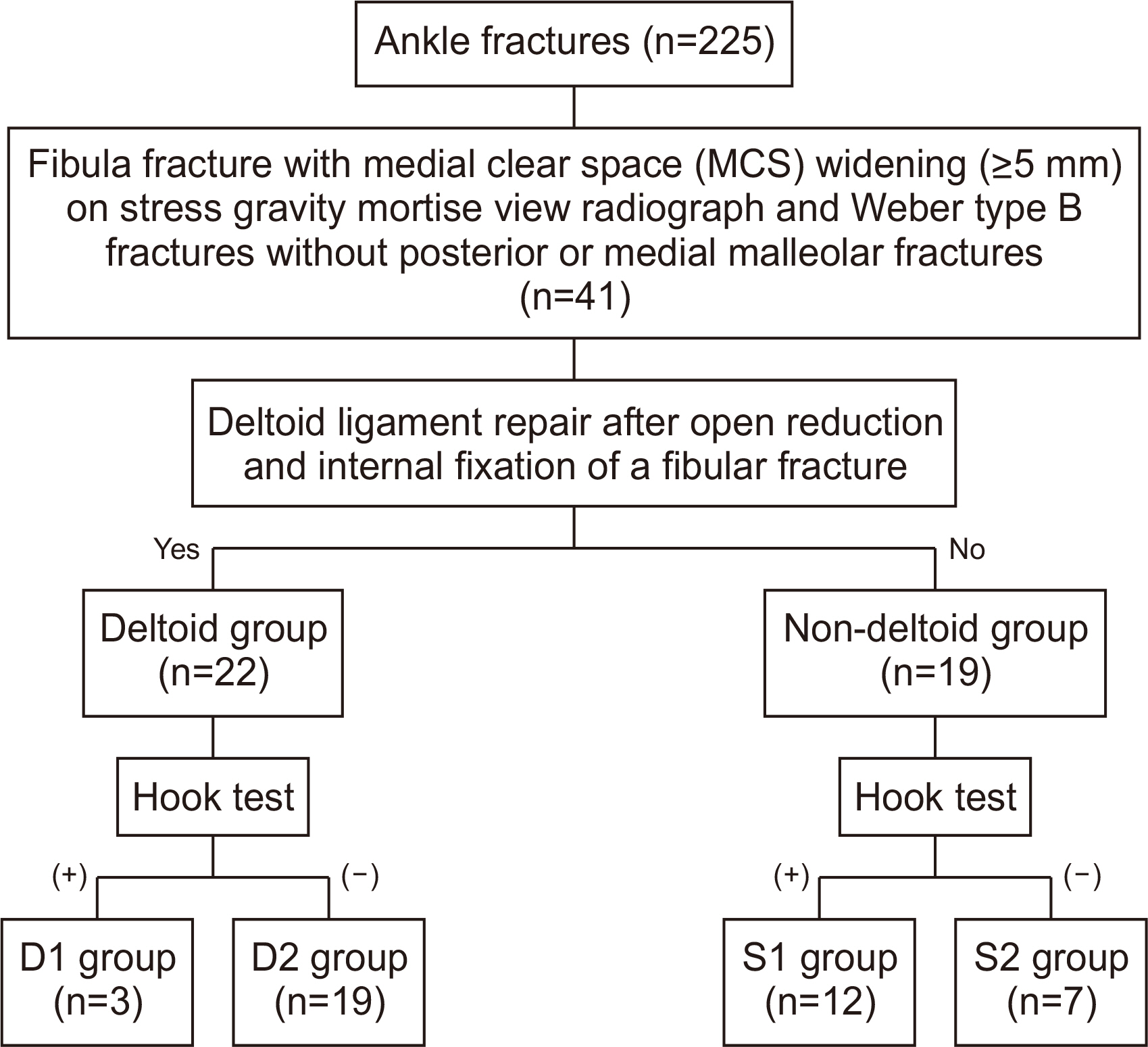
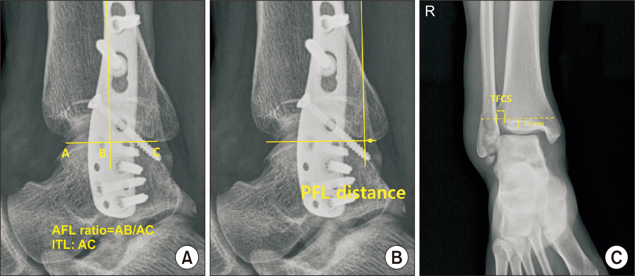
The medical records of 41 patients (41 ankles) who underwent surgery for Weber type B ankle fracture with ruptured deltoid and syndesmotic ligaments were retrospectively analyzed. The mean follow-up duration was 36 months (range 18~65 months). Patients were divided into two groups: those that underwent deltoid ligament repair (the deltoid group) and those who did not (the non-deltoid group). Both groups were also divided into two subgroups, namely, the D1/S1 group, which underwent syndesmotic screw fixation, or the D2/S2 group, which did not. Medial clear space (MCS), tibiofibular clear space (TFCS), anterior fibular line (AFL) ratio, and posterior fibular line (PFL) distance were measured, and visual analog scale (VAS), American Orthopaedic Foot and Ankle Society (AOFAS), and Foot Function Index (FFI) scores were evaluated.
Results
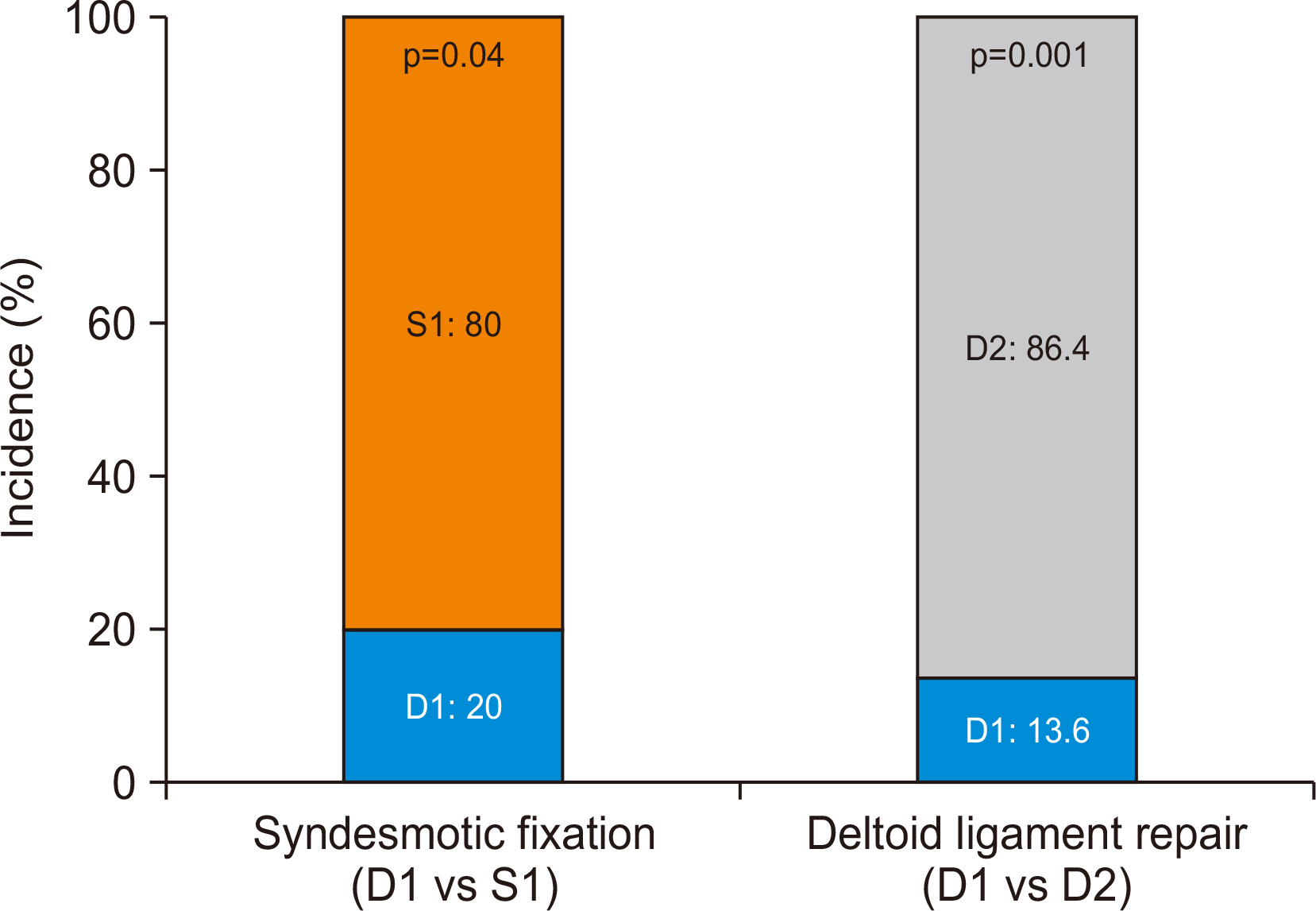
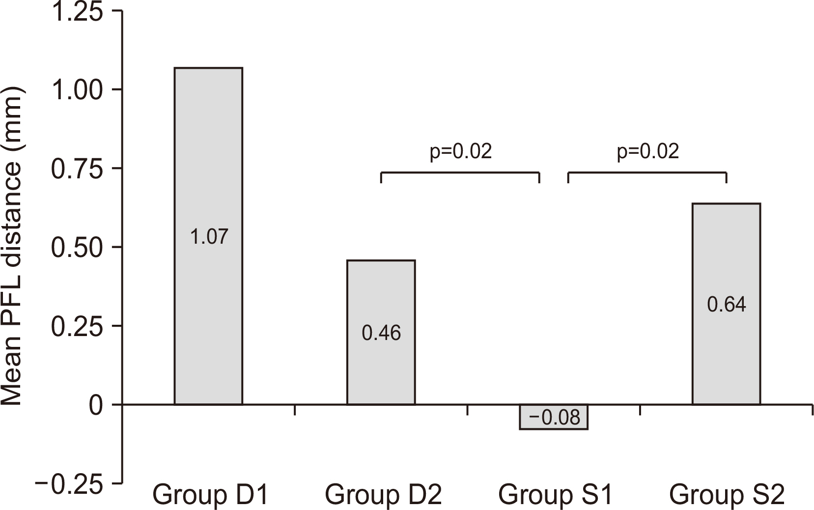
TFCS changed significantly after surgery in the D2 and S1 groups (p=0.01, p=0.03, respectively). Subgroup MCSs, TFCSs, and AFL ratios were not significantly altered by surgery in the four subgroups (p=0.82, p=0.45, p=0.25, respectively). However, postoperative PFL distances were significantly different in the D2 and S1 groups and the S1 and S2 groups (p=0.02, p=0.02, respectively). Mean TFCS decreased significantly after surgery in the D2 and S1 groups. The postoperative VAS, AOFAS scores, and FFI were not significantly different between the subgroups (p=0.44, p=0.40, and p=0.46, respectively).
Conclusion
Deltoid ligament repair seemed to restore ankle stability without addressing syndesmosis in Weber type B ankle fractures with rupture of deltoid and syndesmotic ligaments.
Keyword
Figure
Reference
-
1. Jeong MS, Choi YS, Kim YJ, Kim JS, Young KW, Jung YY. 2014; Deltoid ligament in acute ankle injury: MR imaging analysis. Skeletal Radiol. 43:655–63. doi: 10.1007/s00256-014-1842-5. DOI: 10.1007/s00256-014-1842-5. PMID: 24599341.
Article2. Tornetta P 3rd, Axelrad TW, Sibai TA, Creevy WR. 2012; Treatment of the stress positive ligamentous SE4 ankle fracture: incidence of syndesmotic injury and clinical decision making. J Orthop Trauma. 26:659–61. doi: 10.1097/BOT.0b013e31825cf39c. DOI: 10.1097/BOT.0b013e31825cf39c. PMID: 23100079.3. Brosky T, Nyland J, Nitz A, Caborn DN. 1995; The ankle ligaments: consideration of syndesmotic injury and implications for rehabilitation. J Orthop Sports Phys Ther. 21:197–205. doi: 10.2519/jospt.1995.21.4.197. DOI: 10.2519/jospt.1995.21.4.197. PMID: 7773271.
Article4. Chissell HR, Jones J. 1995; The influence of a diastasis screw on the outcome of Weber type-C ankle fractures. J Bone Joint Surg Br. 77:435–8. doi: 10.1302/0301-620X.77B3.7744931. DOI: 10.1302/0301-620X.77B3.7744931.
Article5. Kennedy JG, Soffe KE, Dalla Vedova P, Stephens MM, O'Brien T, Walsh MG, et al. 2000; Evaluation of the syndesmotic screw in low Weber C ankle fractures. J Orthop Trauma. 14:359–66. doi: 10.1097/00005131-200006000-00010. DOI: 10.1097/00005131-200006000-00010. PMID: 10926245.
Article6. Jones CR, Nunley JA 2nd. 2015; Deltoid ligament repair versus syndesmotic fixation in bimalleolar equivalent ankle fractures. J Orthop Trauma. 29:245–9. doi: 10.1097/BOT.0000000000000220. DOI: 10.1097/BOT.0000000000000220. PMID: 25186845.
Article7. Woo SH, Bae SY, Chung HJ. 2018; Short-term results of a ruptured deltoid ligament repair during an acute ankle fracture fixation. Foot Ankle Int. 39:35–45. doi: 10.1177/1071100717732383. DOI: 10.1177/1071100717732383. PMID: 29078057.
Article8. Michelson JD, Varner KE, Checcone M. Diagnosing deltoid injury in ankle fractures: the gravity stress view. Clin Orthop Relat Res. 2001; (387):178–82. doi: 10.1097/00003086-200106000-00024. DOI: 10.1097/00003086-200106000-00024. PMID: 11400880.9. Schock HJ, Pinzur M, Manion L, Stover M. 2007; The use of gravity or manual-stress radiographs in the assessment of supination-external rotation fractures of the ankle. J Bone Joint Surg Br. 89:1055–9. doi: 10.1302/0301-620X.89B8.19134. DOI: 10.1302/0301-620X.89B8.19134. PMID: 17785745.
Article10. Cotton FJ. 1924; Dislocations and joint-fractures. Ann Surg. 80:286. DOI: 10.1097/00000658-192408000-00013.
Article11. Loizou CL, Sudlow A, Collins R, Loveday D, Smith G. 2017; Radiological assessment of ankle syndesmotic reduction. Foot (Edinb). 32:39–43. doi: 10.1016/j.foot.2017.05.002. DOI: 10.1016/j.foot.2017.05.002. PMID: 28675813.
Article12. Kitaoka HB, Alexander IJ, Adelaar RS, Nunley JA, Myerson MS, Sanders M, et al. 1997; Clinical rating systems for the ankle-hindfoot, midfoot, hallux, and lesser toes. Foot Ankle Int. 18:187–8. doi: 10.1177/107110079701800315. DOI: 10.1177/107110079701800315. PMID: 28799420.
Article13. Budiman-Mak E, Conrad KJ, Roach KE. 1991; The Foot Function Index: a measure of foot pain and disability. J Clin Epidemiol. 44:561–70. doi: 10.1016/0895-4356(91)90220-4. DOI: 10.1016/0895-4356(91)90220-4. PMID: 2037861.
Article14. Earll M, Wayne J, Brodrick C, Vokshoor A, Adelaar R. 1996; Contribution of the deltoid ligament to ankle joint contact characteristics: a cadaver study. Foot Ankle Int. 17:317–24. doi: 10.1177/107110079601700604. DOI: 10.1177/107110079601700604. PMID: 8791077.
Article15. Massri-Pugin J, Lubberts B, Vopat BG, Wolf JC, DiGiovanni CW, Guss D. 2018; Role of the deltoid ligament in syndesmotic instability. Foot Ankle Int. 39:598–603. doi: 10.1177/1071100717750577. DOI: 10.1177/1071100717750577. PMID: 29320936.
Article16. Watanabe K, Kitaoka HB, Berglund LJ, Zhao KD, Kaufman KR, An KN. 2012; The role of ankle ligaments and articular geometry in stabilizing the ankle. Clin Biomech (Bristol, Avon). 27:189–95. doi: 10.1016/j.clinbiomech.2011.08.015. DOI: 10.1016/j.clinbiomech.2011.08.015. PMID: 22000065.
Article17. Klossner O. Late results of operative and non-operative treatment of severe ankle fractures. A clinical study. Acta Chir Scand Suppl. 1962; Suppl 293:1–93.18. Feller R, Borenstein T, Fantry AJ, Kellum RB, Machan JT, Nickisch F, et al. 2017; Arthroscopic quantification of syndesmotic instability in a cadaveric model. Arthroscopy. 33:436–44. doi: 10.1016/j.arthro.2016.11.008. DOI: 10.1016/j.arthro.2016.11.008. PMID: 28160934.
Article19. Burns WC 2nd, Prakash K, Adelaar R, Beaudoin A, Krause W. 1993; Tibiotalar joint dynamics: indications for the syndesmotic screw--a cadaver study. Foot Ankle. 14:153–8. doi: 10.1177/107110079301400308. DOI: 10.1177/107110079301400308. PMID: 8491430.
Article20. Massri-Pugin J, Lubberts B, Vopat BG, Guss D, Hosseini A, DiGiovanni CW. 2017; Effect of sequential sectioning of ligaments on syndesmotic instability in the coronal plane evaluated arthroscopically. Foot Ankle Int. 38:1387–93. doi: 10.1177/1071100717729492. DOI: 10.1177/1071100717729492. PMID: 28884593.
Article21. Chan KB, Lui TH. 2016; Role of ankle arthroscopy in management of acute ankle fracture. Arthroscopy. 32:2373–80. doi: 10.1016/j.arthro.2016.08.016. DOI: 10.1016/j.arthro.2016.08.016. PMID: 27816101.
Article22. Sagi HC, Shah AR, Sanders RW. 2012; The functional consequence of syndesmotic joint malreduction at a minimum 2-year follow-up. J Orthop Trauma. 26:439–43. doi: 10.1097/BOT.0b013e31822a526a. DOI: 10.1097/BOT.0b013e31822a526a. PMID: 22357084.
Article23. Candal-Couto JJ, Burrow D, Bromage S, Briggs PJ. 2004; Instability of the tibio-fibular syndesmosis: have we been pulling in the wrong direction? Injury. 35:814–8. doi: 10.1016/j.injury.2003.10.013. DOI: 10.1016/j.injury.2003.10.013. PMID: 15246807.
Article24. Hermans JJ, Wentink N, Beumer A, Hop WC, Heijboer MP, Moonen AF, et al. 2012; Correlation between radiological assessment of acute ankle fractures and syndesmotic injury on MRI. Skeletal Radiol. 41:787–801. doi: 10.1007/s00256-011-1284-2. DOI: 10.1007/s00256-011-1284-2. PMID: 22012479. PMCID: PMC3368108.
Article25. Schuberth JM, Collman DR, Rush SM, Ford LA. 2004; Deltoid ligament integrity in lateral malleolar fractures: a comparative analysis of arthroscopic and radiographic assessments. J Foot Ankle Surg. 43:20–9. doi: 10.1053/j.jfas.2003.11.005. DOI: 10.1053/j.jfas.2003.11.005. PMID: 14752760.
Article26. Cheung Y, Perrich KD, Gui J, Koval KJ, Goodwin DW. 2009; MRI of isolated distal fibular fractures with widened medial clear space on stressed radiographs: which ligaments are interrupted? AJR Am J Roentgenol. 192:W7–12. doi: 10.2214/AJR.08.1092. DOI: 10.2214/AJR.08.1092. PMID: 19098171. PMCID: PMC5585779.
Article27. Gardner MJ, Demetrakopoulos D, Briggs SM, Helfet DL, Lorich DG. 2006; Malreduction of the tibiofibular syndesmosis in ankle fractures. Foot Ankle Int. 27:788–92. doi: 10.1177/107110070602701005. DOI: 10.1177/107110070602701005. PMID: 17054878.
Article28. Song DJ, Lanzi JT, Groth AT, Drake M, Orchowski JR, Shaha SH, et al. 2014; The effect of syndesmosis screw removal on the reduction of the distal tibiofibular joint: a prospective radiographic study. Foot Ankle Int. 35:543–8. doi: 10.1177/1071100714524552. DOI: 10.1177/1071100714524552. PMID: 24532699.29. Manjoo A, Sanders DW, Tieszer C, MacLeod MD. 2010; Functional and radiographic results of patients with syndesmotic screw fixation: implications for screw removal. J Orthop Trauma. 24:2–6. doi: 10.1097/BOT.0b013e3181a9f7a5. DOI: 10.1097/BOT.0b013e3181a9f7a5. PMID: 20035170.
Article
- Full Text Links
- Actions
-
Cited
- CITED
-
- Close
- Share
- Similar articles
-
- Diagnosis and Management of Suspected Deltoid Injury
- A Clinical Study of Disruption of the Deltoid Ligament Associated with Fractures of Distal Fibula
- The Effect of Syndesmotic Screw of Ankle Fracture with Distal Tibiofibular Diastasis
- Comparative Analysis of Trans-syndesmotic Versus Non-syndesmotic Screw Fixation in Surgical Treatment of Ankle Fracture with Diastasis
- Treatment of the Deltoid Ligament Injury in Ankle Fractures Combined with Distal Tibiofibular Diastasis: retrospective study

