Clin Endosc.
2023 May;56(3):388-390. 10.5946/ce.2023.049.
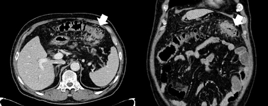
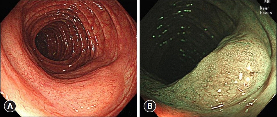
A focally flat-elevated lesion in distal transverse colon resembling a subepithelial tumor
- Affiliations
-
- 1Division of Gastroenterology, Department of Internal Medicine, Daejeon St. Mary’s Hospital, College of Medicine, The Catholic University of Korea, Daejeon, Korea
- KMID: 2542468
- DOI: http://doi.org/10.5946/ce.2023.049
Figure
Reference
-
1. Crump M, Gospodarowicz M, Shepherd FA. Lymphoma of the gastrointestinal tract. Semin Oncol. 1999; 26:324–337.2. Hollie N, Asakrah S. MALT lymphoma of the colon: a clinicopathological review. J Clin Pathol. 2020; 73:378–383.3. Kim MH, Jung JT, Kim EJ, et al. A case of mucosa-associated lymphoid tissue lymphoma of the sigmoid colon presenting as a semipedunculated polyp. Clin Endosc. 2014; 47:192–196.4. Abbas H, Niazi M, Makker J. Mucosa-associated lymphoid tissue (MALT) lymphoma of the colon: a case report and a literature review. Am J Case Rep. 2017; 18:491–497.5. Akasaka R, Chiba T, Dutta AK, et al. Colonic mucosa-associated lymphoid tissue lymphoma. Case Rep Gastroenterol. 2012; 6:569–575.6. Cho J. Basic immunohistochemistry for lymphoma diagnosis. Blood Res. 2022; 57(S1):55–61.
- Full Text Links
- Actions
-
Cited
- CITED
-
- Close
- Share
- Similar articles
-
- A Case of Early Gastric Adenocarcinoma Resembling Subepithelial Tumor
- Adenosquamous Carcinoma in Distal Transverse Colon in a 72-Year-Old Female Patient
- A Case of Malignant Duodenocolic Fistula Diagnosed by Endoscopy
- A Caae of Gastrocolie Fistula Secondary to Benign Gastric Ulcer
- Short-term Clinico-pathological Outcomes of a Laparoscopic Transverse Colectomy for Transverse Colon Cancer


