Percutaneous creation of a choledocho-choledochostomy for intractable iatrogenic bile duct injury
- Affiliations
-
- 1School of Medicine, Duke University, Durham, NC, USA
- 2Department of Radiology, Duke University, Durham, NC, USA
- 3Department of Surgery, Duke University, Durham, NC, USA
- KMID: 2542467
- DOI: http://doi.org/10.5946/ce.2022.098
Figure
-
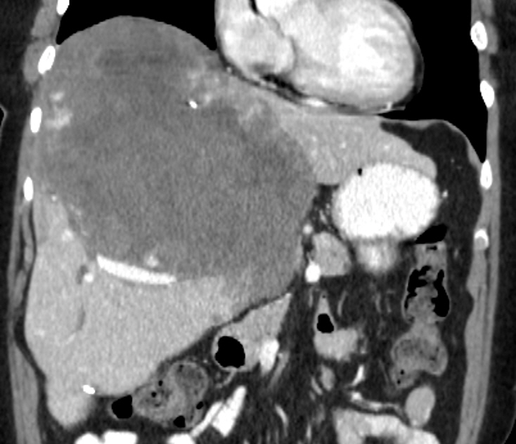
Fig. 1. Coronal reconstruction of computerized tomography with iodinated intravenous contrast demonstrates a large, cavernous hemangioma of the liver prior to extended right partial hepatectomy.

Fig. 2. (A) Contrast injection of the surgical drain (arrowheads) post-operatively reveals a large biloma (black arrow) communicating with the left biliary tree. The tip of the endoscopically placed biliary stent (open arrow) is located in the stump of the left main bile duct. (B) Endoscopic retrograde cholangiopancreatography demonstrates the common hepatic duct (black arrow) and the stump of the left main bile duct (open arrow). The percutaneous biliary drainage catheter (arrowheads) lies within the left biliary tree and terminates in the biloma. (C) Contrast injection of the left biliary tree reveales the isolated left main bile duct (black arrow) terminating at the surgical staple line. The surgical drain was replaced with a 16 French straight drainage catheter (open arrow).
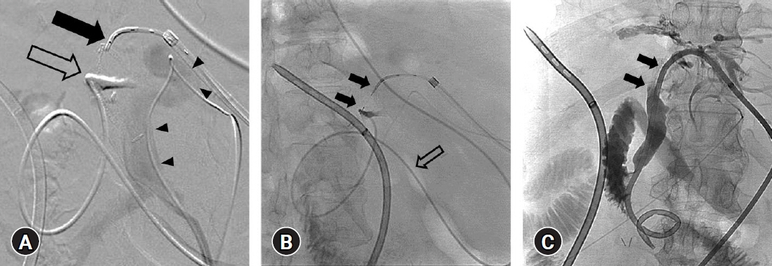
Fig. 3. (A) Digitally subtracted portal venogram through a percutaneous 3 French catheter (arrowheads) demonstrates the safe distance between the left main portal vein and the planned trajectory of the radiofrequency PowerWire (Baylis Medical, black arrow) in the isolated left biliary tree and the nasobiliary drain (open arrow) in the left main duct stump. A safe distance from the hepatic artery was confirmed on the arterial phase of computed tomography with iodinated contrast. (B) The radiofrequency PowerWire (black arrows) passes from the excluded left biliary tree to the stump of the left main duct, opacified with iodinated contrast injected through a nasobiliary drain (open arrow). A 3 French catheter lies within the left portal venous system. (C) A 10 French percutaneous biliary drainage catheter passes from the left biliary tree through the newly created choledocho-choledochostomy (black arrows) and into the duodenum.
Reference
-
1. Günther RW, Schild H, Thelen M. Percutaneous transhepatic biliary drainage: experience with 311 procedures. Cardiovasc Intervent Radiol. 1988; 11:65–71.2. Ball CG, Lillemoe KD. Chapter 113. Prevention and management of bile duct injury. In : Yeo CJ, editor. Shackelford's surgery of the alimentary tract. 2 volume set. 8th ed. Elsevier;2019. p. 1340–1351.3. Sicklick JK, Camp MS, Lillemoe KD, et al. Surgical management of bile duct injuries sustained during laparoscopic cholecystectomy: perioperative results in 200 patients. Ann Surg. 2005; 241:786–792. discussion 793-795.4. Habibollahi P, Benjamin JL, X Bai H, et al. Percutaneous fluoroscopic-guided creation of neoanastomosis for the treatment of biliary occlusions. Cardiovasc Intervent Radiol. 2020; 43:1671–1678.5. McCarthy CJ, Thabet A, Yamada K, et al. Percutaneous creation of biliary-enteric neoanastomosis for anastomotic biliary occlusion following living donor liver transplantation. Liver Transpl. 2017; 23:262–265.6. Robins C, Xiao N, Salem R, et al. Percutaneous biliary neo-anastomosis or neo-duct creation using radiofrequency wires. Cardiovasc Intervent Radiol. 2022; 45:337–343.7. Tocchi A, Mazzoni G, Liotta G, et al. Management of benign biliary strictures: biliary enteric anastomosis vs endoscopic stenting. Arch Surg. 2000; 135:153–157.8. Riaz A, Salem R. Future directions of percutaneous biliary interventions. Semin Intervent Radiol. 2021; 38:373–376.
- Full Text Links
-
- Actions
-
Cited
- CITED
-
- Close
- Share
-
- Similar articles
-
- Interventional Treatment for a Choledocho-Colonic Fistula Due to Bile Duct Injury after Laparoscopic Cholecystectomy: A Case Report
- Transection of Distal Common Bile Duct by Bike Handlebar in a Child
- Successful non-surgical treatment for isolated right anterior section bile duct injury following laparoscopic cholecystectomy: Report of a case
- A Case of Mucin - hypersecreting Tumors of Pancreas and Bile Duct with Inflammatory Choledocho - pancreatic Fistula
- Recent classifications of the common bile duct injury
