J Yeungnam Med Sci.
2023 Apr;40(2):212-217. 10.12701/jyms.2022.00479.
Transient osteoporosis of the hip with a femoral neck fracture during follow-up: a case report
- Affiliations
-
- 1Department of Orthopedic Surgery, Mitsuhashi Hospital, Chiba, Japan
- 2Department of Orthopedic Surgery, Musashino General Hospital, Kawagoe, Japan
- 3Department of Orthopedic Surgery, Sekishindo Hospital, Kawagoe, Japan
- 4Department of Orthopedic Surgery, Nippon Medical School Hospital, Tokyo, Japan
- KMID: 2541950
- DOI: http://doi.org/10.12701/jyms.2022.00479
Abstract
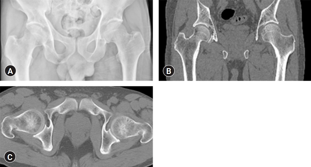
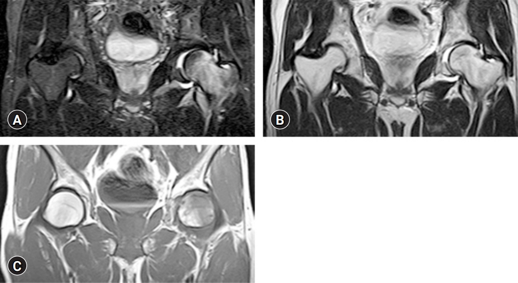
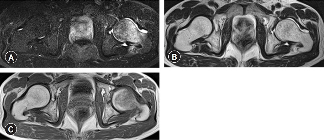
- We report a case of transient osteoporosis of the hip with a femoral neck fracture found during follow-up. A 53-year-old man presented with left hip pain without trauma. The pain did not improve after 2 weeks and he was brought to our hospital by ambulance. Magnetic resonance imaging (MRI) of the left hip joint showed diffuse edema in the bone marrow, which was identified by low signal intensity on T1-weighted images, high signal intensity on T2-weighted images, and increased signal intensity on short tau inversion recovery. This edema extended from the femoral head and neck to the intertrochanteric area. He was diagnosed with transient osteoporosis of the left hip. Rest gradually improved his pain; however, 3 weeks later, his left hip pain worsened without trauma. X-ray, computed tomography, and MRI results of the hip joint demonstrated a left femoral neck fracture, and osteosynthesis was performed. Differential diagnoses included avascular necrosis of the femoral head, infection, complex regional pain syndrome, rheumatoid arthritis, leukemia, and other cancers. Transient osteoporosis of the hip generally has a good prognosis with spontaneous remission within a few months to 1 year. However, a sufficient length of follow-up from condition onset to full recovery is necessary to avoid all probable complications such as fractures.
Keyword
Figure
Reference
-
References
1. Curtiss PH Jr, Kincaid WE. Transitory demineralization of the hip in pregnancy: a report of three cases. J Bone Joint Surg Am. 1959; 41-A:1327–33.2. Van Wagenen K, Pritchard P, Taylor JA. Transient osteoporosis of the hip: a case report. J Can Chiropr Assoc. 2013; 57:116–22.3. Ma FY, Falkenberg M. Case reports: transient osteoporosis of the hip: an atypical case. Clin Orthop Relat Res. 2006; 445:245–9.4. Asadipooya K, Graves L, Greene LW. Transient osteoporosis of the hip: review of the literature. Osteoporos Int. 2017; 28:1805–16.5. Rosen RA. Transitory demineralization of the femoral head. Radiology. 1970; 94:509–12.6. Plenk H Jr, Hofmann S, Eschberger J, Gstettner M, Kramer J, Schneider W, et al. Histomorphology and bone morphometry of the bone marrow edema syndrome of the hip. Clin Orthop Relat Res. 1997; (334):73–84.7. Koo KH, Jeong ST, Jones JP Jr. Borderline necrosis of the femoral head. Clin Orthop Relat Res. 1999; (358):158–65.8. Manara M, Varenna M. A clinical overview of bone marrow edema. Reumatismo. 2014; 66:184–96.9. Orth P, Anagnostakos K. Coagulation abnormalities in osteonecrosis and bone marrow edema syndrome. Orthopedics. 2013; 36:290–300.10. Diwanji SR, Cho YJ, Xin ZF, Yoon TR. Conservative treatment for transient osteoporosis of the hip in middle-aged women. Singapore Med J. 2008; 49:e17–21.11. Daniel RS, Farrar EK, Norton HR, Nussbaum AI. Bilateral transient osteoporosis of the talus in pregnancy. Osteoporos Int. 2009; 20:1973–5.12. Jennings PE, O’Malley BP, Griffin KE, Northover B, Rosenthal FD. Relevance of increased serum thyroxine concentrations associated with normal serum triiodothyronine values in hypothyroid patients receiving thyroxine: a case for “tissue thyrotoxicosis”. Br Med J (Clin Res Ed). 1984; 289:1645–7.13. Dunstan CR, Evans RA, Somers NM. Bone death in transient regional osteoporosis. Bone. 1992; 13:161–5.14. Flores-Robles BJ, Sanz-Sanz J, Sanabria-Sanchinel AA, Huntley-Pascual D, Andréu Sánchez JL, Campos Esteban J, et al. Zoledronic acid treatment in primary bone marrow edema syndrome. J Pain Palliat Care Pharmacother. 2017; 31:52–6.15. Emad Y, Ragab Y, El-Shaarawy N, Rasker JJ. Transient osteoporosis of the hip, complete resolution after treatment with alendronate as observed by MRI description of eight cases and review of the literature. Clin Rheumatol. 2012; 31:1641–7.16. Kibbi L, Touma Z, Khoury N, Arayssi T. Oral bisphosphonates in treatment of transient osteoporosis. Clin Rheumatol. 2008; 27:529–32.17. Schapira D, Braun Moscovici Y, Gutierrez G, Nahir AM. Severe transient osteoporosis of the hip during pregnancy: successful treatment with intravenous biphosphonates. Clin Exp Rheumatol. 2003; 21:107–10.18. Yagishita K, Jinno T, Koga D, Kato T, Enomoto M, Kato T, et al. Transient osteoporosis of the hip treated with hyperbaric oxygen therapy: a case series. Undersea Hyperb Med. 2016; 43:847–54.19. Bashaireh KM, Aldarwish FM, Al-Omari AA, Albashaireh MA, Hajjat M, Al-Ebbini MA, et al. Transient osteoporosis of the hip: risk and therapy. Open Access Rheumatol. 2020; 12:1–8.20. Hadji P, Boekhoff J, Hahn M, Hellmeyer L, Hars O, Kyvernitakis I. Pregnancy-associated transient osteoporosis of the hip: results of a case-control study. Arch Osteoporos. 2017; 12:11.
- Full Text Links
- Actions
-
Cited
- CITED
-
- Close
- Share
- Similar articles
-
- Osteoporotic Hip Fracture: How We Make Better Results?
- The Relationship between the Variation of the femoral neckshaft angle according to Age and the Fracture of the Hip
- Central Fracture-dislocation of the Hip Combined with Ipsilateral Femoral Neck Fracture: A Case Report
- Cementless Bipolar Hemiarthroplasty for Hip Fracture in Patients More than Seventy Years Old with Osteoporosis
- Primary Total Hip Replacement in the Lower Limb Amputees


