Accuracy of three-dimensional periodontal ligament models generated using cone-beam computed tomography at different resolutions for the assessment of periodontal bone loss
- Affiliations
-
- 1Department of Orthodontics, National Engineering Laboratory for Digital and Material Technology of Stomatology; Beijing Key Laboratory of Digital Stomatology, Peking University School and Hospital of Stomatology, Beijing, China
- 2Department of Periodontology, National Engineering Laboratory for Digital and Material Technology of Stomatology; Beijing Key Laboratory of Digital Stomatology, Peking University School and Hospital of Stomatology, Beijing, China
- 3Department of Oral and Maxillofacial Surgery, National Engineering Laboratory for Digital and Material Technology of Stomatology; Beijing Key Laboratory of Digital Stomatology, Peking University School and Hospital of Stomatology, Beijing, China
- 4Center of Digital Dentistry, Peking University School and Hospital of Stomatology; National Engineering Laboratory for Digital and Material Technology of Stomatology; Research Center of Engineering and Technology for Digital Dentistry of Ministry of Health; Beijing Key Laboratory of Digital Stomatology, Peking University School and Hospital of Stomatology, Beijing, China
- KMID: 2540947
- DOI: http://doi.org/10.4041/kjod22.120
Abstract
Objective
To develop a method for generating three-dimensional (3D) digital models of the periodontal ligament (PDL) using 3D cone-beam computed tomography (CBCT) reconstruction and to evaluate the accuracy and agreement of the 3D PDL models in the measurement of periodontal bone loss.
Methods
CBCT data collected from four patients with skeletal Class III malocclusion prior to periodontal surgery were reconstructed at three voxel sizes (0.2 mm, 0.25 mm, and 0.3 mm), and 3D tooth and alveolar bone models were generated to obtain digital PDL models for the maxillary and mandibular anterior teeth. Linear measurements of the alveolar bone crest obtained during periodontal surgery were compared with the digital measurements for assessment of the accuracy of the digital models. The agreement and reliability of the digital PDL models were analyzed using intra- and interexaminer correlation coefficients and Bland–Altman plots.
Results
Digital models of the maxillary and mandibular anterior teeth, PDL, and alveolar bone of the four patients were successfully established. Relative to the intraoperative measurements, linear measurements obtained from the 3D digital models were accurate, and there were no significant differences among different voxel sizes at different sites. High diagnostic coincidence rates were found for the maxillary anterior teeth. The digital models showed high intra- and interexaminer agreement.
Conclusions
Digital PDL models generated by 3D CBCT reconstruction can provide accurate and useful information regarding the alveolar crest morphology and facilitate reproducible measurements. This could assist clinicians in the evaluation of periodontal prognosis and establishment of an appropriate orthodontic treatment plan.
Figure
-
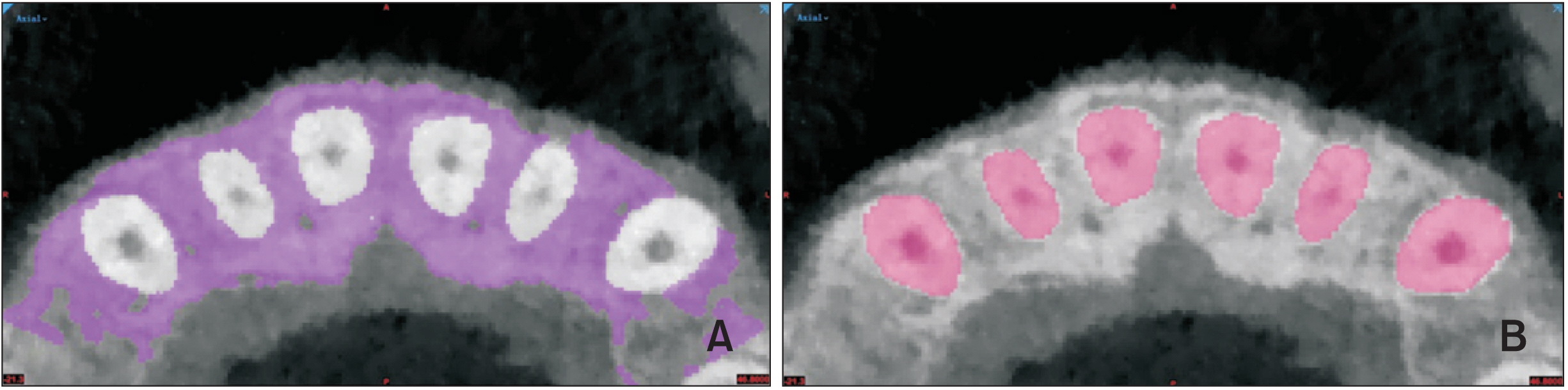
Figure 1 Process of alveolar bone and tooth segmentation in Mimics 19.0 software (Materialise, Leuven, Belgium). A, Manual alveolar bone segmentation. B, Manual tooth segmentation.

Figure 2 Digital tooth, bone, and soft tissue models in Geomagic (Geomagic, Cary, NC, USA). A, Digital tooth models with an intraoral scan superimposed over the crowns. B, Digital tooth and bone models. C, Digital tooth and bone models generated from cone-beam computed tomography datasets and a soft tissue model derived from an intraoral scan.

Figure 3 Digital linear measurements. The measurment referece lines at midlabial, distobracket, and distolabial sites are shown (white dotted line). A, Vertical bone level measurements (black arrow line) at mesiolabial, mesiobracket, midlabial, distobracket, and distolabial sites. B, Bone–bracket distance measurements (black arrow line) at mesiobracket, midlabial, and distobracket sites.
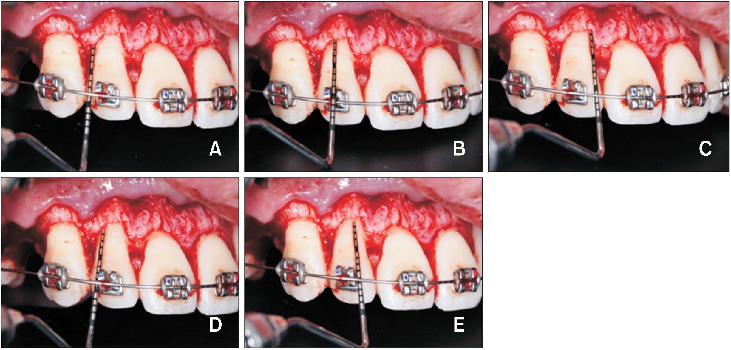
Figure 4 Intraoperative linear measurements. A–C, Linear measurements at the distolabial (A), midlabial (B), and mesiolabial (C) sites. D, E, Linear measurements at the distobracket (D) and mesiobracket (E) sites.

Figure 5 Establishment of digital periodontal ligament (PDL) models. A, Tooth and alveolar bone digital models. B, The alveolar bone crests are drawn on the tooth models. C, The curves of the alveolar bone crests were extracted. D, The tooth models are separated along the curves of the alveolar bone crests. E, The digital PDL models. F, Computation of the PDL area.

Figure 6 Bland–Altman plots of intraoperative and digital linear measurements with different cone-beam computed tomography (CBCT) voxel sizes. The difference against the mean and the limits of agreement are shown. A–C, Vertical bone level (VBL) measurements for the maxillary anterior teeth with different CBCT voxel sizes. D–F, Bone–bracket distance (BBD) measurements for the maxillary anterior teeth with different CBCT voxel sizes. G–I, VBL measurements for the mandibular anterior teeth with different CBCT voxel sizes. J–L, BBD measurements for the mandibular anterior teeth with different CBCT voxel sizes.

Figure 7 Bland–Altman plots of periodontal ligament (PDL) area measurements by examiner 1 and examiner 2.
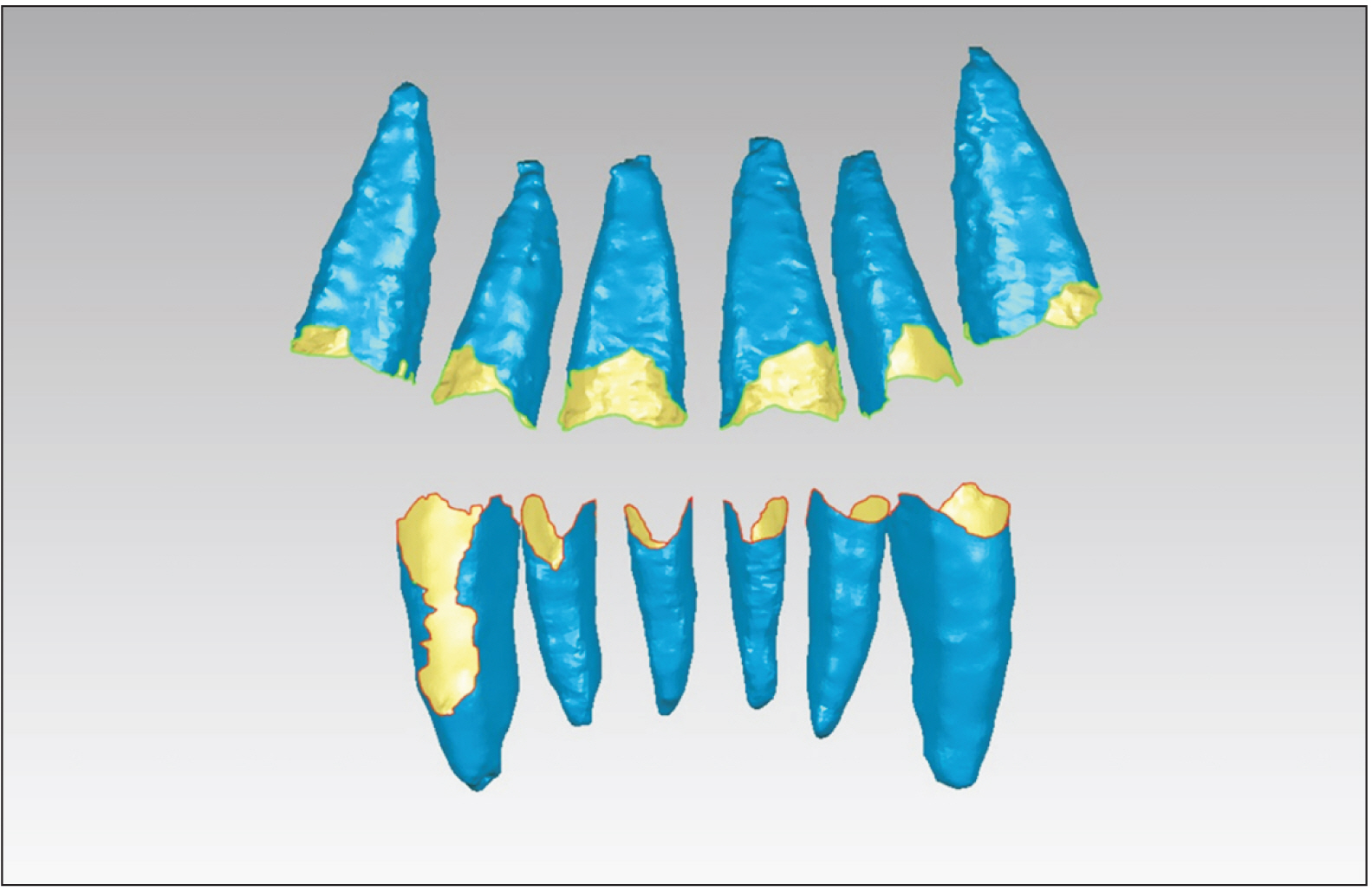
Figure 8 Periodontal ligament models generated for a representative case. The periodontal ligament models of maxillary and mandibular anterior teeth for one of the four patients are shown in Geomagic (Geomagic, Cary, NC, USA).
Reference
-
1. Beertsen W, McCulloch CA, Sodek J. 1997; The periodontal ligament: a unique, multifunctional connective tissue. Periodontol 2000. 13:20–40. DOI: 10.1111/j.1600-0757.1997.tb00094.x. PMID: 9567922.2. de Jong T, Bakker AD, Everts V, Smit TH. 2017; The intricate anatomy of the periodontal ligament and its development: lessons for periodontal regeneration. J Periodontal Res. 52:965–74. DOI: 10.1111/jre.12477. PMID: 28635007.3. Jiang N, Guo W, Chen M, Zheng Y, Zhou J, Kim SG, et al. 2016; Periodontal ligament and alveolar bone in health and adaptation: tooth movement. Front Oral Biol. 18:1–8. DOI: 10.1159/000351894. PMID: 26599112. PMCID: PMC4662052.4. Proffit WR, Fields HW, Larson B, Sarver DM. 2018. Contemporary orthodontics. 6th ed. Elsevier;Philadelphia: DOI: 10.1053/j.sodo.2018.10.005.5. Park SB, An SY, Han WJ, Park JT. 2017; Three-dimensional measurement of periodontal surface area for quantifying inflammatory burden. J Periodontal Implant Sci. 47:154–64. DOI: 10.5051/jpis.2017.47.3.154. PMID: 28680711. PMCID: PMC5494310.6. Lee S, Hwang S, Jang W, Choi YJ, Chung CJ, Kim KH. 2018; Assessment of lower incisor alveolar bone width using cone-beam computed tomography images in skeletal Class III adults of different vertical patterns. Korean J Orthod. 48:349–56. DOI: 10.4041/kjod.2018.48.6.349. PMID: 30450327. PMCID: PMC6234113.7. Kook YA, Kim G, Kim Y. 2012; Comparison of alveolar bone loss around incisors in normal occlusion samples and surgical skeletal class III patients. Angle Orthod. 82:645–52. DOI: 10.2319/070111-424.1. PMID: 22129151. PMCID: PMC8845546.8. Fleiner J, Hannig C, Schulze D, Stricker A, Jacobs R. 2013; Digital method for quantification of circumferential periodontal bone level using cone beam CT. Clin Oral Investig. 17:389–96. DOI: 10.1007/s00784-012-0715-3. PMID: 22431146.9. Hong HH, Hong A, Huang YF, Liu HL. 2018; Incompatible amount of 3-D and 2-D periodontal attachments on micro-CT scanned premolars. PLoS One. 13:e0193894. Erratum in: PLoS One 2018;13:e0209206. DOI: 10.1371/journal.pone.0209206. PMID: 30532186. PMCID: PMC6287807. PMID: 1693ad5ce4a447f69625152477eab30b.10. Hong HH, Chang CC, Hong A, Liu HL, Wang YL, Chang SH, et al. 2017; Decreased amount of supporting alveolar bone at single-rooted premolars is under estimated by 2D examinations. Sci Rep. 8:45774. DOI: 10.1038/srep45774. PMID: 28367999. PMCID: PMC5377944.11. Klock KS, Gjerdet NR, Haugejorden O. 1993; Periodontal attachment loss assessed by linear and area measurements in vitro. J Clin Periodontol. 20:443–7. DOI: 10.1111/j.1600-051X.1993.tb00386.x. PMID: 8349835.12. Gu Y, Tang Y, Zhu Q, Feng X. 2016; Measurement of root surface area of permanent teeth with root variations in a Chinese population-a micro-CT analysis. Arch Oral Biol. 63:75–81. DOI: 10.1016/j.archoralbio.2015.12.001. PMID: 26723016.13. Al-Rawi B, Hassan B, Vandenberge B, Jacobs R. 2010; Accuracy assessment of three-dimensional surface reconstructions of teeth from cone beam computed tomography scans. J Oral Rehabil. 37:352–8. DOI: 10.1111/j.1365-2842.2010.02065.x. PMID: 20180895.14. Wang Y, He S, Guo Y, Wang S, Chen S. 2013; Accuracy of volumetric measurement of simulated root resorption lacunas based on cone beam computed tomography. Orthod Craniofac Res. 16:169–76. DOI: 10.1111/ocr.12016. PMID: 23419069.15. Tasanapanont J, Apisariyakul J, Wattanachai T, Sriwilas P, Midtbø M, Jotikasthira D. 2017; Comparison of 2 root surface area measurement methods: 3-dimensional laser scanning and cone-beam computed tomography. Imaging Sci Dent. 47:117–22. DOI: 10.5624/isd.2017.47.2.117. PMID: 28680848. PMCID: PMC5489667.16. Palkovics D, Mangano FG, Nagy K, Windisch P. 2020; Digital three-dimensional visualization of intrabony periodontal defects for regenerative surgical treatment planning. BMC Oral Health. 20:351. DOI: 10.1186/s12903-020-01342-w. PMID: 33261592. PMCID: PMC7709443. PMID: 9abcedf320a6445da164dc6f9aa2b879.17. Tayman MA, Kamburoğlu K, Küçük Ö, Ateş FSÖ, Günhan M. 2019; Comparison of linear and volumetric measurements obtained from periodontal defects by using cone beam-CT and micro-CT: an in vitro study. Clin Oral Investig. 23:2235–44. DOI: 10.1007/s00784-018-2665-x. PMID: 30284102.18. Mito T, Sato K, Mitani H. 2002; Cervical vertebral bone age in girls. Am J Orthod Dentofacial Orthop. 122:380–5. DOI: 10.1067/mod.2002.126896. PMID: 12411883.19. Vasir NS, Thompson RT, Davies TM. 1991; Dental and skeletal changes following sagittal split osteotomy for correction of mandibular prognathism. Eur J Orthod. 13:134–42. DOI: 10.1093/ejo/13.2.134. PMID: 2055252.20. Forst D, Nijjar S, Flores-Mir C, Carey J, Secanell M, Lagravere M. 2014; Comparison of in vivo 3D cone-beam computed tomography tooth volume measurement protocols. Prog Orthod. 15:69. DOI: 10.1186/s40510-014-0069-2. PMID: 25534123. PMCID: PMC4274349.21. Jing WD, Jiao J, Xu L, Hou JX, Li XT, Wang XX, et al. 2020; Periodontal soft- and hard-tissue changes after augmented corticotomy in Chinese adult patients with skeletal Angle Class III malocclusion: a non-randomized controlled trial. J Periodontol. 91:1419–28. DOI: 10.1002/JPER.19-0522. PMID: 32149391.22. Scarfe WC, Azevedo B, Pinheiro LR, Priaminiarti M, Sales MAO. 2017; The emerging role of maxillofacial radiology in the diagnosis and management of patients with complex periodontitis. Periodontol 2000. 74:116–39. DOI: 10.1111/prd.12193. PMID: 28429477.23. Kim DM, Bassir SH. 2017; When is cone-beam computed tomography imaging appropriate for diagnostic inquiry in the management of inflammatory periodontitis? An American Academy of Periodontology best evidence review. J Periodontol. 88:978–98. DOI: 10.1902/jop.2017.160505. PMID: 28967334.24. Choi IGG, Cortes ARG, Arita ES, Georgetti MAP. 2018; Comparison of conventional imaging techniques and CBCT for periodontal evaluation: a systematic review. Imaging Sci Dent. 48:79–86. DOI: 10.5624/isd.2018.48.2.79. PMID: 29963478. PMCID: PMC6015929.25. Spin-Neto R, Gotfredsen E, Wenzel A. 2013; Impact of voxel size variation on CBCT-based diagnostic outcome in dentistry: a systematic review. J Digit Imaging. 26:813–20. DOI: 10.1007/s10278-012-9562-7. PMID: 23254628. PMCID: PMC3705012.26. Maret D, Telmon N, Peters OA, Lepage B, Treil J, Inglèse JM, et al. 2012; Effect of voxel size on the accuracy of 3D reconstructions with cone beam CT. Dentomaxillofac Radiol. 41:649–55. DOI: 10.1259/dmfr/81804525. PMID: 23166362. PMCID: PMC3528196.27. Dong T, Yuan L, Liu L, Qian Y, Xia L, Ye N, et al. 2019; Detection of alveolar bone defects with three different voxel sizes of cone-beam computed tomography: an in vitro study. Sci Rep. 9:8146. DOI: 10.1038/s41598-019-44675-5. PMID: 31148581. PMCID: PMC6544761.28. Sang YH, Hu HC, Lu SH, Wu YW, Li WR, Tang ZH. 2016; Accuracy assessment of three-dimensional surface reconstructions of in vivo teeth from cone-beam computed tomography. Chin Med J (Engl). 129:1464–70. DOI: 10.4103/0366-6999.183430. PMID: 27270544. PMCID: PMC4910372. PMID: fbc5a617cf2f4d249b6cc807b0b034a7.29. Patcas R, Müller L, Ullrich O, Peltomäki T. 2012; Accuracy of cone-beam computed tomography at different resolutions assessed on the bony covering of the mandibular anterior teeth. Am J Orthod Dentofacial Orthop. 141:41–50. DOI: 10.1016/j.ajodo.2011.06.034. PMID: 22196184.30. Sun Z, Smith T, Kortam S, Kim DG, Tee BC, Fields H. 2011; Effect of bone thickness on alveolar bone-height measurements from cone-beam computed tomography images. Am J Orthod Dentofacial Orthop. 139:e117–27. DOI: 10.1016/j.ajodo.2010.08.016. PMID: 21300222.31. Wood R, Sun Z, Chaudhry J, Tee BC, Kim DG, Leblebicioglu B, et al. 2013; Factors affecting the accuracy of buccal alveolar bone height measurements from cone-beam computed tomography images. Am J Orthod Dentofacial Orthop. 143:353–63. DOI: 10.1016/j.ajodo.2012.10.019. PMID: 23452969.32. de-Azevedo-Vaz SL, Vasconcelos Kde F, Neves FS, Melo SL, Campos PS, Haiter-Neto F. 2013; Detection of periimplant fenestration and dehiscence with the use of two scan modes and the smallest voxel sizes of a cone-beam computed tomography device. Oral Surg Oral Med Oral Pathol Oral Radiol. 115:121–7. DOI: 10.1016/j.oooo.2012.10.003. PMID: 23217543.33. Icen M, Orhan K, Şeker Ç, Geduk G, Cakmak Özlü F, Cengiz Mİ. 2020; Comparison of CBCT with different voxel sizes and intraoral scanner for detection of periodontal defects: an in vitro study. Dentomaxillofac Radiol. 49:20190197. DOI: 10.1259/dmfr.20190197. PMID: 32134338. PMCID: PMC7333464.34. Pauwels R, Faruangsaeng T, Charoenkarn T, Ngonphloy N, Panmekiate S. 2015; Effect of exposure parameters and voxel size on bone structure analysis in CBCT. Dentomaxillofac Radiol. 44:20150078. DOI: 10.1259/dmfr.20150078. PMID: 26054572. PMCID: PMC4628422.35. Harris BT, Montero D, Grant GT, Morton D, Llop DR, Lin WS. 2017; Creation of a 3-dimensional virtual dental patient for computer-guided surgery and CAD-CAM interim complete removable and fixed dental prostheses: a clinical report. J Prosthet Dent. 117:197–204. DOI: 10.1016/j.prosdent.2016.06.012. PMID: 27666493.36. Lim SW, Moon RJ, Kim MS, Oh MH, Lee KM, Hwang HS, et al. 2020; Construction reproducibility of a composite tooth model composed of an intraoral-scanned crown and a cone-beam computed tomography-scanned root. Korean J Orthod. 50:229–37. DOI: 10.4041/kjod.2020.50.4.229. PMID: 32632042. PMCID: PMC7369385.37. Lee RJ, Weissheimer A, Pham J, Go L, de Menezes LM, Redmond WR, et al. 2015; Three-dimensional monitoring of root movement during orthodontic treatment. Am J Orthod Dentofacial Orthop. 147:132–42. DOI: 10.1016/j.ajodo.2014.10.010. PMID: 25533080.38. Lee RJ, Pham J, Choy M, Weissheimer A, Dougherty HL Jr, Sameshima GT, et al. 2014; Monitoring of typodont root movement via crown superimposition of single cone-beam computed tomography and consecutive intraoral scans. Am J Orthod Dentofacial Orthop. 145:399–409. DOI: 10.1016/j.ajodo.2013.12.011. PMID: 24582031.39. Wang T, Pei X, Luo F, Jia L, Qin H, Cheng X, et al. 2017; Evaluation of tooth root surface area using a three-dimensional scanning technique and cone beam computed tomographic reconstruction in vitro. Arch Oral Biol. 84:13–8. DOI: 10.1016/j.archoralbio.2017.07.014. PMID: 28934648.40. Kuralt M, Gašperšič R, Fidler A. 2021; The precision of gingival recession measurements is increased by an automated curvature analysis method. BMC Oral Health. 21:505. DOI: 10.1186/s12903-021-01858-9. PMID: 34620155. PMCID: PMC8499415. PMID: 49aef6cf0c0c40a7b638220944d155a8.41. Pini-Prato G, Franceschi D, Cairo F, Nieri M, Rotundo R. 2010; Classification of dental surface defects in areas of gingival recession. J Periodontol. 81:885–90. DOI: 10.1902/jop.2010.090631. PMID: 20450362.42. Maret D, Peters OA, Galibourg A, Dumoncel J, Esclassan R, Kahn JL, et al. 2014; Comparison of the accuracy of 3-dimensional cone-beam computed tomography and micro-computed tomography reconstructions by using different voxel sizes. J Endod. 40:1321–6. DOI: 10.1016/j.joen.2014.04.014. PMID: 25146011.43. Gu Y, Zhu Q, Tang Y, Zhang Y, Feng X. 2017; Measurement of root surface area of permanent teeth in a Chinese population. Arch Oral Biol. 81:26–30. DOI: 10.1016/j.archoralbio.2017.04.015. PMID: 28460250.44. Damstra J, Fourie Z, Huddleston Slater JJ, Ren Y. 2010; Accuracy of linear measurements from cone-beam computed tomography-derived surface models of different voxel sizes. Am J Orthod Dentofacial Orthop. 137:16.e1–6. discussion 16–7. DOI: 10.1016/j.ajodo.2009.06.016. PMID: 20122425.
- Full Text Links
-
- Actions
-
Cited
- CITED
-
- Close
- Share
-
- Similar articles
-
- Comparison of conventional imaging techniques and CBCT for periodontal evaluation: A systematic review
- Optimizing the reconstruction filter in cone-beam CT to improve periodontal ligament space visualization: An in vitro study
- Commentary on "Reliability of two different presurgical preparation methods for implant dentistry based on panoramic radiography and cone-beam computed tomography in cadavers"
- The accuracy of the imaging reformation of cone beam computed tomography for the assessment of bone defect healing
- Quantification of Microstructures in Mice Alveolar Bone using Micro-computed tomography (microCT)
