J Liver Cancer.
2023 Mar;23(1):189-201. 10.17998/jlc.2023.03.11.
Current status of ultrasonography in national cancer surveillance program for hepatocellular carcinoma in South Korea: a large-scale multicenter study
- Yoo SH
 1
1 - Kim SS2
- Kim SG3
- Kwon JH1
- Lee HA4
- Seo YS5
- Jung YK6
- Yim HJ6
- Song DS7
- Kang SH6
- Kim MY8
- Ahn YH2
- Han J2
- Kim YS3
- Chang Y9
- Jeong SW9
- Jang JY9
- Yoo JJ3
- Affiliations
-
- 1Department of Internal Medicine, Incheon St. Mary’s Hospital, The Catholic University of Korea, Incheon, Korea
- 2Department of Internal Medicine, Ajou University Hospital, Ajou University School of Medicine, Suwon, Korea
- 3Department of Gastroenterology and Hepatology, Soonchunhyang University College of Medicine, Bucheon, Korea
- 4Department of Internal Medicine, Ewha Womans University College of Medicine, Seoul, Korea
- 5Department of Internal Medicine, Korea University Anam Hospital, Korea University College of Medicine, Seoul, Korea
- 6Department of Internal Medicine, Korea University Ansan Hospital, Ansan, Korea
- 7Department of Internal Medicine, St. Vincent`s Hospital, The Catholic University of Korea, Suwon, Korea
- 8Department of Internal Medicine, Wonju Severance Christian Hospital, Yonsei University Wonju College of Medicine, Wonju, Korea
- 9Division of Gastroenterology and Hepatology, Department of Internal Medicine, Soonchunhyang University College of Medicine, Seoul, Korea
- KMID: 2540831
- DOI: http://doi.org/10.17998/jlc.2023.03.11
Abstract
- Background
/Aim: Abdominal ultrasonography (USG) is recommended as a surveillance test for high-risk groups for hepatocellular carcinoma (HCC). This study aimed to analyze the current status of the national cancer surveillance program for HCC in South Korea and investigate the effects of patient-, physician-, and machine-related factors on HCC detection sensitivity.
Methods
This multicenter retrospective cohort study collected surveillance USG data from the high-risk group for HCC (liver cirrhosis or chronic hepatitis B or C >40 years of age) at eight South Korean tertiary hospitals in 2017.
Results
In 2017, 45 experienced hepatologists or radiologists performed 8,512 USG examinations. The physicians had a mean 15.0±8.3 years of experience; more hepatologists (61.4%) than radiologists (38.6%) participated. Each USG scan took a mean 12.2±3.4 minutes. The HCC detection rate by surveillance USG was 0.3% (n=23). Over 27 months of follow-up, an additional 135 patients (0.7%) developed new HCC. The patients were classified into three groups based on timing of HCC diagnosis since the 1st surveillance USG, and no significant intergroup difference in HCC characteristics was noted. HCC detection was significantly associated with patient-related factors, such as old age and advanced fibrosis, but not with physician- or machine-related factors.
Conclusions
This is the first study of the current status of USG as a surveillance method for HCC at tertiary hospitals in South Korea. It is necessary to develop quality indicators and quality assessment procedures for USG to improve the detection rate of HCC.
Figure
-

Figure 1. Three aspects of the surveillance test. BMI, body mass index.
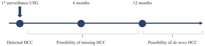
Figure 2. Brief schematic of hepatocellular carcinoma classification. USG, ultrasound; HCC, hepatocellular carcinoma.

Reference
-
References
1. European Association for the Study of the Liver. EASL clinical practice guidelines: management of hepatocellular carcinoma. J Hepatol. 2018; 69:182–236.2. Singal AG, Lampertico P, Nahon P. Epidemiology and surveillance for hepatocellular carcinoma: new trends. J Hepatol. 2020; 72:250–261.
Article3. Tzartzeva K, Obi J, Rich NE, Parikh ND, Marrero JA, Yopp A, et al. Surveillance imaging and alpha fetoprotein for early detection of hepatocellular carcinoma in patients with cirrhosis: a metaanalysis. Gastroenterology. 2018; 154:1706–1718.e1.
Article4. Yoon JS, Lee HA, Kim HY, Sinn DH, Lee DH, Hong SK, et al. Hepatocellular carcinoma in Korea: an analysis of the 2015 Korean nationwide cancer registry. J Liver Cancer. 2021; 21:58–68.
Article5. Esfeh JM, Hajifathalian K, Ansari-Gilani K. Sensitivity of ultrasound in detecting hepatocellular carcinoma in obese patients compared to explant pathology as the gold standard. Clin Mol Hepatol. 2020; 26:54–59.
Article6. Barclay RL, Vicari JJ, Doughty AS, Johanson JF, Greenlaw RL. Colonoscopic withdrawal times and adenoma detection during screening colonoscopy. N Engl J Med. 2006; 355:2533–2541.
Article7. Korean Liver Cancer Association (KLCA); National Cancer Center (NCC). 2022 KLCA-NCC Korea practice guidelines for the management of hepatocellular carcinoma. J Liver Cancer. 2022 Dec 9. doi: 10.17998/jlc.2022.11.07. [Epub ahead of print].8. Marrero JA, Kulik LM, Sirlin CB, Zhu AX, Finn RS, Abecassis MM, et al. Diagnosis, staging, and management of hepatocellular carcinoma: 2018 practice guidance by the American Association for the Study of Liver Diseases. Hepatology. 2018; 68:723–750.
Article9. Kokudo N, Takemura N, Hasegawa K, Takayama T, Kubo S, Shimada M, et al. Clinical practice guidelines for hepatocellular carcinoma: the Japan Society of Hepatology 2017 (4th JSH-HCC guidelines) 2019 update. Hepatol Res. 2019; 49:1109–1113.
Article10. Omata M, Cheng AL, Kokudo N, Kudo M, Lee JM, Jia J, et al. AsiaPacific clinical practice guidelines on the management of hepatocellular carcinoma: a 2017 update. Hepatol Int. 2017; 11:317–370.
Article11. Kwon JW, Tchoe HJ, Lee J, Suh JK, Lee JH, Shin S. The impact of national surveillance for liver cancer: results from real-world setting in Korea. Gut Liver. 2020; 14:108–116.
Article12. Sohn W, Lee YS, Lee JG, An J, Jang ES, Lee DH, et al. A survey of liver cancer specialists’ views on the National Liver Cancer screening program in Korea. J Liver Cancer. 2020; 20:53–59.
Article13. da Silva PH, Gomes MM, de Matos CAL, de Souza E Silva IS, Gonzalez AM, Torres US, et al. HCC detection on surveillance US: comparing focused liver protocol using US LI-RADS technical guidelines to a general complete abdominal US protocol. J Ultrasound Med. 2021; 40:2487–2495.14. Singal AG, Li X, Tiro J, Kandunoori P, Adams-Huet B, Nehra MS, et al. Racial, social, and clinical determinants of hepatocellular carcinoma surveillance. Am J Med. 2015; 128:90.e1–e7.
Article15. Mehta NJ, Celik AD, Peters MG. Screening for hepatocellular carcinoma: what is missing? Hepatol Commun. 2016; 1:18–22.
Article16. Nathani P, Gopal P, Rich N, Yopp A, Yokoo T, John B, et al. Hepatocellular carcinoma tumour volume doubling time: a systematic review and meta-analysis. Gut. 2021; 70:401–407.
Article17. Kim DM, Park SK, Park SG. A study on the performance evaluation criteria and methods of abdominal ultrasound devices based on international standards. Safety. 2021; 7:31.
Article18. Tao X, Li J, Gu Y, Ma L, Xu W, Wang R, et al. A national quality improvement program on ultrasound department in China: a controlled cohort study of 1297 public hospitals. Int J Environ Res Public Health. 2022; 20:397.
Article19. Gomaa AI, Khan SA, Toledano MB, Waked I, Taylor-Robinson SD. Hepatocellular carcinoma: epidemiology, risk factors and pathogenesis. World J Gastroenterol. 2008; 14:4300–4308.
Article20. Fujiwara N, Friedman SL, Goossens N, Hoshida Y. Risk factors and prevention of hepatocellular carcinoma in the era of precision medicine. J Hepatol. 2018; 68:526–549.
Article21. Tanaka H. Current role of ultrasound in the diagnosis of hepatocellular carcinoma. J Med Ultrason (2001). 2020; 47:239–255.
Article22. Simmons O, Fetzer DT, Yokoo T, Marrero JA, Yopp A, Kono Y, et al. Predictors of adequate ultrasound quality for hepatocellular carcinoma surveillance in patients with cirrhosis. Aliment Pharmacol Ther. 2017; 45:169–177.
Article23. Singal A, Volk ML, Waljee A, Salgia R, Higgins P, Rogers MA, et al. Meta-analysis: surveillance with ultrasound for early-stage hepatocellular carcinoma in patients with cirrhosis. Aliment Pharmacol Ther. 2009; 30:37–47.
Article24. Lupsor-Platon M, Serban T, Silion AI, Tirpe GR, Tirpe A, Florea M. Performance of ultrasound techniques and the potential of artificial intelligence in the evaluation of hepatocellular carcinoma and non-alcoholic fatty liver disease. Cancers (Basel). 2021; 13:790.
Article25. Kim DH, Choi JI. Current status of image-based surveillance in hepatocellular carcinoma. Ultrasonography. 2021; 40:45–56.
Article26. Del Poggio P, Olmi S, Ciccarese F, Di Marco M, Rapaccini GL, Benvegnù L, et al. Factors that affect efficacy of ultrasound surveillance for early stage hepatocellular carcinoma in patients with cirrhosis. Clin Gastroenterol Hepatol. 2014; 12:1927–1933.e2.
Article27. Sumida Y, Yoneda M, Seko Y, Ishiba H, Hara T, Toyoda H, et al. Surveillance of hepatocellular carcinoma in nonalcoholic fatty liver disease. Diagnostics (Basel). 2020; 10:579.
Article28. Plaz Torres MC, Bodini G, Furnari M, Marabotto E, Zentilin P, Strazzabosco M, et al. Surveillance for hepatocellular carcinoma in patients with non-alcoholic fatty liver disease: universal or selective? Cancers (Basel). 2020; 12:1422.
Article29. Choi EJ, Kim YJ. Liquid biopsy for early detection and therapeutic monitoring of hepatocellular carcinoma. J Liver Cancer. 2022; 22:103–114.
Article30. Kim YY, An C, Kim DY, Aljoqiman KS, Choi JY, Kim MJ. Failure of hepatocellular carcinoma surveillance: inadequate echogenic window and macronodular parenchyma as potential culprits. Ultrasonography. 2019; 38:311–320.
Article31. An C, Choi YA, Choi D, Paik YH, Ahn SH, Kim MJ, et al. Growth rate of early-stage hepatocellular carcinoma in patients with chronic liver disease. Clin Mol Hepatol. 2015; 21:279–286.
Article
- Full Text Links
-
- Actions
-
Cited
- CITED
-
- Close
- Share
-
- Similar articles
-
- Current status of image-based surveillance in hepatocellular carcinoma
- National Cancer Screening Program for Hepatocellular Carcinoma
- The Usefulness and Current Status of the Screening Program for Early Diagnosis of Hepatocellular Carcinoma
- Surveillance of hepatocellular carcinoma: is only ultrasound enough?
- Current Status and Future Directions of Hepatocellular Carcinoma Surveillance Test Based on Cost-effective Analysis
