Clin Endosc.
2023 Jan;56(1):132-134. 10.5946/ce.2022.298.
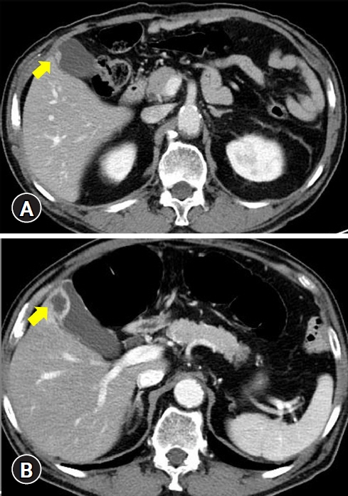
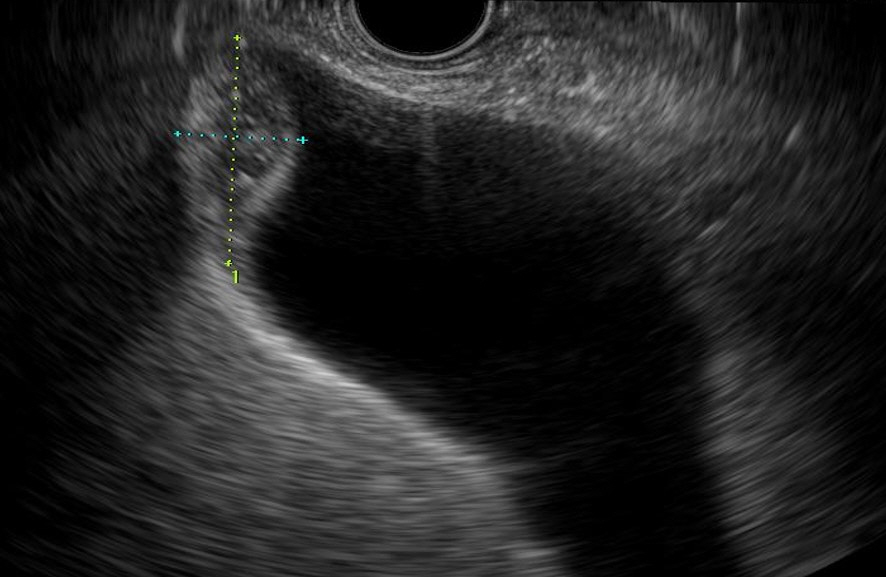
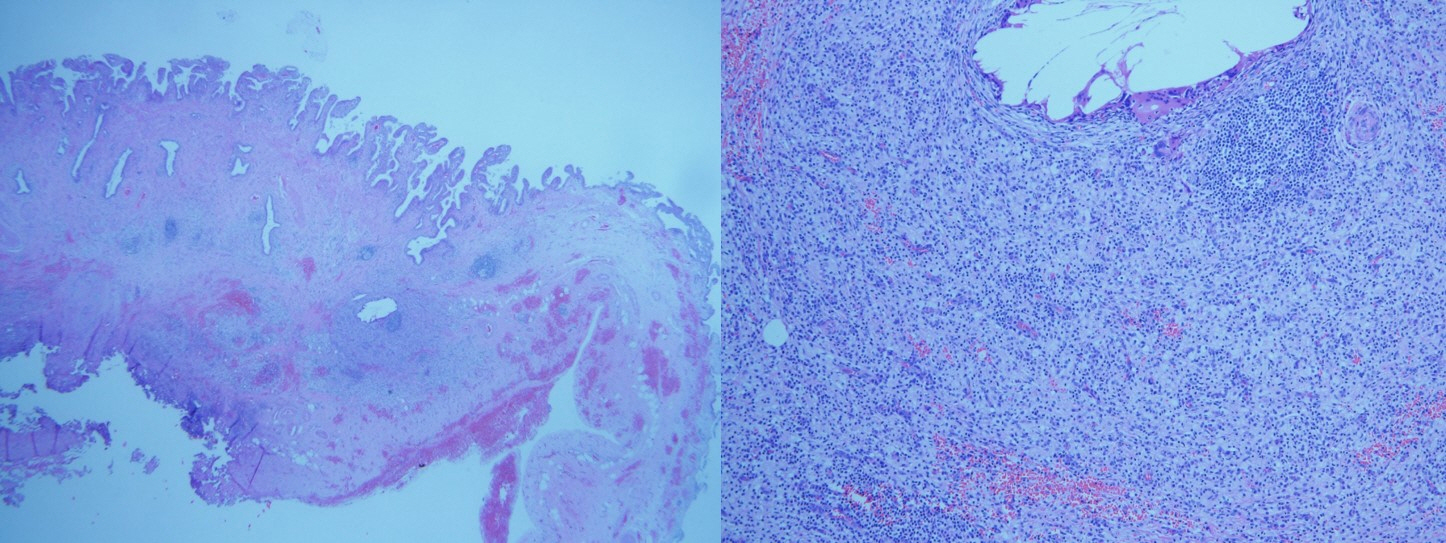
A rapidly growing round mass in the gallbladder
- Affiliations
-
- 1Division of Gastroenterology, Department of Internal Medicine, Dankook University College of Medicine, Cheonan, Korea
- 2Division of Gastroenterology and Hepatology, Department of Internal Medicine, Soonchunhyang University Cheonan Hospital, Soonchunhyang University School of Medicine, Cheonan, Korea
- KMID: 2538763
- DOI: http://doi.org/10.5946/ce.2022.298
Figure
Reference
-
1. Goodman ZD, Ishak KG. Xanthogranulomatous cholecystitis. Am J Surg Pathol. 1981; 5:653–659.2. Hanada K, Nakata H, Nakayama T, et al. Radiologic findings in xanthogranulomatous cholecystitis. AJR Am J Roentgenol. 1987; 148:727–730.3. Agarwal AK, Kalayarasan R, Javed A, et al. Mass-forming xanthogranulomatous cholecystitis masquerading as gallbladder cancer. J Gastrointest Surg. 2013; 17:1257–1264.4. Makimoto S, Takami T, Hatano K, et al. Xanthogranulomatous cholecystitis: a review of 31 patients. Surg Endosc. 2021; 35:3874–3880.5. Shuto R, Kiyosue H, Komatsu E, et al. CT and MR imaging findings of xanthogranulomatous cholecystitis: correlation with pathologic findings. Eur Radiol. 2004; 14:440–446.6. Tanaka K, Katanuma A, Hayashi T, et al. Role of endoscopic ultrasound for gallbladder disease. J Med Ultrason. 2021; 48:187–198.
- Full Text Links
- Actions
-
Cited
- CITED
-
- Close
- Share
- Similar articles
-
- Left-sided Gallbladder: 2 cases
- Laparoscopic Cholecystectomy in Patients with a Left-sided Gallbladder
- A Case of Rhabdomyosarcoma Presenting a Rapidly Growing Thyroid Mass Showing Cytological Features Mimic Anaplastic Thyroid Carcinoma
- Preduodenal Portal Vein and Left Sided Gallbladder in Hepatolithiasis Patient: A case report
- A Case of Squamous Cell Carcinoma of Gallbladder after Laparoscopic Cholecystectomy


