Correction of late adolescent skeletal Class III using the Alt-RAMEC protocol and skeletal anchorage
- Affiliations
-
- 1Department of Orthodontics, Faculty of Dentistry, Süleyman Demirel University, Isparta, Turkey
- 2Department of Oral and Maxillofacial Surgery, Faculty of Dentistry, Süleyman Demirel University, Isparta, Turkey
- KMID: 2538713
- DOI: http://doi.org/10.4041/kjod21.337
Abstract
- This case report describes skeletal anchorage-supported maxillary protraction performed with the Alternate Rapid Maxillary Expansion and Constriction (AltRAMEC) protocol over a treatment duration of 14 months in a 16-year-old female patient who was in the late growth-development period. Miniplates were applied to the patient's aperture piriformis area to apply force from the protraction appliance. After 9 weeks of following the Alt-RAMEC protocol, miniplates were used to transfer a unilateral 500-g protraction force to a Petit-type face mask. A significant improvement was observed in the soft tissue profile in measurements made both cephalometrically and in three dimensional photographs. Subsequently, the second phase of fixed orthodontic treatment was started and the treatment was completed with the retention phase. Following treatment completion, occlusion, smile esthetics, and soft tissue profile improved significantly in response to orthopedic and orthodontic treatment.
Keyword
Figure
-

Figure 1 Facial and intraoral photographs taken at the initial visit (age: 16 years, 7 months).
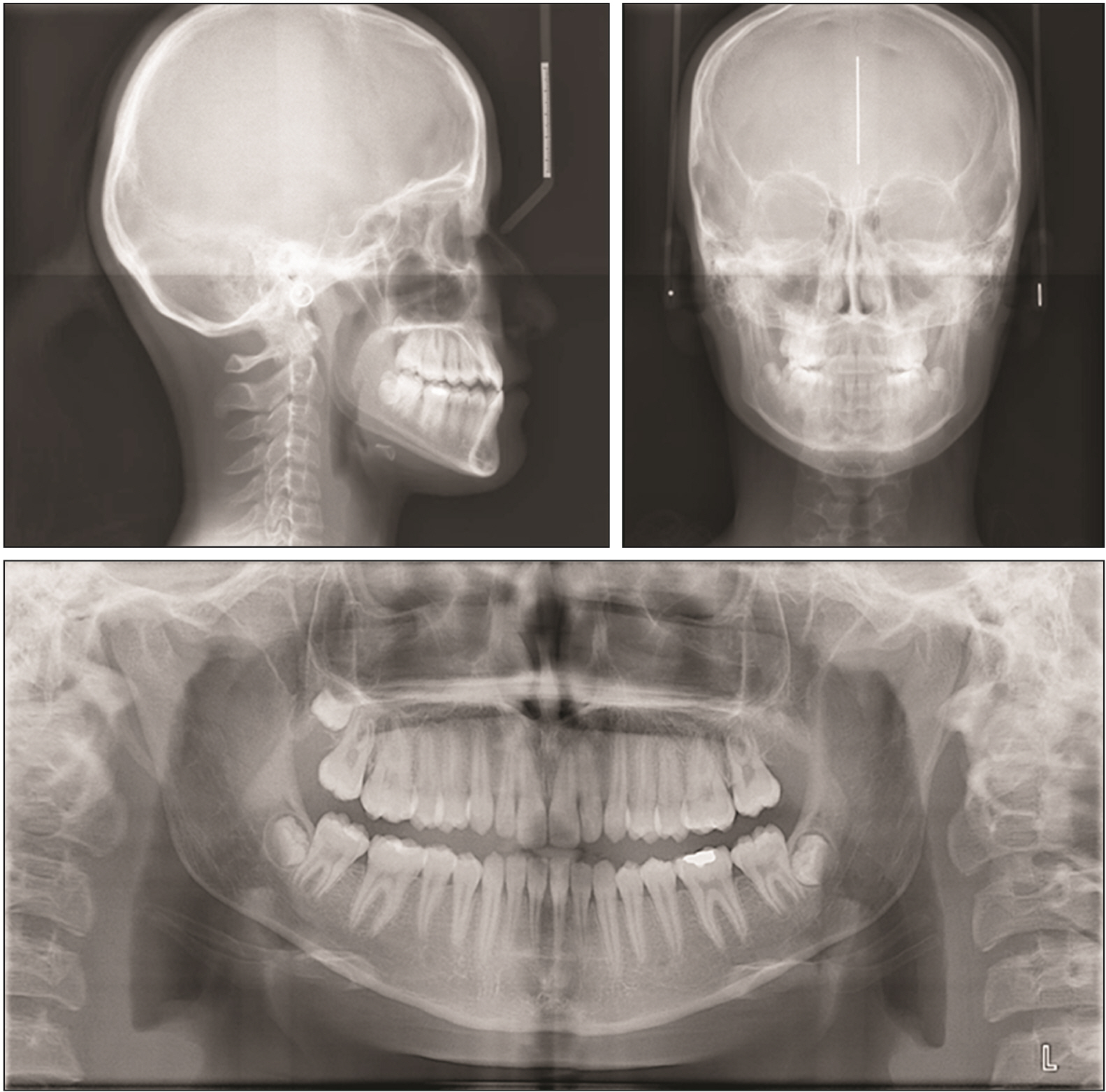
Figure 2 Lateral and posteroanterior cephalographs, and panoramic radiograph taken at the initial visit (age: 16 years, 7 months).

Figure 3 Growth-development period (hand-wrist and cervical vertebrae) at the patient’s initial visit.
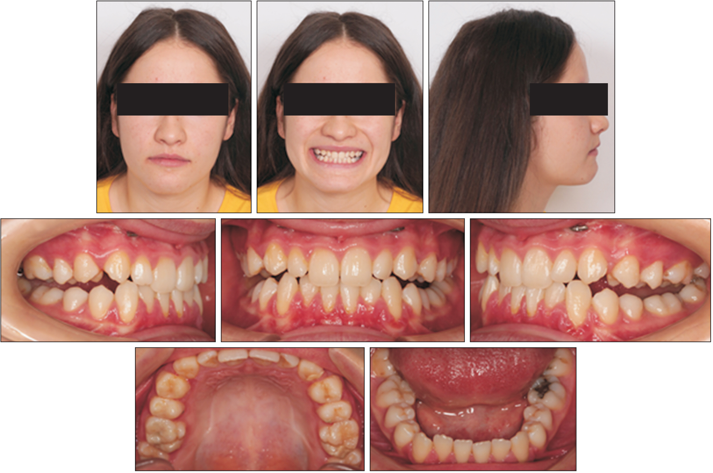
Figure 4 Post-protraction lateral cephalograph and panoramic radiograph.
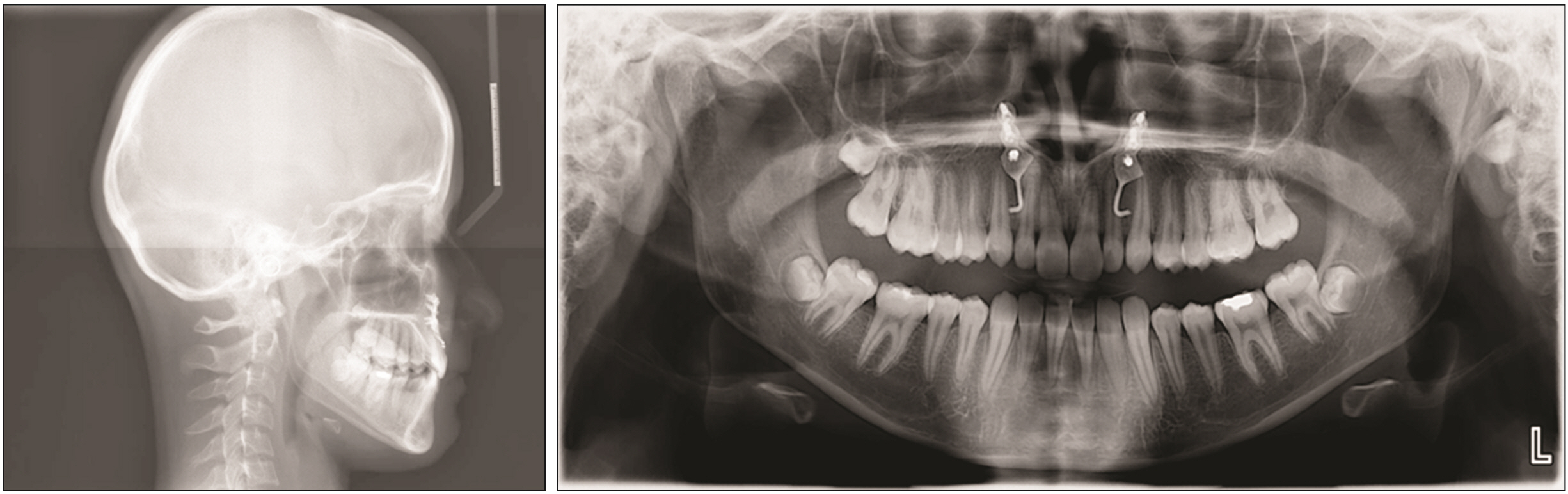
Figure 5 Post-protraction facial and intraoral photographs.

Figure 6 Treatment stages. Alt-RAMEC, alternate rapid maxillary expansion and constriction; RME, rapid maxillary expansion.
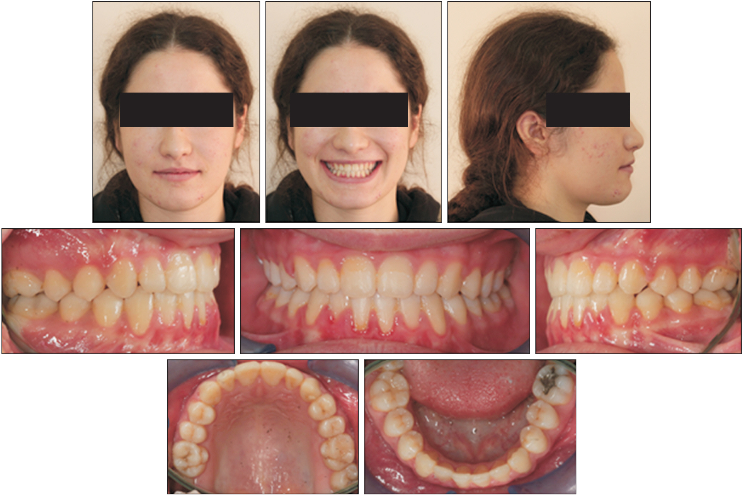
Figure 7 Facial and intraoral photographs taken at the debond visit (age: 17 years, 9 months).

Figure 8 Lateral and posteroanterior cephalographs, and panoramic radiograph taken at the debond visit (age: 17 years, 9 months).
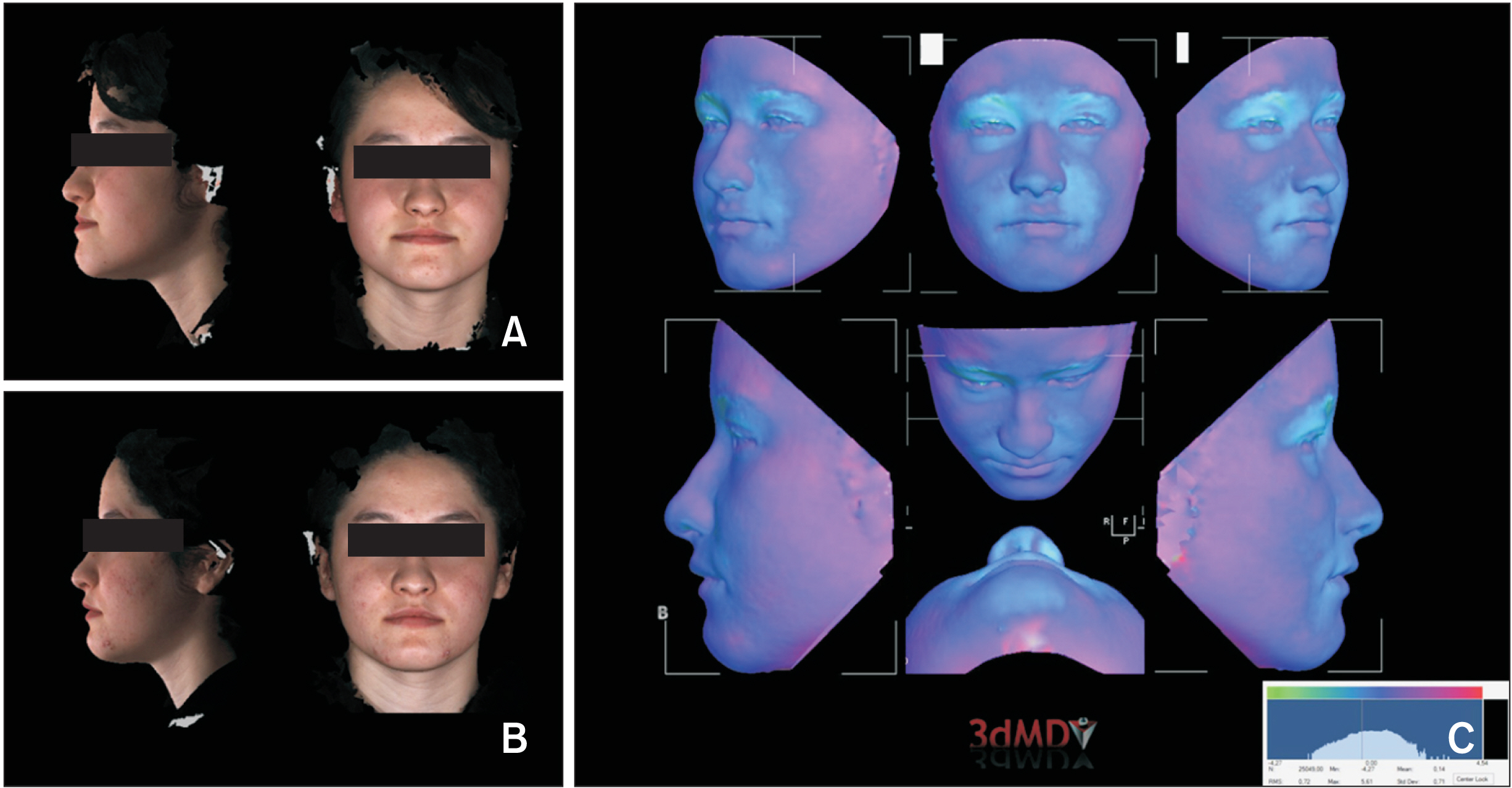
Figure 9 Initial (A) and post-protraction (B) three dimensional (3D) photographs and (C) 3D volumetric tracing on the 3D photographs.

Figure 10 Facial and intraoral photographs taken 6 months after debonding (age: 18 years, 3 months).
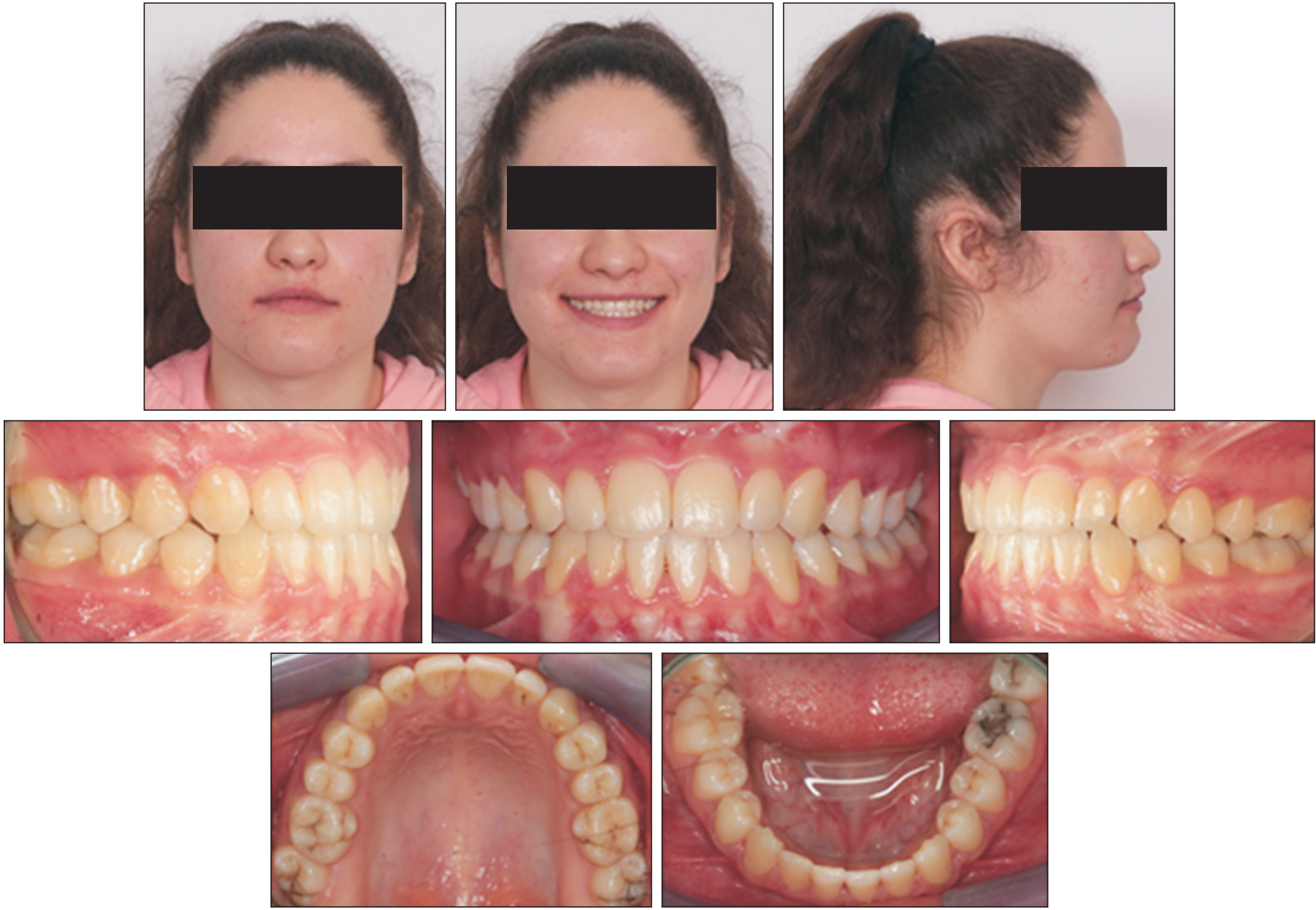
Figure 11 Facial and intraoral photographs taken 2 years after debonding (age: 19 years, 9 months).
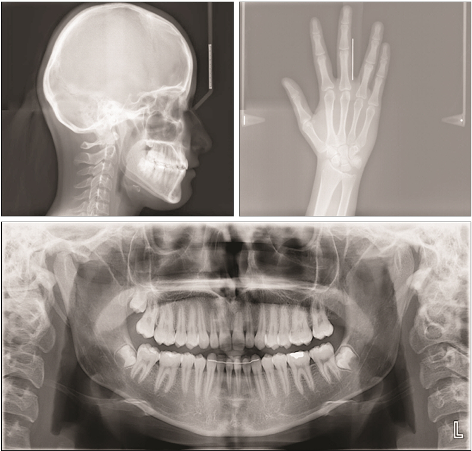
Figure 12 Lateral cephalograph, hand wrist radiograph, and panoramic radiograph taken 2 years after debonding (age: 19 years, 9 months).
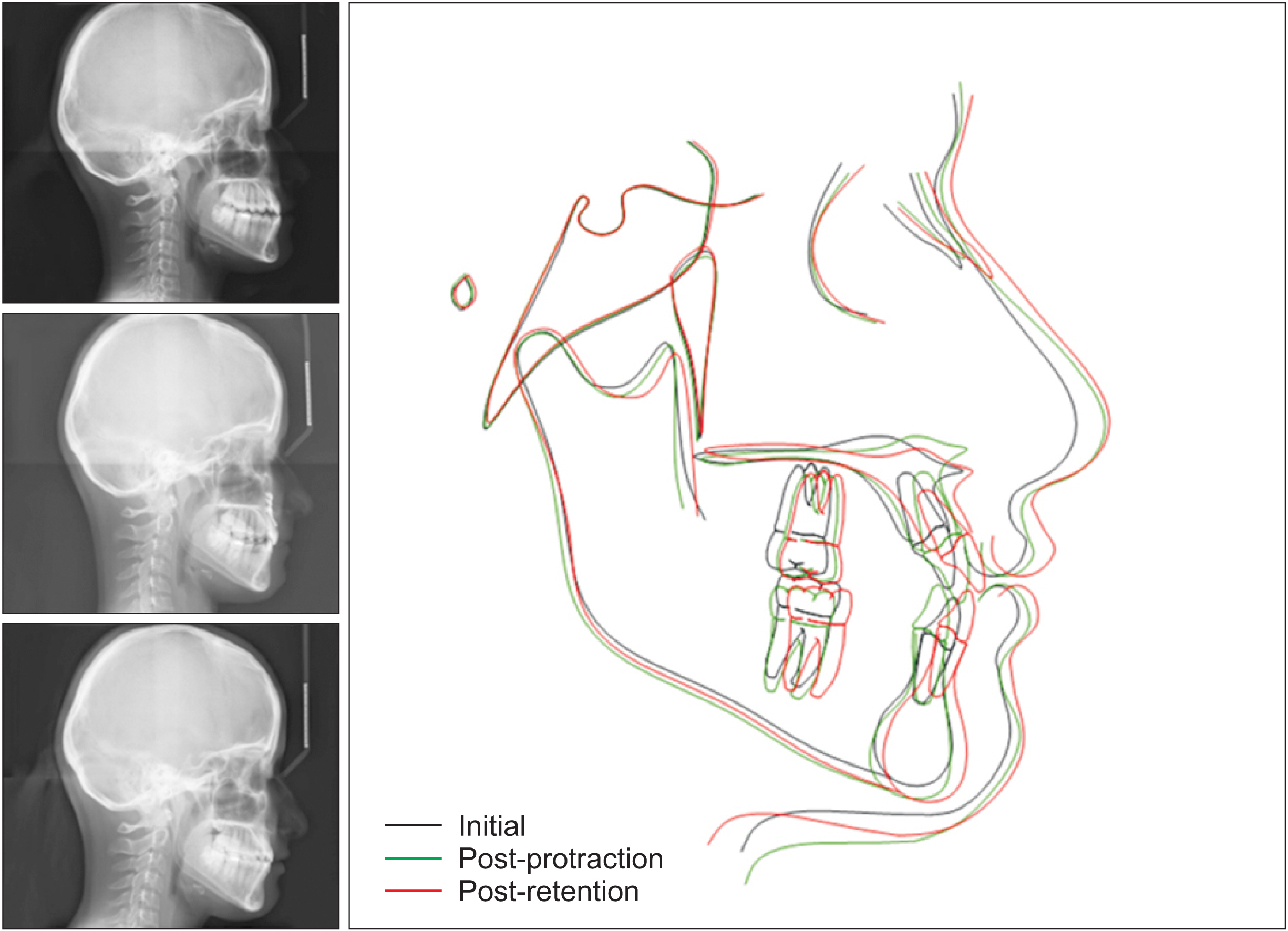
Figure 13 Cephalometric superimpositions.
Reference
-
1. Proffit WR, Fields HW, Sarver DM. 2007. Contemporary orthodontics. 4th ed. Mosby Elsevier;St. Louis:2. Ngan PW, Deguchi T, Roberts EW. 2014. Orthodontic treatment of class III malocclusion. Bentham Science Publishers;DOI: 10.2174/97816080549161140101.3. Westwood PV, McNamara JA Jr, Baccetti T, Franchi L, Sarver DM. 2003; Long-term effects of Class III treatment with rapid maxillary expansion and facemask therapy followed by fixed appliances. Am J Orthod Dentofacial Orthop. 123:306–20. DOI: 10.1067/mod.2003.44. PMID: 12637903.4. Nanda R. 2015. Esthetics and biomechanics in orthodontics. 2nd ed. Elsevier/Saunders;St Louis: DOI: 10.1067/mod.2003.44.5. Cha BK, Choi DS, Ngan P, Jost-Brinkmann PG, Kim SM, Jang IS. 2011; Maxillary protraction with miniplates providing skeletal anchorage in a growing Class III patient. Am J Orthod Dentofacial Orthop. 139:99–112. DOI: 10.1016/j.ajodo.2009.06.025. PMID: 21195283.
Article6. Buyukcavus MH, Kale B, Aydemir B. 2020; Comparison of treatment effects of different maxillary protraction methods in skeletal class III patients. Orthod Craniofac Res. 23:445–54. DOI: 10.1111/ocr.12389. PMID: 32406170.
Article7. Fakharian M, Bardideh E, Abtahi M. 2019; Skeletal Class III malocclusion treatment using mandibular and maxillary skeletal anchorage and intermaxillary elastics: a case report. Dental Press J Orthod. 24:52–9. DOI: 10.1590/2177-6709.24.5.052-059.oar. PMID: 31721947. PMCID: PMC6833933.
Article8. Büyükçavuş MH. 2019; Alternate Rapid Maxillary Expansion and Constriction (Alt-RAMEC) protocol: a comprehensive literature review. Turk J Orthod. 32:47–51. DOI: 10.5152/TurkJOrthod.2019.18021. PMID: 30944900. PMCID: PMC6436906.
Article9. Pithon MM, Santos NL, Santos CR, Baião FC, Pinheiro MC, Matos M Neto, et al. 2016; Is alternate rapid maxillary expansion and constriction an effective protocol in the treatment of Class III malocclusion? A systematic review. Dental Press J Orthod. 21:34–42. DOI: 10.1590/2177-6709.21.6.034-042.oar. PMID: 28125138. PMCID: PMC5278931.
Article10. Kaya D, Kocadereli I, Kan B, Tasar F. 2011; Effects of facemask treatment anchored with miniplates after alternate rapid maxillary expansions and constrictions; a pilot study. Angle Orthod. 81:639–46. DOI: 10.2319/081010-473.1. PMID: 21299407. PMCID: PMC8919738.
Article11. Yilmaz HN, Garip H, Satilmis T, Kucukkeles N. 2015; Corticotomy-assisted maxillary protraction with skeletal anchorage and Class III elastics. Angle Orthod. 85:48–57. DOI: 10.2319/121513-921.1. PMID: 24913740. PMCID: PMC8634805.
Article12. Nevzatoğlu S, Küçükkeleş N. 2014; Long-term results of surgically assisted maxillary protraction vs regular facemask. Angle Orthod. 84:1002–9. DOI: 10.2319/120913-905.1. PMID: 24654941. PMCID: PMC8638485.
Article13. Meazzini MC, Zappia LB, Tortora C, Autelitano L, Tintinelli R. 2019; Short- and long-term effects of late maxillary advancement with the Liou-Alt-RAMEC protocol in unilateral cleft lip and palate. Cleft Palate Craniofac J. 56:159–67. DOI: 10.1177/1055665618772395. PMID: 29702006.
Article14. Ganesh G, Tripathi T, Rai P. 2020; Orthopaedic and orthodontic treatment with hyrax, Class III elastics on mandibular miniplates, maxillary mini-implants in a Class III adolescent: a case report. Int Orthod. 18:827–38. DOI: 10.1016/j.ortho.2020.06.001. PMID: 32654977.
Article15. Shi H, Ge HS, Chen LY, Li ZH. 2020; Meta-analysis of the efficacy of bone anchorage and maxillary facemask protraction devices in treating skeletal class III malocclusion in adolescents. Hua Xi Kou Qiang Yi Xue Za Zhi. 38:69–74. Chinese. DOI: 10.1016/j.ortho.2020.06.001.16. Park JH, Emamy M, Lee SH. 2019; Adult skeletal Class III correction with camouflage orthodontic treatment. Am J Orthod Dentofacial Orthop. 156:858–69. DOI: 10.1016/j.ajodo.2018.07.029. PMID: 31784020.
Article17. Merwin D, Ngan P, Hagg U, Yiu C, Wei SH. 1997; Timing for effective application of anteriorly directed orthopedic force to the maxilla. Am J Orthod Dentofacial Orthop. 112:292–9. DOI: 10.1016/S0889-5406(97)70259-2. PMID: 9294359.
Article18. Rodríguez de Guzmán-Barrera J, Sáez Martínez C, Boronat-Catalá M, Montiel-Company JM, Paredes-Gallardo V, Gandía-Franco JL, et al. 2017; Effectiveness of interceptive treatment of class III malocclusions with skeletal anchorage: a systematic review and meta-analysis. PLoS One. 12:e0173875. DOI: 10.1371/journal.pone.0173875. PMID: 28328995. PMCID: PMC5362089.
Article
- Full Text Links
-
- Actions
-
Cited
- CITED
-
- Close
- Share
-
- Similar articles
-
- A study of the calcification of the second and the third molars in skeletal Class II and III malocclusions
- Class III nonsurgical treatment using indirect skeletal anchorage: A case report
- Directional forces using skeletal anchorage for treatment of skeletal Class II div.1 malocclusion
- Does surgically assisted maxillary protraction with skeletal anchorage and Class III elastics affect the pharyngeal airway? A retrospective, long-term study
- Maxillary protraction treatment of skeletal Class III children using miniplate anchorage
