Diagnosis of Bowel Endometriosis Using Endoscopic Ultrasound-guided Fine Needle Aspiration
- Affiliations
-
- 1Department of Gastroenterology, Centro Hospitalar Tondela-Viseu, E.P.E., Viseu, Portugal
- 2Department of Gastroenterology, Centro Hospitalar e Universitário de Coimbra, Coimbra, Portugal
- KMID: 2538688
- DOI: http://doi.org/10.4166/kjg.2022.104
Abstract
- Endometriosis is a relatively common gynecological condition in women of reproductive age. The rectosigmoid region is the most commonly affected segment when the gastrointestinal tract is involved. A differential diagnosis of colorectal neoplasia is difficult because of the similar clinical, endoscopic, and radiology findings. A 42-year-old female presented with abdominal distention and was subsequently diagnosed with a large bowel obstruction in the rectum. A temporary colostomy was performed, and endoscopy revealed a rectal mass obstructing the rectum. The biopsy showed normal mucosa, and it was difficult to exclude rectal malignancies even after the imaging workup. Endoscopic ultrasound demonstrated a hypoechoic lesion below the rectal mucosa, and fine needle aspiration confirmed the diagnosis of bowel endometriosis. Bowel endometriosis is a challenging diagnosis. Endoscopic ultrasound-guided fine-needle aspiration is useful for acquiring adequate samples for histological confirmation and a definitive diagnosis of bowel endometriosis.
Keyword
Figure
-
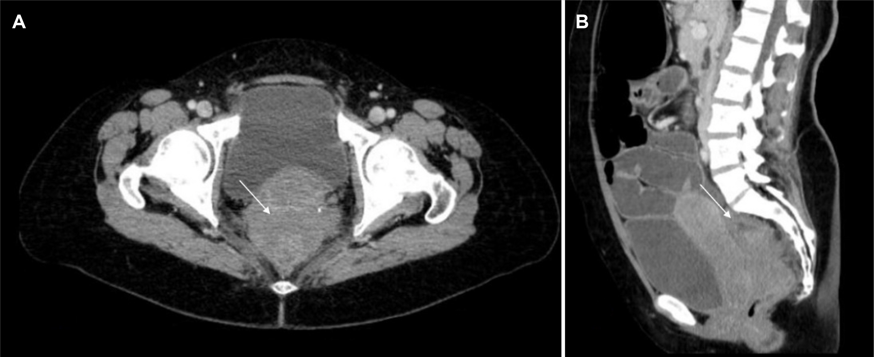
Fig. 1 Pelvic CT transversal (A) and coronal (B) images revealing a stenotic lesion in the upper rectum (arrow), without a clear margin from the cervix, with upstream large and small bowel distension.
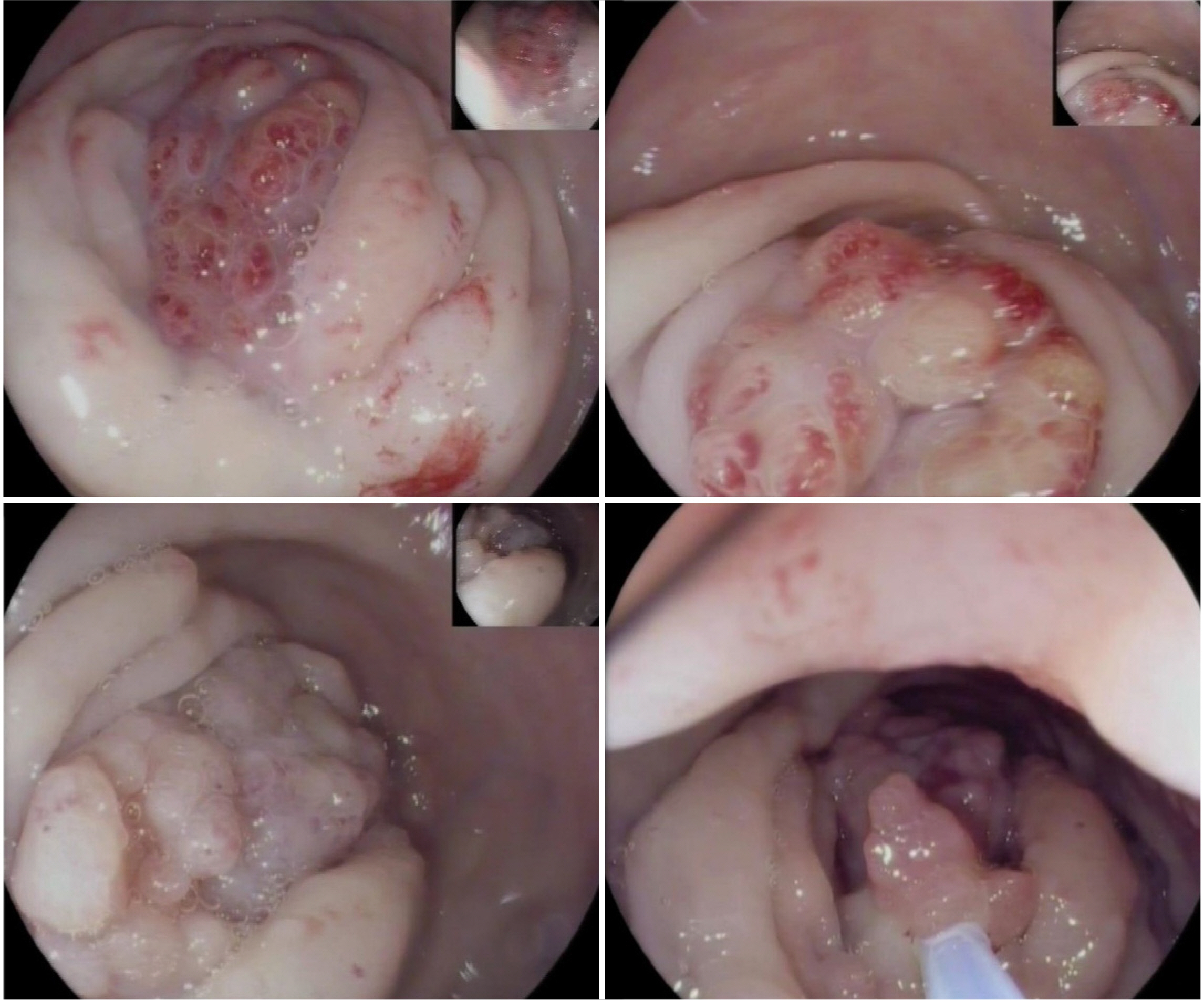
Fig. 2 Rectosigmoidoscopy showing a pseudopolypoid lesion causing a rectal obstruction.

Fig. 3 Pelvic MRI T1 (A) and T2 weighted in transversal (B) and sagittal (C) view demonstrating a stenotic lesion (arrows) in the upper rectum and its anatomical relations with the uterus.
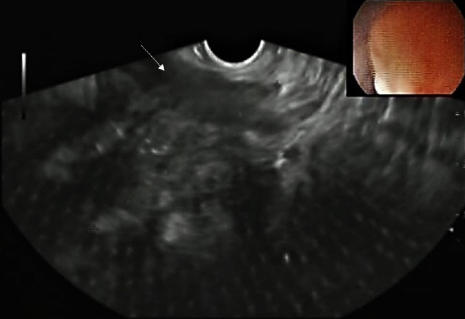
Fig. 4 Rectal EUS (linear probe) revealing an irregularly shaped hypoechoic and heterogeneous lesion (arrow) extending into the muscular layer of the rectal wall.
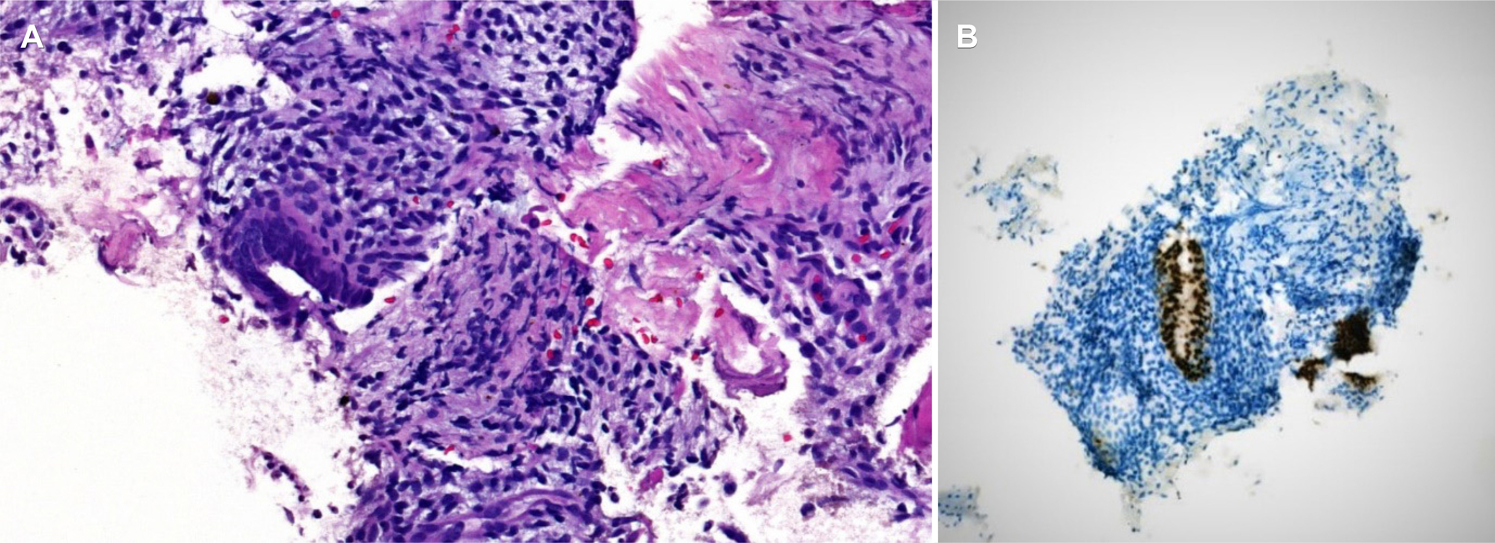
Fig. 5 Histopathology showing endometrial-type epithelium in the colonic mucosa (A: H&E stain), reactive to CK7 (B).
Reference
-
1. Parasar P, Ozcan P, Terry KL. 2017; Endometriosis: epidemiology, diagnosis and clinical management. Curr Obstet Gynecol Rep. 6:34–41. DOI: 10.1007/s13669-017-0187-1. PMID: 29276652. PMCID: PMC5737931.
Article2. Giudice LC, Kao LC. 2004; Endometriosis. Lancet. 364:1789–1799. DOI: 10.1016/S0140-6736(04)17403-5. PMID: 15541453.
Article3. Habib N, Centini G, Lazzeri L, et al. 2020; Bowel endometriosis: current perspectives on diagnosis and treatment. Int J Womens Health. 12:35–47. DOI: 10.2147/IJWH.S190326. PMID: 32099483. PMCID: PMC6996110.4. Roman H. FRIENDS group (French coloRectal Infiltrating ENDometriosis Study group). A national snapshot of the surgical management of deep infiltrating endometriosis of the rectum and colon in France in 2015: a multicenter series of 1135 cases. J Gynecol Obstet Hum Reprod. 2017; 46:159–165. DOI: 10.1016/j.jogoh.2016.09.004. PMID: 28403973.
Article5. Abrão MS, Petraglia F, Falcone T, Keckstein J, Osuga Y, Chapron C. 2015; Deep endometriosis infiltrating the recto-sigmoid: critical factors to consider before management. Hum Reprod Update. 21:329–339. DOI: 10.1093/humupd/dmv003. PMID: 25618908.
Article6. De Ceglie A, Bilardi C, Blanchi S, et al. 2008; Acute small bowel obstruction caused by endometriosis: a case report and review of the literature. World J Gastroenterol. 14:3430–3434. DOI: 10.3748/wjg.14.3430. PMID: 18528943. PMCID: PMC2716600.
Article7. Wolthuis AM, Meuleman C, Tomassetti C, D'Hooghe T, de Buck van Overstraeten A, D'Hoore A. 2014; Bowel endometriosis: colorectal surgeon's perspective in a multidisciplinary surgical team. World J Gastroenterol. 20:15616–15623. DOI: 10.3748/wjg.v20.i42.15616. PMID: 25400445. PMCID: PMC4229526.
Article8. Adam A, Narayanan M, Hachem C. 2018; Endoscopic appearance and management of recto-sigmoid endometriosis: case report. Gastroenterology Res. 11:326–328. DOI: 10.14740/gr1049w. PMID: 30116434. PMCID: PMC6089590.
Article9. Khashper A, Addley HC, Abourokbah N, Nougaret S, Sala E, Reinhold C. 2012; T2-hypointense adnexal lesions: an imaging algorithm. Radiographics. 32:1047–1064. DOI: 10.1148/rg.324115180. PMID: 22786993.
Article10. Tomiguchi J, Miyamoto H, Ozono K, et al. 2017; Preoperative diagnosis of intestinal endometriosis by magnifying colonoscopy and target biopsy. Case Rep Gastroenterol. 11:494–499. DOI: 10.1159/000475751. PMID: 29033768. PMCID: PMC5624264.
Article11. Schröder J, Löhnert M, Doniec JM, Dohrmann P. 1997; Endoluminal ultrasound diagnosis and operative management of rectal endometriosis. Dis Colon Rectum. 40:614–617. DOI: 10.1007/BF02055389. PMID: 9152194.
Article12. Shi B, Sun B, Zhao Q, Zhang X. 2020; EUS diagnosis of rectal endometriosis. VideoGIE. 6:105–107. DOI: 10.1016/j.vgie.2020.10.014. PMID: 33884342. PMCID: PMC7859544.
Article13. Alborzi S, Rasekhi A, Shomali Z, et al. 2018; Diagnostic accuracy of magnetic resonance imaging, transvaginal, and transrectal ultrasonography in deep infiltrating endometriosis. Medicine (Baltimore). 97:e9536. DOI: 10.1097/MD.0000000000009536. PMID: 29465552. PMCID: PMC5842011.
Article14. James TW, Fan YC, Schiff LD, Gangarosa LM. 2019; Lower endoscopic ultrasound in preoperative evaluation of rectosigmoid endometriosis. Endosc Int Open. 7:E837–E840. DOI: 10.1055/a-0901-7259. PMID: 31198849. PMCID: PMC6561764.
Article15. Miwa T, Iwashita T, Aiba M, et al. 2020; Endoscopic ultrasound-guided fine needle aspiration for the diagnosis of bowel endometriosis: a case report. Med Ultrason. 22:243–246. DOI: 10.11152/mu-2000. PMID: 32190855.
Article16. Pishvaian AC, Ahlawat SK, Garvin D, Haddad NG. 2006; Role of EUS and EUS-guided FNA in the diagnosis of symptomatic rectosigmoid endometriosis. Gastrointest Endosc. 63:331–335. DOI: 10.1016/j.gie.2005.06.019. PMID: 16427951.
Article17. Hara K, Yamao K, Ohashi K, et al. 2003; Endoscopic ultrasonography and endoscopic ultrasound-guided fine-needle aspiration biopsy for the diagnosis of lower digestive tract disease. Endoscopy. 35:966–969. DOI: 10.1055/s-2003-43473. PMID: 14606022.
Article18. Roseau G, Dumontier I, Palazzo L, et al. 2000; Rectosigmoid endometriosis: endoscopic ultrasound features and clinical implications. Endoscopy. 32:525–530. DOI: 10.1055/s-2000-9008. PMID: 10917184.
Article19. Kishimoto K, Kawashima K, Moriyama I, et al. 2020; Sigmoid endometriosis diagnosed preoperatively using endoscopic ultrasound-guided fine-needle aspiration. Clin J Gastroenterol. 13:158–163. DOI: 10.1007/s12328-019-01046-x. PMID: 31549336.
Article20. Moawad GN, Klebanoff JS, Habib N, Bendifallah S. 2021; Colorectal endometriosis: ample data without definitive recommendations. Facts Views Vis Obgyn. 13:3–7. DOI: 10.52054/FVVO.13.1.006. PMID: 33889855. PMCID: PMC8051188.
Article
- Full Text Links
-
- Actions
-
Cited
- CITED
-
- Close
- Share
-
- Similar articles
-
- Fine-Needle Biopsy: Should This Be the First Choice in Endoscopic Ultrasound-Guided Tissue Acquisition?
- Endoscopic Ultrasound-Fine Needle Aspiration versus Core Biopsy for the Diagnosis of Subepithelial Tumors
- How Can We Get the Best Results with Endoscopic Ultrasound-Guided Fine Needle Aspiration?
- Role of Repeated Endoscopic Ultrasound-Guided Fine Needle Aspiration for Inconclusive Initial Cytology Result
- Procore and Flexible 19 Gauge Needle Can Replace Trucut Biopsy Needle?
