Variant muscle fibers connecting the orbicularis oculi to the orbicularis oris: case report
- Affiliations
-
- 1Oral Medicine Research Center, Fukuoka Gakuen, Fukuoka, Japan
- 2Department of Oral and Maxillofacial Anatomy, Graduate School of Medical and Dental Sciences, Tokyo Medical and Dental University, Tokyo, Japan
- 3Department of Neurosurgery, Tulane Center for Clinical Neurosciences, Tulane University School of Medicine, New Orleans, LA, USA
- 4Department of Neurology, Tulane Center for Clinical Neurosciences, Tulane University School of Medicine, New Orleans, LA, USA
- 5Dental and Oral Medical Center, Kurume University School of Medicine, Kurume, Fukuoka, Japan
- 6Division of Gross and Clinical Anatomy, Department of Anatomy, Kurume University School of Medicine, Kurume, Fukuoka, Japan
- 7Department of Anatomical Sciences, St. George’s University, St. George’s, Grenada, West Indies, LA, USA
- 8Department of Structural & Cellular Biology, Tulane University School of Medicine, New Orleans, LA, USA
- 9Department of Surgery, Tulane University School of Medicine, New Orleans, LA, USA
- 10Department of Anatomy, Daegu Catholic University School of Medicine, Daegu, Korea
- KMID: 2537469
- DOI: http://doi.org/10.5115/acb.22.108
Abstract
- The orbicularis oculi (OOc) is a sphincteric muscle of the eyelids, whereas contraction of the orbicularis oris (OOr), another sphincteric muscle, causes narrowing of the lips. Facial muscle fibers normally blend with adjacent muscles. However, muscle fibers connecting the various facial muscles that have different actions and that are located at distant sites, such as the OOc and the OOr have been rarely reported. Herein, we report a rare case of connecting fibers between the inferior margin of the OOc and the OOr. These connecting fibers were blended with the OOr between the inserting fibers of the levator labii superioris and levator anguli oris. Contraction of such variant muscles might affect typical facial expressions.
Figure
-
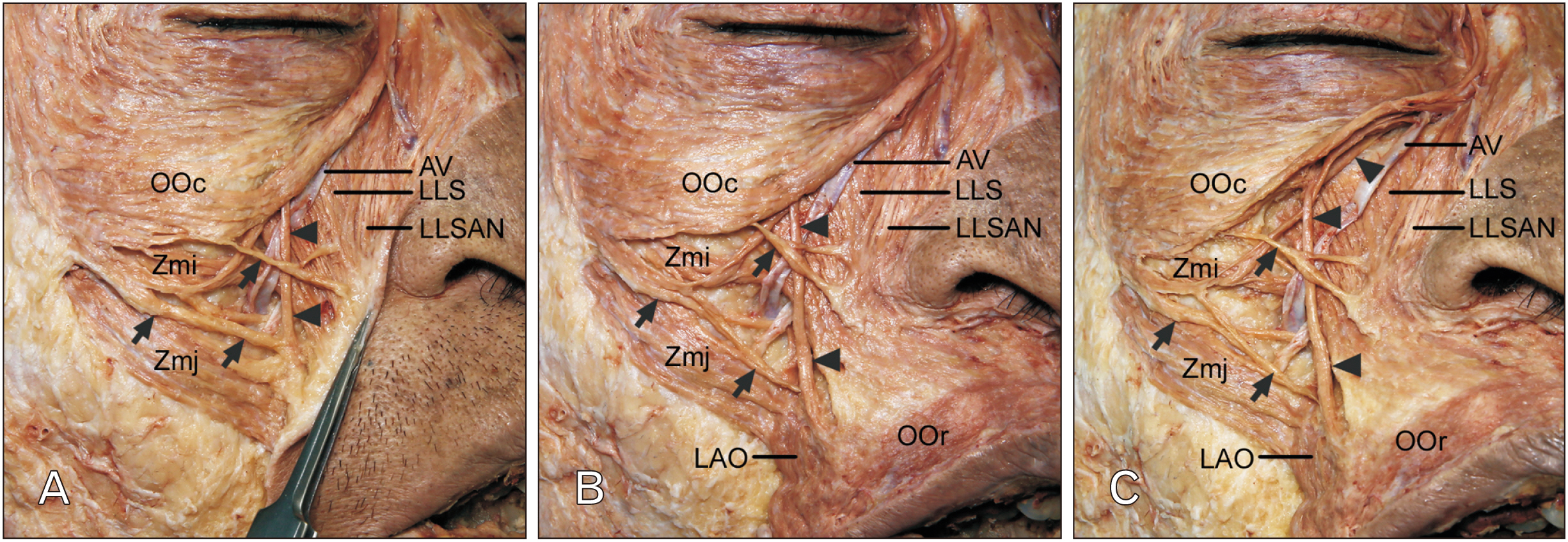
Fig. 1 Connecting fibers between the inferior margin of the OOc and OOr. (A) At the middle of the inferior margin of the OOc, the connecting fibers (arrowheads) descended perpendicularly and passed beneath the site between the middle and the lower thirds of the nasolabial fold. Adjacent to the connecting fibers, there were several fibers (arrows) from the OOc attaching along the nasolabial fold. Skin just lateral to the nasolabial fold was removed and the remaining skin was reflected to reveal the courses and attachments of the connecting fibers and the adjacent extending fibers from the OOc. (B) The connecting fibers (arrowheads) blended with the OOr between the inserting fibers of the LLS and LAO. The several fibers (arrows) from the OOc were toward the LLSAN, LLS, and Zmi. The remaining skin was removed to expose the entire muscles in the face. (C) The extending fibers of the OOc that divided the connecting fibers (arrowheads) were attached to the maxilla just above the origin site of the LLS. The inferior margin of the OOc were reflected superiorly to reveal the course and attachment of the connecting fibers. AV, angular vein; LAO, levator anguli oris; LLS, levator labii superioris; LLSAN, lavetor labii superioris alaeque nasi; OOc, orbicularis oculi; OOr, orbicularis oris; Zmi, zygomaticus minor; Zmj, zygomaticus major.
Reference
-
References
1. Standring S. 2020. Gray's anatomy: the anatomical basis of clinical practice. 42nd ed. Elsevier;London:2. Choi DY, Hur MS, Youn KH, Kim J, Kim HJ, Kim SS. 2014; Clinical anatomic considerations of the zygomaticus minor muscle based on the morphology and insertion pattern. Dermatol Surg. 40:858–63. DOI: 10.1111/dsu.0000000000000063. PMID: 25006853.3. Hur MS, Youn KH, Kim HJ. 2018; New insight regarding the zygomaticus minor as related to cosmetic facial injections. Clin Anat. 31:974–80. DOI: 10.1002/ca.23272. PMID: 30194870.4. Hur MS, O J, Yang HM, Kwon HJ, Lee S, Lim HS, Lim SY, Oh CS. 2020; Heights and spatial relationships of the facial muscles acting on the nasolabial fold by dissection and three-dimensional microcomputed tomography. PLoS One. 15:e0237043. DOI: 10.1371/journal.pone.0237043. PMID: 32750081. PMCID: PMC7402499. PMID: 16c7bda3b1234161886d07c76a25f1b4.5. Iwanaga J, Singh V, Takeda S, Ogeng'o J, Kim HJ, Moryś J, Ravi KS, Ribatti D, Trainor PA, Sañudo JR, Apaydin N, Sharma A, Smith HF, Walocha JA, Hegazy AMS, Duparc F, Paulsen F, Del Sol M, Adds P, Louryan S, Fazan VPS, Boddeti RK, Tubbs RS. 2022; Standardized statement for the ethical use of human cadaveric tissues in anatomy research papers: recommendations from Anatomical Journal Editors-in-Chief. Clin Anat. 35:526–8. DOI: 10.1002/ca.23849. PMID: 35218594.6. Hollinshead WH. 1982. Anatomy for surgeons: Vol. 1. The head and neck. 3rd ed. Harper & Row;New York:7. Iwanaga J, Hur MS, Kikuta S, Ibaragi S, Tubbs RS. 2021; Extended crossing fibers of the mentalis muscle attaching to the contralateral mandible. Anat Cell Biol. 54:522–4. DOI: 10.5115/acb.21.127. PMID: 34465670. PMCID: PMC8693138.8. Hur MS, Lee S, Jung HS, Schneider RA. 2022; Anatomical connections among the depressor supercilii, levator labii superioris alaeque nasi, and inferior fibers of orbicularis oculi: implications for variation in human facial expressions. PLoS One. 17:e0264148. DOI: 10.1371/journal.pone.0264148. PMID: 35231048. PMCID: PMC8887774. PMID: 009306069e2b40cf8d0ed46f2a9bdf13.9. Woodburne RT, Burkel WE. 1994. Essentials of human anatomy. 9th ed. Oxford University Press;New York:10. Pessa JE, Brown F. 1992; Independent effect of various facial mimetic muscles on the nasolabial fold. Aesthetic Plast Surg. 16:167–71. DOI: 10.1007/BF00450609. PMID: 1570780.11. Snider CC, Amalfi AN, Hutchinson LE, Sommer NZ. 2017; New insights into the anatomy of the midface musculature and its implications on the nasolabial fold. Aesthetic Plast Surg. 41:1083–90. DOI: 10.1007/s00266-017-0889-9. PMID: 28508263.12. Kwon HJ, O J, Cho TH, Choi YJ, Yang HM. 2020; The nasolabial fold: a micro-computed tomography study. Plast Reconstr Surg. 145:71–9. DOI: 10.1097/PRS.0000000000006328. PMID: 31577657.13. Morris H. 1947. Human anatomy: a complete systematic treatise. 10th ed. Blakiston;Philadelphia:14. Sinnatamby CS. 2011. Last's anatomy: regional and applied. 12th ed. Churchill Livingstone/Elsevier;New York:15. Iwanaga J, Singh V, Ohtsuka A, Hwang Y, Kim HJ, Moryś J, Ravi KS, Ribatti D, Trainor PA, Sañudo JR, Apaydin N, Şengül G, Albertine KH, Walocha JA, Loukas M, Duparc F, Paulsen F, Del Sol M, Adds P, Hegazy A, Tubbs RS. 2021; Acknowledging the use of human cadaveric tissues in research papers: recommendations from Anatomical Journal Editors. Clin Anat. 34:2–4. DOI: 10.1002/ca.23671. PMID: 32808702.
- Full Text Links
-
- Actions
-
Cited
- CITED
-
- Close
- Share
-
- Similar articles
-
- New Operative Technique for Blepharoptosis using Fronto-Orbicularis Oculi Muscle Advancement
- The effects of facial denervation on facial muscles and bones in growing rabbits
- Orbicularis oris muscle reconstruction and cheiloplasty with Z-plasty in a patient with a transverse facial cleft
- Reconstruction of Philtral Column with Overlapping of Orbicularis Oris Muscle Flap in Secondary Cleft Lip Nose Deformity
- THE SELECTIVE USING OF MUSCLE FLAPS AROUND EYE FOR THE CORRECTION OF BLEPHAROPTOSIS AND ITS COMPLICATIONS
