Posttraumatic single-stage ear reconstruction with a contralateral temporoparietal free flap: a case report
- Affiliations
-
- 1Department of Plastic and Reconstructive Surgery, Yonsei University College of Medicine, Seoul, Korea
- 2Department of Plastic and Reconstructive Surgery, Gangnam Severance Hospital, Seoul, Korea
- KMID: 2536226
- DOI: http://doi.org/10.12790/ahm.22.0061
Abstract
- In addition to the inherent difficulties of ear reconstruction, including its three-dimensional, symmetrical, and bilateral nature, posttraumatic ear reconstruction is even more challenging because of the destruction of the adjacent soft tissues and vessels following trauma. In severe cases, ipsilateral reconstruction becomes especially difficult. In the case herein, we present posttraumatic single-stage ear reconstruction with a contralateral temporoparietal fascial free flap using a branch of a facial artery as the recipient vessel. Posttraumatic ear reconstruction should be performed after considering the extent of tissue damage, the available treatment options, and the patient’s preferences. In challenging posttraumatic ear reconstruction cases that involve a lack of soft tissue and vessels on the ipsilateral side, a contralateral temporoparietal fascial free flap using a facial artery as the recipient vessel should be considered as a treatment option.
Keyword
Figure
-

Fig. 1. Patient’s photograph before operation. Previously operated anterolateral thigh flap and the posttraumatic scar was noted at the frontotemporal area. (A) Front view. (B) Lateral view.

Fig. 2. Elevated temporoparietal fascia flap with superficial temporal artery.
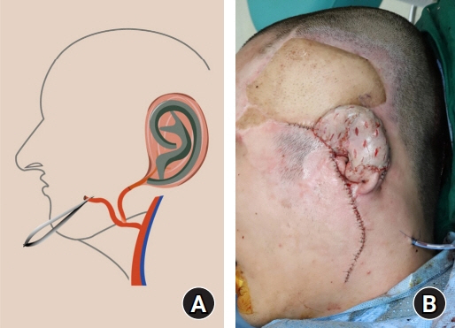
Fig. 3. (A) Polyethylene ear framework was covered with the fascia flap and laid on desired position. Pedicle was laid caudal to the external auditory meatus, through the subcutaneous tunnel beneath the remnant ear lobule. Microanastomosis was performed between the free flap perforator and facial artery. (B) Immediate postoperative photograph (lateral view).
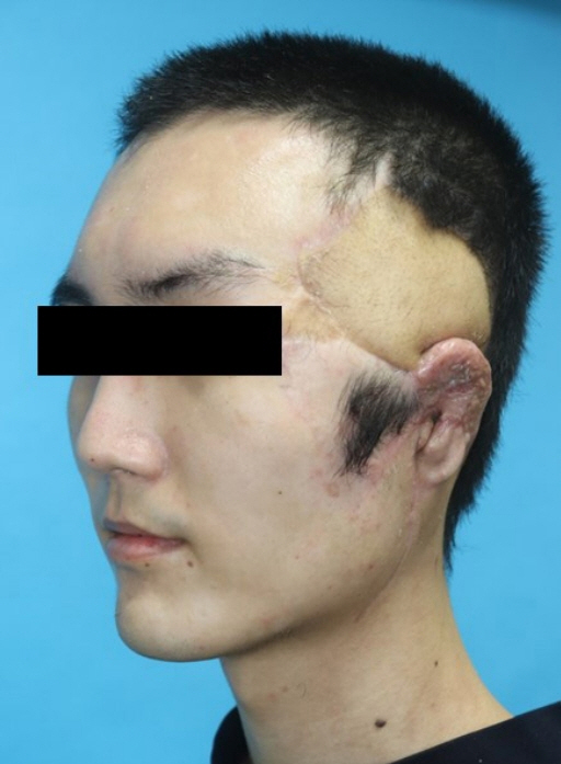
Fig. 4. A photograph of patient 1 month after the operation. The flap was well-taken without complication.
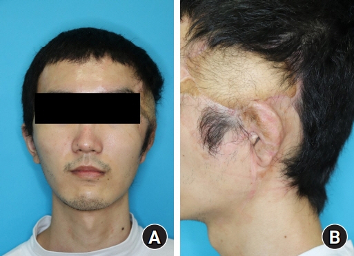
Fig. 5. A photograph of patient 9 months after the operation. (A) Front view. (B) Lateral view.
Reference
-
References
1. Gault D. Post traumatic ear reconstruction. J Plast Reconstr Aesthet Surg. 2008; 61 Suppl 1:S5–12.
Article2. Luo X, Yang J, Yang Q, Wang X. Classification and reconstruction of posttraumatic ear deformity. J Craniofac Surg. 2012; 23:654–7.
Article3. da Silva JC, Guimarães Filho W, de Oliveira Araújo BG. Ear reconstruction after traumatic injuries. Rev Bras Cir Plást. 2011; 26:428–32.4. Romo T, Fozo MS, Sclafani AP. Microtia reconstruction using a porous polyethylene framework. Facial Plast Surg. 2000; 16:15–22.
Article5. Frodel JL, Lee S. The use of high-density polyethylene implants in facial deformities. Arch Otolaryngol Head Neck Surg. 1998; 124:1219–23.
Article6. Tanzer RC. Total reconstruction of the auricle: the evolution of a plan of treatment. Plast Reconstr Surg. 1971; 47:523–33.7. Park C, Suk Roh T. Total ear reconstruction in the devascularized temporoparietal region: I. Use of the contralateral temporoparietal fascial free flap. Plast Reconstr Surg. 2001; 108:1145–53.
Article8. Bhandari PS. Total ear reconstruction in post burn deformity. Burns. 1998; 24:661–70.
Article9. Brent B, Byrd HS. Secondary ear reconstruction with cartilage grafts covered by axial, random, and free flaps of temporoparietal fascia. Plast Reconstr Surg. 1983; 72:141–52.
Article10. Pearl RA, Sabbagh W. Reconstruction following traumatic partial amputation of the ear. Plast Reconstr Surg. 2011; 127:621–9.
Article
- Full Text Links
-
- Actions
-
Cited
- CITED
-
- Close
- Share
-
- Similar articles
-
- Two Cases of Ear Reconstruction Using Prefabricated Radial Forearm Free Flap
- Long-term Follow-up of Reconstruction of the Hand with a Temporoparietal Fascial Free Flap
- Occurrence of contralateral breast cancer in a BRCA-positive breast cancer patient who underwent free TRAM flap reconstruction: a case report
- Reconstruction of a temporal scalp defect without ipsilateral donor vessel possibilities using a local transposition flap and a latissimus dorsi free flap anastomosed to the contralateral side: a case report
- PREFABRICATION OF AURICLE WITH ABDOMINAL FLAP AND CARTILAGE IN RABBIT
