Safety and Effectiveness of the Novel Catheter 3.0 System for Diagnostic Cerebral Angiography: A Pilot Study
- Affiliations
-
- 1Department of Radiology, Research Institute of Radiology, Asan Medical Center, University of Ulsan College of Medicine, Seoul, Korea
- 2Biomedical Engineering Research Center, Asan Institute for Life Sciences, Asan Medical Center, Seoul, Korea
- 3Department of Radiologic Technology, Asan Medical Center, University of Ulsan College of Medicine, Seoul, Korea
- KMID: 2535949
- DOI: http://doi.org/10.5469/neuroint.2022.00248
Abstract
- Purpose
The purpose of this study was to evaluate the safety and effectiveness of a new angiographic system (Catheter 3.0 system) using a 5 French (Fr), large-bore angiography catheter, a 0.032-inch stiff guidewire, and a continuous flushing system in diagnostic cerebral angiography.
Materials and Methods
This retrospective study included 30 consecutive patients who underwent transfemoral cerebral angiography using the Catheter 3.0 system from October 2019 to March 2020. As the control group, we included 30 consecutive patients examined before the Catheter 3.0 system was introduced. Procedural outcomes, including technical success, procedure time, dose metrics, procedure-related complications, and image quality were reviewed and analyzed.
Results
All transfemoral cerebral angiographies were performed for a diagnosis of unruptured intracranial aneurysms. The Catheter 3.0 system showed a significantly shorter fluoroscopy time (6.2 vs. 9.7 minutes, P=0.008) and lower fluoroscopy dose (387.2 vs. 614.4, P=0.002) compared with the conventional 4-Fr catheter system. The Catheter 3.0 system also showed better results in terms of procedural time (21.0 vs. 22.5 minutes, P=0.072) and technical success rate (98.1% vs. 94.0%, P=0.078), although a statistical significance was not reached. The complication rate and qualitative assessment of the digital subtraction angiography (DSA) image quality were similar between the two groups.
Conclusion
The Catheter 3.0 system using a 5 Fr catheter with a large inner diameter was convenient, effective, and safe compared with the conventional system in diagnostic cerebrovascular angiography.
Figure
-

Fig. 1. Components and configuration of the Catheter 3.0 system. Grafia: Sungjin-Hitech, Suwon, Korea, Anguis: Sungjin-Hitech, Suwon, Korea.
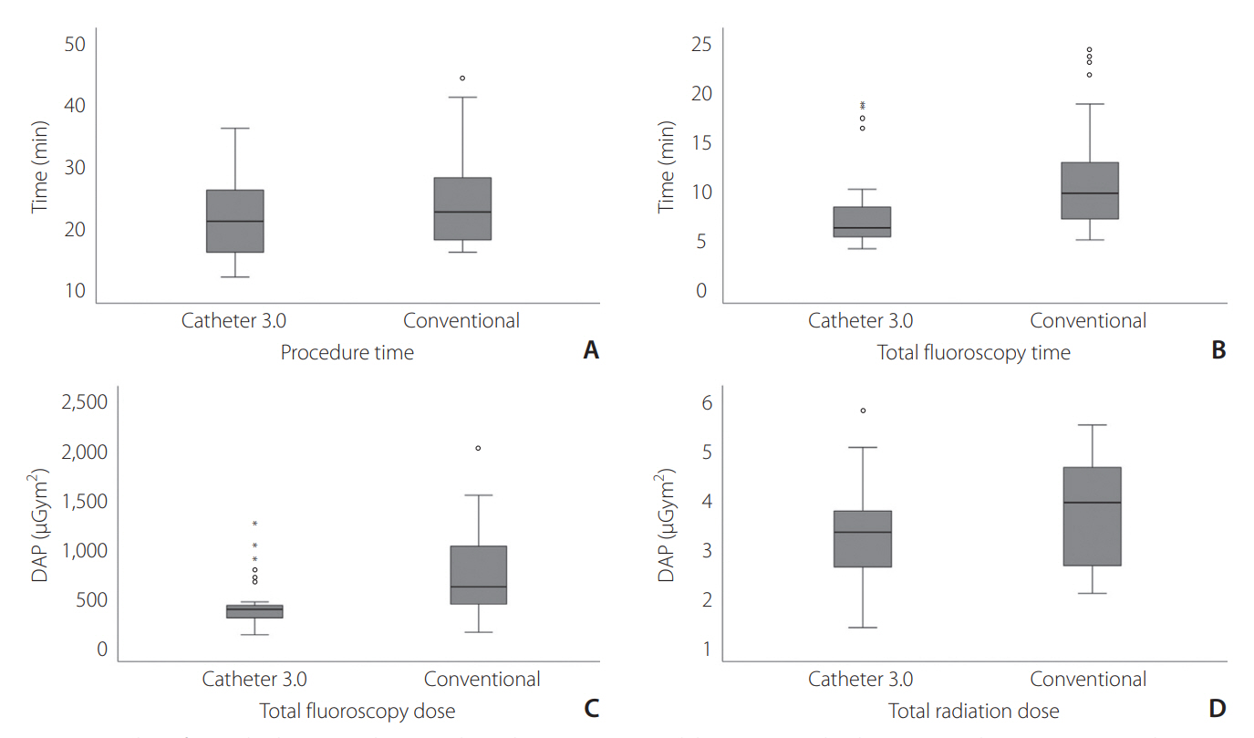
Fig. 2. Box plots of procedural outcomes between the Catheter 3.0 system and the conventional catheter system, demonstrating procedure time (A), total fluoroscopy time (B), total fluoroscopy dose (C), and total radiation dose (D). The top of the box represents the 75th percentile, the bottom of the box represents the 25th percentile, and the line in the middle represents the 50th percentile. Circles and asterisks represent the “outlier” and “extreme values”, respectively.
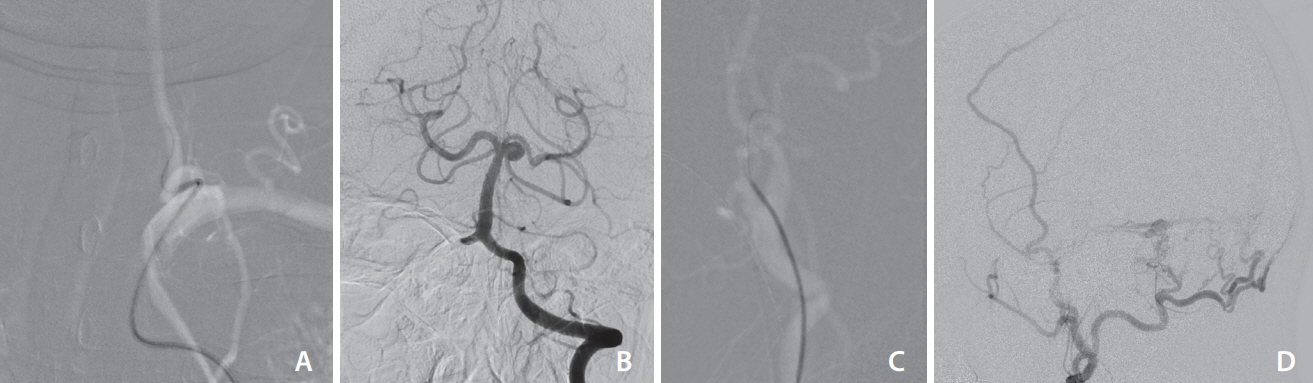
Fig. 3. Representative cases using the Catheter 3.0 system in tortuous vessels. (A) A patient had a marked tortuosity in the supraaortic neck vessel, which prevented stable and safe catheterization of the left vertebral artery. The guidewire leading in front of the catheter prevented the wedging of the catheter tip against the vessel wall and kickback of the catheter during contrast injection. (B) Despite flow competition from the contralateral vertebral artery, the aneurysm at the superior cerebellar artery origin of the basilar artery was well-visualized on the left vertebral arteriogram. (C) Catheterization of the left external carotid artery with marked tortuosity. The guidewire that entered the occipital artery guided the catheter toward the occipital artery without dislodging during contrast injection. (D) Good opacification of the feeders of the sigmoid sinus dural arteriovenous fistula arising from the occipital and middle meningeal arteries.
Reference
-
1. Alakbarzade V, Pereira AC. Cerebral catheter angiography and its complications. Pract Neurol. 2018; 18:393–398.
Article2. Dowd CF. Cerebral angiography: techniques and practice. In : Hetts SW, Cooke DL, editors. Handbook of clinical neurology. Amsterdam: Elsevier;2021. p. 107–119.3. Kwon B, Song Y, Hwang SM, Choi JH, Maeng J, Lee DH. Injection of contrast media using a large-bore angiography catheter with a guidewire in place: physical factors influencing injection pressure in cerebral angiography. Interv Neuroradiol. 2021; 27:558–565.
Article4. Ahn SH, Prince EA, Dubel GJ. Basic neuroangiography: review of technique and perioperative patient care. Semin Intervent Radiol. 2013; 30:225–233.
Article5. Shin JH. Cerebral angiography: the first small step for the neurointerventionist. Neurointervention. 2020; 15:101–103.
Article6. de Raadt A, Warrens MJ, Bosker RJ, Kiers HAL. A comparison of reliability coefficients for ordinal rating scales. J Classif. 2021; 38:519–543.7. Lee HJ, Yang PS, Lee SB, Yi JS, Ryu SY, Kim TW, et al. The influence of flush methods on transfemoral catheter cerebral angiography: continuous flush versus intermittent flush. J Vasc Interv Radiol. 2016; 27:651–657.
Article8. Wojak JC, Abruzzo TA, Bello JA, Blackham KA, Hirsch JA, Jayaraman MV, et al. Quality improvement guidelines for adult diagnostic cervicocerebral angiography: update cooperative study between the Society of Interventional Radiology (SIR), American Society of Neuroradiology (ASNR), and Society of NeuroInterventional Surgery (SNIS). J Vasc Interv Radiol. 2015; 26:1596–1608.
Article9. Thiex R, Norbash AM, Frerichs KU. The safety of dedicated-team catheter-based diagnostic cerebral angiography in the era of advanced noninvasive imaging. AJNR Am J Neuroradiol. 2010; 31:230–234.
Article10. Fifi JT, Meyers PM, Lavine SD, Cox V, Silverberg L, Mangla S, et al. Complications of modern diagnostic cerebral angiography in an academic medical center. J Vasc Interv Radiol. 2009; 20:442–447.
Article11. Kaufmann TJ, Huston J 3rd, Mandrekar JN, Schleck CD, Thielen KR, Kallmes DF. Complications of diagnostic cerebral angiography: evaluation of 19,826 consecutive patients. Radiology. 2007; 243:812–819.
Article12. Dawkins AA, Evans AL, Wattam J, Romanowski CA, Connolly DJ, Hodgson TJ, et al. Complications of cerebral angiography: a prospective analysis of 2,924 consecutive procedures. Neuroradiology. 2007; 49:753–759.
Article13. Cooperative Study between the ASNR ASITN, and the SCVIR. Quality improvement guidelines for adult diagnostic neuroangiography. Cooperative study between the ASNR, ASITN, and the SCVIR. American Society of Neuroradiology. American Society of Interventional and Therapeutic Neuroradiology. Society of Cardiovascular and Interventional Radiology. AJNR Am J Neuroradiol. 2000; 21:146–150.14. Cloft HJ, Jensen ME, Kallmes DF, Dion JE. Arterial dissections complicating cerebral angiography and cerebrovascular interventions. AJNR Am J Neuroradiol. 2000; 21:541–545.
- Full Text Links
-
- Actions
-
Cited
- CITED
-
- Close
- Share
-
- Similar articles
-
- A Study on the Pilot Fatigue Measurement Methods for Fatigue Risk Management
- Transient Cortical Blindness following Cerebral Angiography : Case Report
- Phase III Clinical Trial Comparing Iopamidol 250 mgI/mL and Iopamidol 300mgI/mL in Cerebral Angiography: Multicenteric, Randomized and Double Blind Study
- Comparative Analysis of Magnetic Resonance Angiography and 4-Vessel Angiography in the Diagnosis of Pediatric Moyamoya Disease
- Infraoptic Anterior Cerebral Artery Arising from Contralateral Internal Carotid Artery: Case Report
