A beginner’s guide to peripheral nerve ultrasound
- Affiliations
-
- 1Department of Neurology, School of Medicine, Catholic University of Daegu, Daegu, Korea
- KMID: 2535717
- DOI: http://doi.org/10.14253/acn.2022.24.2.46
Abstract
- Ultrasonography is currently being developed as a tool for evaluating peripheral neuropathy. It is one of the painless and least-invasive methods of medical diagnostic testing that yields anatomic views of the nerves and their surrounding structures. Here I first describe the equipment settings and technique for nerve ultrasound along with typical sonographic findings for normal nerves. I then address frequently used parameters for nerve measurements that facilitate diagnoses of focal and generalized neuropathies.
Figure
-
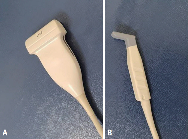
Fig. 1. The two types of ultrasound transducers specialized for musculoskeletal applications: linear array (A) and hockey stick (B).

Fig. 2. Normal sonographic anatomy of the median nerve at different levels. (A) The elbow was flexed to 80-90° with the forearm in full supination in a seated position. (B) Transverse image shows the median nerve (arrow) at the proximal tunnel (at the scaphoid-pisiform level). (C) The median nerve (arrow) lies between the flexor digitorum profundus and the flexor digitorum superficialis at the forearm. (D) The median nerve (arrow) lies close to the brachial artery (open arrowhead) at the distal third of the upper arm. BB, biceps brachii; Br, brachialis; H, humerus; fdp, flexor digitorum profundus; fds, flexor digitorum superficialis; fcr, flexor carpi radialis; S, scaphoid; P, pisiform; u, ulnar nerve; a, ulnar artery
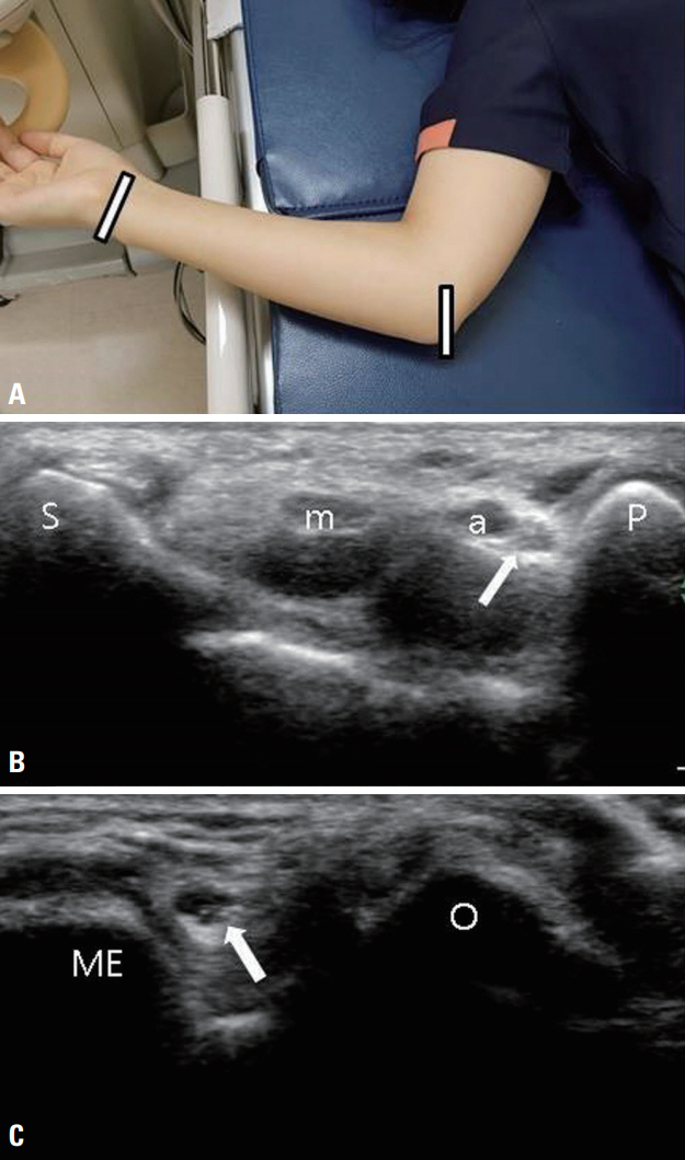
Fig. 3. Normal sonographic anatomy of the ulnar nerve at different levels. (A) The elbow was flexed at 90° in a supine position. (B) Transverse image shows the ulnar nerve (arrow) at the proximal tunnel (at the scaphoid-pisiform level). The ulnar artery and nerve run lateral to the pisiform. (C) Transverse image shows the ulnar nerve (arrow) at the medial epicondyle-olecranon level. m, median nerve; a, ulnar artery; S, scaphoid; P, pisiform; O, olecranon; ME, medial epicondyle.
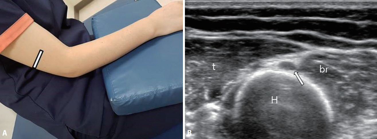
Fig. 4. Normal sonographic anatomy of the radial nerve at the spiral groove. (A) The elbow was flexed moderately with the forearm pronated in a seated position. (B) The radial nerve (arrow) lies in close contact with the humerus. t, triceps; br, brachialis; H, humerus.
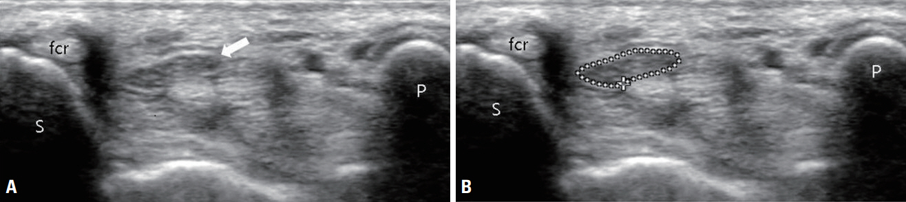
Fig. 5. Normal median nerve. (A) Transverse cross-sectional view of the median nerve (arrow) at the distal wrist crease in a healthy participant. (B) The tracing method performed just inside the hyperechoic rim for measuring the cross-sectional area of the median nerve. fcr, flexor carpi radialis tendon; S, scaphoid; P, pisiform.
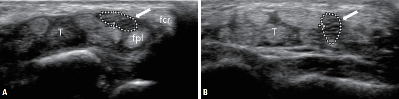
Fig. 6. Ultrasonographic images of the median nerve with full mobility. (A) At rest, the median nerve (arrow) is the most-superficial structure. (B) The median nerve (arrow) dives deep into the flexor tendon when the fingers and wrist are flexed. T, tendon; fpl, flexor pollicis longus; fcr, flexor carpi radialis.
Reference
-
1. Walker FO, Cartwright MS, Wiesler ER, Caress J. Ultrasound of nerve and muscle. Clin Neurophysiol. 2004; 115:495–507.2. Suk JI, Walker FO, Cartwright MS. Ultrasonography of peripheral nerves. Curr Neurol Neurosci Rep. 2013; 13:328.3. Silvestri E, Martinoli C, Derchi LE, Bertolotto M, Chiaramondia M, Rosenberg I. Echotexture of peripheral nerves: correlation between US and histologic findings and criteria to differentiate tendons. Radiology. 1995; 197:291–296.4. Duncan I, Sullivan P, Lomas F. Sonography in the diagnosis of carpal tunnel syndrome. AJR Am J Roentgenol. 1999; 173:681–684.5. Roll SC, Case-Smith J, Evans KD. Diagnostic accuracy of ultrasonography vs. electromyography in carpal tunnel syndrome: a systematic review of literature. Ultrasound Med Biol. 2011; 37:1539–1553.6. Beekman R, Visser LH, Verhagen WI. Ultrasonography in ulnar neuropathy at the elbow: a critical review. Muscle Nerve. 2011; 43:627–635.7. Seok JI, Lee SB, Bae CB. Ultrasonographic findings of the normal nerves in common entrapment site; cross-sectional area reference value and normal variant. J Korean Neurol Assoc. 2015; 33:8–12.8. Beekman R, Visser LH. Sonography in the diagnosis of carpal tunnel syndrome: a critical review of the literature. Muscle Nerve. 2003; 27:26–33.9. Beekman R, Van Der Plas JP, Uitdehaag BM, Schellens RL, Visser LH. Clinical, electrodiagnostic, and sonographic studies in ulnar neuropathy at the elbow. Muscle Nerve. 2004; 30:202–208.10. Yoon JS, Walker FO, Cartwright MS. Ultrasonographic swelling ratio in the diagnosis of ulnar neuropathy at the elbow. Muscle Nerve. 2008; 38:1231–1235.11. Hobson-Webb LD, Massey JM, Juel VC, Sanders DB. The ultrasonographic wrist-to-forearm median nerve area ratio in carpal tunnel syndrome. Clin Neurophysiol. 2008; 119:1353–1357.12. Cho JM, Yoon JS, Kim SJ, Park BK, Lee GH, Jeong JS. Feasibility of ultrasonographic area ratio of median nerve in the diagnosis of carpal tunnel syndrome in Korea. J Korean Acad Rehabil Med. 2009; 33:627–631.13. Bathala L, Kumar P, Kumar K, Shaik A, Visser LH. Normal values of median nerve cross-sectional area obtained by ultrasound along its course in the arm with electrophysiological correlations, in 100 Asian subjects. Muscle Nerve. 2014; 49:284–286.14. Mallouhi A, Pülzl P, Trieb T, Piza H, Bodner G. Predictors of carpal tunnel syndrome: accuracy of gray-scale and color Doppler sonography. AJR Am J Roentgenol. 2006; 186:1240–1245.15. Martinoli C, Bianchi S, Zamorani MP, Zunzunegui JL, Derchi LE. Ultrasound of the elbow. Eur J Ultrasound. 2001; 14:21–27.16. Boom J, Visser LH. Quantitative assessment of nerve echogenicity: comparison of methods for evaluating nerve echogenicity in ulnar neuropathy at the elbow. Clin Neurophysiol. 2012; 123:1446–1453.17. Watanabe T, Ito H, Sekine A, Katano Y, Nishimura T, Kato Y, et al. Sonographic evaluation of the peripheral nerve in diabetic patients: the relationship between nerve conduction studies, echo intensity, and cross-sectional area. J Ultrasound Med. 2010; 29:697–708.18. Tagliafico A, Tagliafico G, Martinoli C. Nerve density: a new parameter to evaluate peripheral nerve pathology on ultrasound. Preliminary study. Ultrasound Med Biol. 2010; 36:1588–1593.19. Ghasemi-Esfe AR, Khalilzadeh O, Vaziri-Bozorg SM, Jajroudi M, Shakiba M, Mazloumi M, et al. Color and power Doppler US for diagnosing carpal tunnel syndrome and determining its severity: a quantitative image processing method. Radiology. 2011; 261:499–506.20. Sernik RA, Abicalaf CA, Pimentel BF, Braga-Baiak A, Braga L, Cerri GG. Ultrasound features of carpal tunnel syndrome: a prospective case-control study. Skeletal Radiol. 2008; 37:49–53.21. Erel E, Dilley A, Greening J, Morris V, Cohen B, Lynn B. Longitudinal sliding of the median nerve in patients with carpal tunnel syndrome. J Hand Surg Br. 2003; 28:439–443.22. Wang Y, Filius A, Zhao C, Passe SM, Thoreson AR, An KN, et al. Altered median nerve deformation and transverse displacement during wrist movement in patients with carpal tunnel syndrome. Acad Radiol. 2014; 21:472–480.23. Seok JI, Lee DK. Physiological factors influencing median nerve mobility in normal subjects. Muscle Nerve. 2016; 54:883–886.24. Ozturk E, Sonmez G, Colak A, Sildiroglu HO, Mutlu H, Senol MG, et al. Sonographic appearances of the normal ulnar nerve in the cubital tunnel. J Clin Ultrasound. 2008; 36:325–329.
- Full Text Links
-
- Actions
-
Cited
- CITED
-
- Close
- Share
-
- Similar articles
-
- Normal Sonographic Peripheral Nerve Cross-Sectional Area Based on the Results of Meta-Analysis Studies
- Usefulness of Ultrasound for Detecting Suspected Peripheral Nerve Lesions in Diagnosis of Peripheral Neuropathy : Case Report and Brief Review of the Literature
- Diagnostic Usefulness of Neuromuscular Ultrasound in Anatomical Localization of Peripheral Nerve Injury: Detailed Lesion Localization Using Neuromuscular Ultrasound in a Patient with Traumatic Ulnar Nerve Injury at the Hand
- Neuromuscular Ultrasound in Wrist and Hand
- Ultrasound Guided Nerve Block at Vertebra and Lower Extremity
