Anesth Pain Med.
2022 Oct;17(4):397-403. 10.17085/apm.22170.
Detection of paradoxical carbon dioxide gas embolism with opening of patent foramen ovale by perioperative transesophageal echocardiography during laparoscopic hepatectomy - A case report -
- Affiliations
-
- 1Department of Anesthesiology and Pain Medicine, Daegu Fatima Hospital, Daegu, Korea
- KMID: 2535338
- DOI: http://doi.org/10.17085/apm.22170
Abstract
- Background
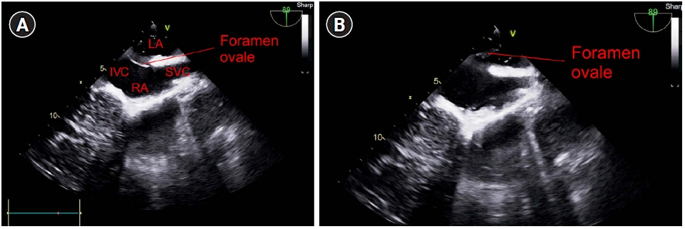
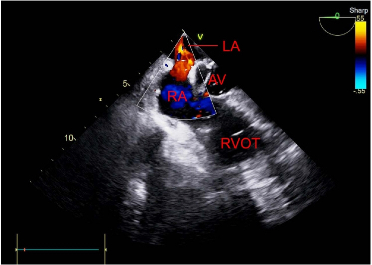
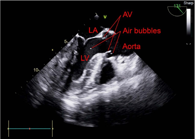
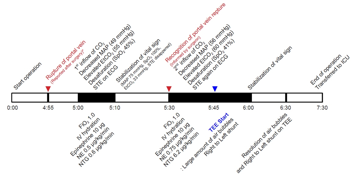
Due to its various advantages, laparoscopic surgery is preferred over laparotomy in patients who require hepatic resection. Carbon dioxide embolism —which occurs approximately ten times more often in laparoscopic hepatectomy than in general laparoscopic surgery—presents with insignificant symptoms and may be overlooked. Case: A 70-year-old male with hepatic cell carcinoma underwent laparoscopic hepatectomy. Though his vital signs were stable during the initiation of surgery, they became unstable during the procedure. The surgeon detected portal vein rupture, and transesophageal echocardiography was subsequently performed. A large amount of gas in the heart chamber and paradoxical embolism through a patent foramen ovale due to a right-to-left shunt were observed. We treated the symptoms, and the surgery was completed without any further issues.
Conclusions
Active use of transesophageal echocardiography to identify and monitor heart functions during a suspected carbon dioxide embolism can significantly reduce morbidity and mortality associated with that embolism.
Keyword
Figure
Reference
-
1. Hu J, Or BH, Hu K, Wang ML. Comparison of the post-operative outcomes and survival of laparoscopic versus open resections for gastric gastrointestinal stromal tumors: a multi-center prospective cohort study. Int J Surg. 2016; 33 Pt A:65–71.
Article2. Jobin LM. Carbon dioxide embolisms during laparoscopic surgery. Nurse Anesth Capstones. 2019; 26.3. Perugini RA, Callery MP. Complications of laparoscopic surgery. Surgical treatment: evidence-based and problem-oriented. In : Holzheimer RG, Mannick JA, editors. Munich: Zuckschwerdt;2001, p 2.4. Otsuka Y, Katagiri T, Ishii J, Maeda T, Kubota Y, Tamura A, et al. Gas embolism in laparoscopic hepatectomy: what is the optimal pneumoperitoneal pressure for laparoscopic major hepatectomy? J Hepatobiliary Pancreat Sci. 2013; 20:137–40.
Article5. Tranchart H, O'Rourke N, Van Dam R, Gaillard M, Lainas P, Sugioka A, et al. Bleeding control during laparoscopic liver resection: a review of literature. J Hepatobiliary Pancreat Sci. 2015; 22:371–8.
Article6. Park CH, Lee JY, Kim YC, Kim SH, Jung KH, Kim MG, et al. Detection of carbon dioxide embolism using transesophageal echocardiography during thoracoscopic sympathicotomy. Korean J Anesthesiol. 2006; 50:173–8.
Article7. Lee JY, Jo YS, Na SJ, Lee KE, Kim YD. A case of cerebral air embolism after removal of subclavian venous catheter. J Korean Neurol Assoc. 2005; 23:712–4.8. Nagelhout JJ, Plaus KL. Nurse anesthesia. 5th ed. St. Louis (MO): Elsevier/Saunders;2014.9. Park EY, Kwon JY, Kim KJ. Carbon dioxide embolism during laparoscopic surgery. Yonsei Med J. 2012; 53:459–66.
Article10. Hatano Y, Murakawa M, Segawa H, Nishida Y, Mori K. Venous air embolism during hepatic resection. Anesthesiology. 1990; 73:1282–5.
Article11. Gordy S, Rowell S. Vascular air embolism. Int J Crit Illn Inj Sci. 2013; 3:73–6.
Article12. Couture P, Boudreault D, Derouin M, Allard M, Lepage Y, Girard D, et al. Venous carbon dioxide embolism in pigs: an evaluation of end-tidal carbon dioxide, transesophageal echocardiography, pulmonary artery pressure, and precordial auscultation as monitoring modalities. Anesth Analg. 1994; 79:867–73.13. Shulman D, Aronson HB. Capnography in the early diagnosis of carbon dioxide embolism during laparoscopy. Can Anaesth Soc J. 1984; 31:455–9.
Article14. Guggiari M, Lechat P, Garen-Colonne C, Fusciardi J, Viars P. Early detection of patent foramen ovale by two-dimensional contrast echocardiography for prevention of paradoxical air embolism during sitting position. Anesth Analg. 1988; 67:192–4.
Article15. Wilmshurst PT, de Belder MA. Patent foramen ovale in adult life. Br Heart J. 1994; 71:209–12.
Article
- Full Text Links
- Actions
-
Cited
- CITED
-
- Close
- Share
- Similar articles
-
- Thrombus in Transit within a Patent Foramen Ovale: Gone with the Cough!
- A Case of Paradoxical Renal Embolism through Patent Foramen Ovale
- CT Diagnosis of Paradoxical Embolism via a Patent Foramen Ovale in a Patient with a Pulmonary Embolism and Prominent Eustachian Valve
- A Case of Impending Paradoxical Embolus in a Patient with Acute Pulmonary Embolism
- Renal infarction caused by paradoxical embolism through a patent foramen ovale


