J Korean Assoc Oral Maxillofac Surg.
2022 Oct;48(5):315-317. 10.5125/jkaoms.2022.48.5.315.
Osseous metaplasia of the palate: a case report
- Affiliations
-
- 1Department of Oral and Maxillofacial Surgery, Dankook University Dental Hospital, Cheonan, Korea
- KMID: 2534813
- DOI: http://doi.org/10.5125/jkaoms.2022.48.5.315
Abstract
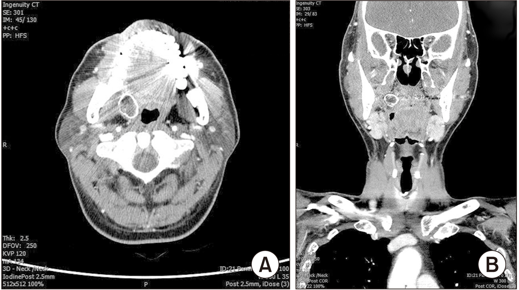
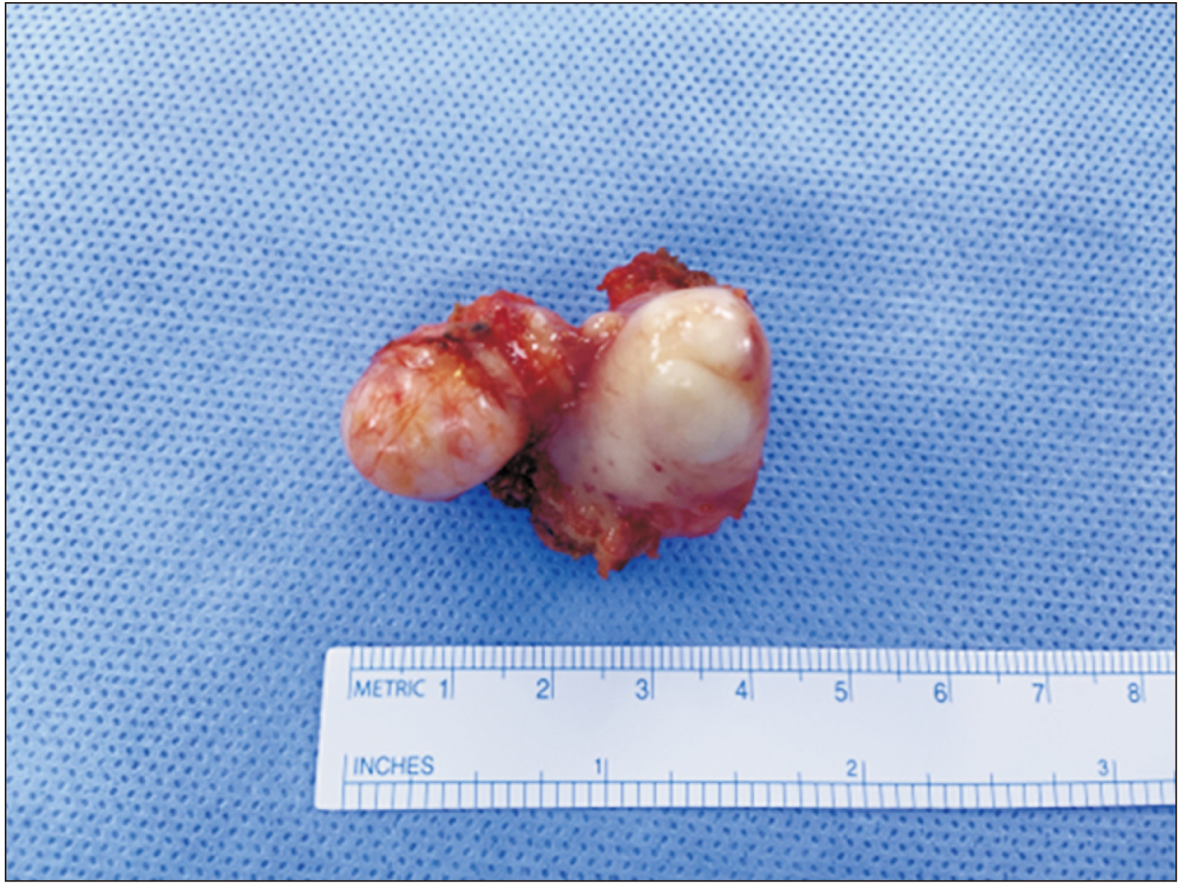
- Osseous metaplasia is defined as the formation of lamellar bone inside soft tissue structures where bone normally does not exist. It results from the transformation of non-osseous connective tissue into mature bone. This condition is rare in the oral and maxillofacial region. We report a case of osseous metaplasia of the maxilla, a rare benign tumor in an uncommon region. A 60-year-old male patient visited our clinic complaining of foreign body sensation and asymptomatic swelling on the right palatal side. However, he did not experience pain and reported no local trauma that he could remember. Intra-oral examination revealed an exophytic lesion on the right palatal portion. On computed tomography, there was a round hard-tissue mass approximately 2 cm in diameter on the right palate area. The mass was biopsied and diagnosed as an osseous metaplasia. We review the clinical, radiographic, and histologic features and common causes of osseous metaplasia and report a rare case of osseous metaplasia of the palate.
Keyword
Figure
Reference
-
References
1. Hong SH, Lee YB, Jung YS, Jung HD. 2016; Heterotopic ossification. J Int Soc Simul Surg. 3:84–6. https://doi.org/10.18204/JISSiS.2016.3.2.084. DOI: 10.18204/JISSiS.2016.3.2.084. PMID: 26135077. PMCID: PMC6948799.
Article2. Chun JS, Hong R, Kim JA. 2013; Osseous metaplasia with mature bone formation of the thyroid gland: three case reports. Oncol Lett. 6:977–9. https://doi.org/10.3892/ol.2013.1475. DOI: 10.3892/ol.2013.1475. PMID: 24137448. PMCID: PMC3796393.
Article3. Lee JY, Lee HA, Kwon HM, Na SH, Hwang JY, Lee DH. 2012; A case of endometrial osseous metaplasia treated by hysteroscopic operation. Korean J Obstet Gynecol. 55:361–5. https://doi.org/10.5468/KJOG.2012.55.5.361. DOI: 10.5468/KJOG.2012.55.5.361.
Article4. Cutright DE. 1972; Osseous and chondromatous metaplasia caused by dentures. Oral Surg Oral Med Oral Pathol. 34:625–33. https://doi.org/10.1016/0030-4220(72)90346-5. DOI: 10.1016/0030-4220(72)90346-5. PMID: 4506720.
Article5. Maitra S, Gupta D, Radojkovic M, Sood S. 2009; Osseous metaplasia of the maxillary sinus with formation of a well-developed haversian system and bone marrow. Ear Nose Throat J. 88:1115–20. DOI: 10.1177/014556130908800907. PMID: 19750475.
Article6. Bennett JH, Jones J, Speight PM. 1993; Odontogenic squamous cell carcinoma with osseous metaplasia. J Oral Pathol Med. 22:286–8. https://doi.org/10.1111/j.1600-0714.1993.tb01073.x. DOI: 10.1111/j.1600-0714.1993.tb01073.x. PMID: 8117349.
Article7. Vencio EF, Alencar RC, Zancope E. 2007; Heterotopic ossification in the anterior maxilla: a case report and review of the literature. J Oral Pathol Med. 36:120–2. https://doi.org/10.1111/j.1600-0714.2007.00467.x. DOI: 10.1111/j.1600-0714.2007.00467.x. PMID: 17238976.
Article
- Full Text Links
- Actions
-
Cited
- CITED
-
- Close
- Share
- Similar articles
-
- Lipogranuloma with Osseous Metaplasia in the Breast That Developed after "Bu-Hwang" Oriental Medicine Treatment
- Endometrial Osseous Metaplasia: Sonographic Findings
- A case of endometrial osseous metaplasia of a referred endometria l cancer patient
- A Case of Endometrial Osseous Metaplasia
- Pulmonary Alveolar Proteinosis accompanied by Osseous Metaplasia: A case report


