Isolated oculomotor nerve palsy secondary to non-aneurysmal subarachnoid hemorrhage
- Affiliations
-
- 1Department of Neurosurgery, Drexel University College of Medicine, Philadelphia, PA, USA
- 2Department of Neurosurgery, David Geffen School of Medicine at UCLA, University of California Los Angeles, Los Angeles, CA, USA
- 3Department of Neurosurgery, Global Neurosciences Institute, Chester, PA, USA
- KMID: 2533679
- DOI: http://doi.org/10.7461/jcen.2022.E2021.06.007
Abstract
- We present a case series of two patients who developed unilateral cranial nerve III (CNIII) palsy following non-aneurysmal SAH (NASAH). Subarachnoid hemorrhage (SAH) can present with various signs and symptoms. Early diagnosis is paramount to determine treatment course. Thus, clinicians must be aware of the variable clinical presentations of this condition. Two patients were admitted to a single institution for SAH. Patient 1, 52-year-old male, presented with headache, left eye ptosis, and painless diplopia. A non-contrast head computed tomography (CT) demonstrated a SAH within the left sylvian fissure and blood surrounding the mesencephalon and falx. Patient 2, 70-year-old male, presented with mild headache, acute onset of blurry vision, and right eye ptosis. A non-contrast head CT demonstrated a diffuse SAH predominantly in the Sylvian and suprasellar cisterns. Patients were admitted to the neuro intensive care unit and underwent diagnostic angiograms to identify possible aneurysms. Magnetic resonance imaging and angiograms for both patients were negative. Patients were managed with best medical therapy and followed up in the outpatient setting. Unilateral CNIII palsy in the setting of NASAH was identified in both patients. Diagnostic angiograms were negative for aneurysms; therefore, SAH were determined to be spontaneous. We propose that unilateral CNIII palsy is a possible sign of NASAH.
Keyword
Figure
-
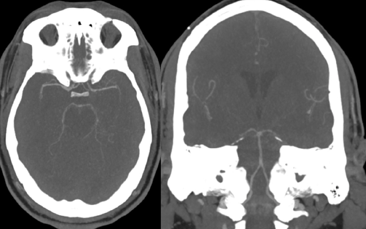
Fig. 1. CT Angiography of the posterior circulation demonstrating no evidence of AVM or aneurysm. CT, computed tomography; AVM, arteriovenous malformations
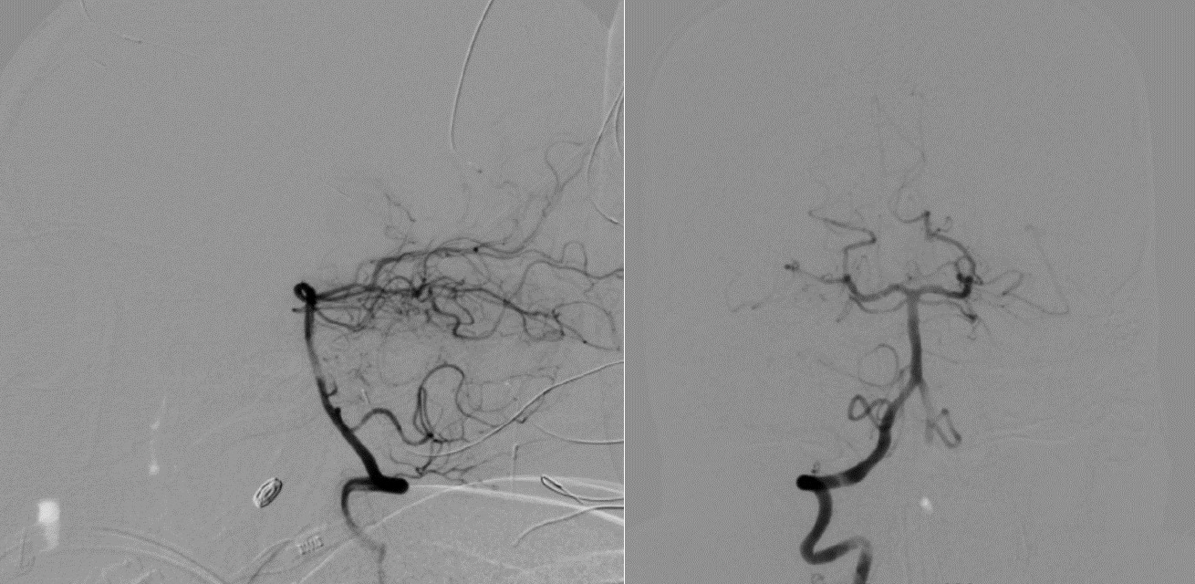
Fig. 2. Digital subtraction angiogram of the posterior circulation demonstrating no evidence of AVM or aneurysm (Patient 1). AVM, arteriovenous malformations
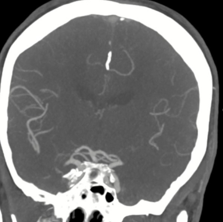
Fig. 3. CTA demonstrating no evidence of AVM or aneurysm. CTA, computed tomography angiogram; AVM, arteriovenous malformations
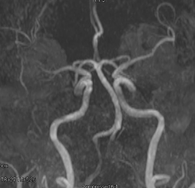
Fig. 4. MRA demonstrating no evidence of AVM or aneurysm. MRA, magnetic resonance angiogram; AVM, arteriovenous malformations
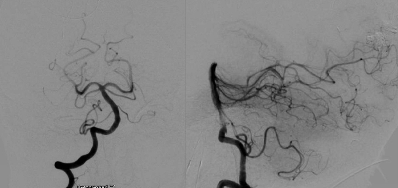
Fig. 5. Digital subtraction angiogram of the posterior circulation demonstrating no evidence of AVM or aneurysm (Patient 2). AVM, arteriovenous malformations
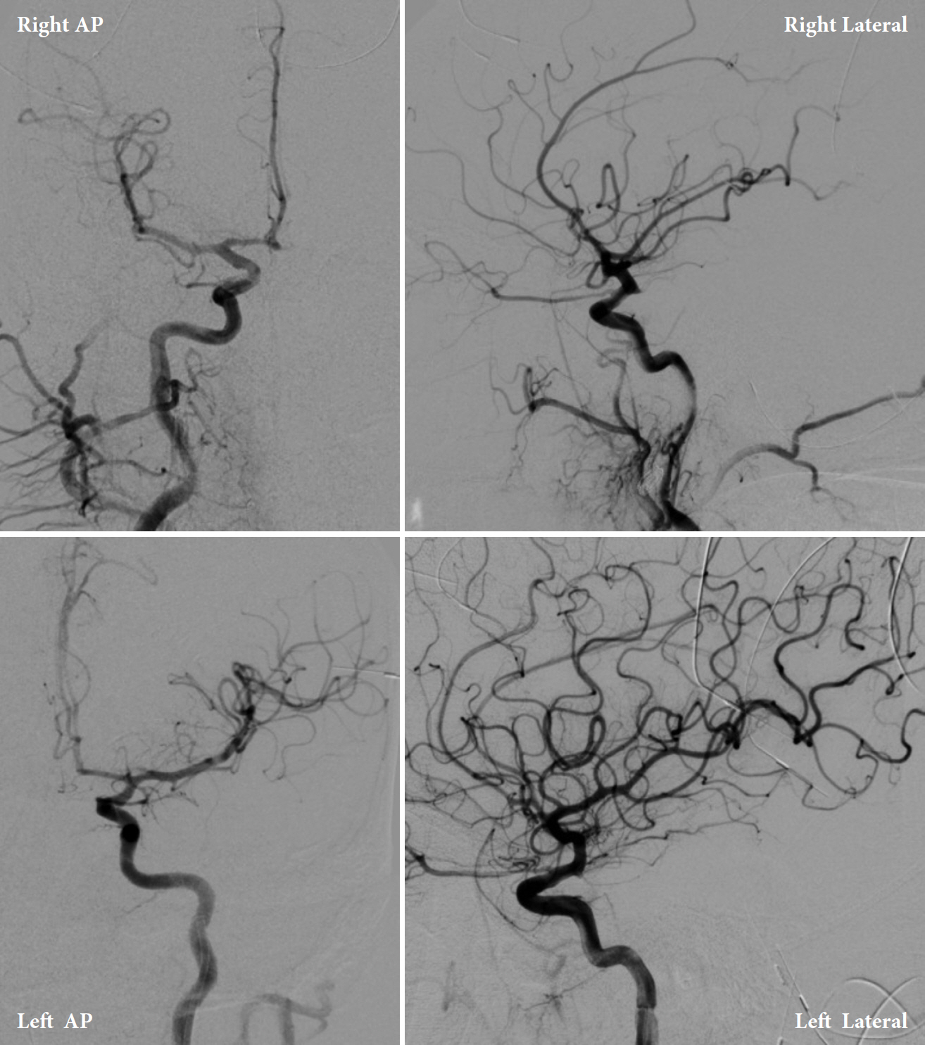
Fig. 6. Patient 1. internal carotid artery angiogram. AP, anteroposterior
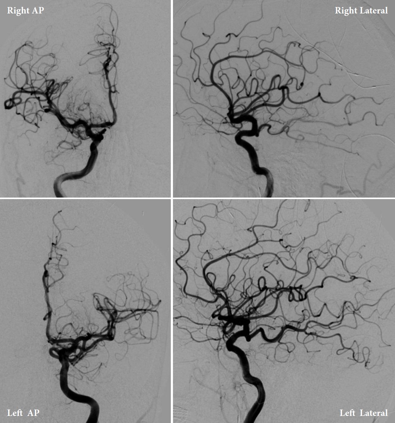
Fig. 7. Patient 2. internal carotid artery angiogram. AP, anteroposterior
Reference
-
1. Abbatemarco JR, Yacoub HA. Isolated cranial nerve-III palsy secondary to perimesencephalic subarachnoid hemorrhage. Case Rep Neurol Med. 2016; 2016:6319548.
Article2. Bartleson JD, Trautmann JC, Sundt TM Jr. Minimal oculomotor nerve paresis secondary to unruptured intracranial aneurysm. Arch Neurol. 1986; Oct. 43(10):1015–20.
Article3. Biousse V, Newman NJ. Third nerve palsies. Semin Neurol. 2000; 20(1):55–74.
Article4. Brismar J, Sundbärg G. Subarachnoid hemorrhage of unknown origin: prognosis and prognostic factors. J Neurosurg. 1985; Sep. 63(3):349–54.
Article5. Chen PR, Amin-Hanjani S, Albuquerque FC, McDougall C, Zabramski JM, Spetzler RF. Outcome of oculomotor nerve palsy from posterior communicating artery aneurysms: Comparison of clipping and coiling. Neurosurgery. 2006; Jun. 58(6):1040–6.
Article6. Coelho LG, Costa JM, Silva EI. Non-aneurysmal spontaneous subarachnoid hemorrhage: Perimesencephalic versus non-perimesencephalic. Rev Bras Ter Intensiva. 2016; Jun. 28(2):141–6.
Article7. Cornejo R, Romero C, Ugalde D, Bustos P, Diaz G, Galvez R, et al. High-volume hemofiltration and prone ventilation in subarachnoid hemorrhage complicated by severe acute respiratory distress syndrome and refractory septic shock. Rev Bras Ter Intensiva. 2014; Apr-Jun. 26(2):193–9.
Article8. du Mesnil de Rochemont R, Heindel W, Wesselmann C, Krüger K, Lanfermann H, Ernestus RI, et al. Nontraumatic subarachnoid hemorrhage: Value of repeat angiography. Radiology. 1997; Mar. 202(3):798–800.
Article9. Gottlob I, Catalano RA, Reinecke RD. Surgical management of oculomotor nerve palsy. Am J Ophthalmol. 1991; Jan. 111(1):71–6.
Article10. Hou X, Li C, Yang D, Li D, Zeng L, Mei Y. Diffusion tensor imaging tractography detecting isolated oculomotor paralysis caused by pituitary apoplexy. Neurologist. 2020; Nov. 25(6):157–61.
Article11. Ildan F, Tuna M, Erman T, Göçer AI, Cetinalp E. Prognosis and prognostic factors in nonaneurysmal perimesencephalic hemorrhage: A follow-up study in 29 patients. Surg Neurol. 2002; Mar. 57(3):160–5.12. Inamasu J, Nakamura Y, Saito R, Kuroshima Y, Ohba S, Ichikizaki K. Early resolution of third nerve palsy following endovascular treatment of a posterior communicating artery aneurysm. J Neuroophthalmol. 2002; Mar. 22(1):12–4.
Article13. Indraswari F, Mukharesh L, Burger KM, Leon Guerrero CR. Cases of stroke presenting with an isolated third nerve palsy. Stroke. 2021; Jan. 52(2):e58–60.
Article14. Jacobson DM, Trobe JD. The emerging role of magnetic resonance angiography in the management of patients with third cranial nerve palsy. Am J Ophthalmol. 1999; Jul. 128(1):94–6.
Article15. Kamat AA, Tizzard S, Mathew B. Painful third nerve palsy in a patient with perimesencephalic subarachnoid haemorrhage. Br J Neurosurg. 2005; Jun. 19(3):247–50.
Article16. Lee AG, Onan HW, Brazis PW, Prager TC. An imaging guide to the evaluation of third cranial nerve palsies. Strabismus. 1999; Sep. 7(3):153–68.
Article17. Levy RL, Geist CE, Miller NR. Isolated oculomotor palsy following minor head trauma. Neurology. 2005; Jul. 65(1):169.
Article18. Lustbader JM, Miller NR. Painless, pupil-sparing but otherwise complete oculomotor nerve paresis caused by basilar artery aneurysm. Arch Ophthalmology. 1988; May. 106(5):583–4.
Article19. Modi PA, Arsiwalla T. Cranial Nerve III Palsy. Treasure Island, FL: StatPearls Publishing;2020.20. Raza HK, Chen H, Chansysouphanthong T, Cui G. The aetiologies of the unilateral oculomotor nerve palsy: a review of the literature. Somatosens Mot Res. 2018; 35(3-4):229–39.
Article21. Rinkel GJ, Wijdicks EF, Hasan D, Kienstra GE, Franke CL, Hageman LM, et al. Outcome in patients with subarachnoid haemorrhage and negative angiography according to pattern of haemorrhage on computed tomography. Lancet. 1991; Oct. 338(8773):964–8.
Article22. Sadamasa N, Sano N, Takeda N, Yoshida K, Narumi O, Chin M, et al. Perimesencephalic nonaneurysmal subarachnoid hemorrhage with unilateral third cranial nerve palsy: two case reports. Neurol Med Chir (Tokyo). 2012; 52(12):918–20.23. Schwartz TH, Solomon RA. Perimesencephalic nonaneurysmal subarachnoid hemorrhage: Review of the literature. Neurosurgery. 1996; Sep. 39(3):433–40.
Article24. Singh AK, Agarwal P, Baiswar R. Isolated oculomotor nerve palsy - A rare initial manifestation of tuberculous meningitis. J Assoc Physicians India. 2020; Nov. 68(11):73–4.25. Tatter SB, Crowell RM, Ogilvy CS. Aneurysmal and microaneurysmal “angiogram-negative” subarachnoid hemorrhage. Neurosurgery. 1995; Jul. 37(1):48–55.
Article26. Topcuoglu MA, Ogilvy CS, Carter BS, Buonanno FS, Koroshetz WJ, Singhal AB. Subarachnoid hemorrhage without evident cause on initial angiography studies: Diagnostic yield of subsequent angiography and other neuroimaging tests. J Neurosurg. 2003; Jun. 98(6):1235–40.
Article27. Van Calenbergh F, Plets C, Goffin J, Velghe L. Nonaneurysmal subarachnoid hemorrhage: Prevalence of perimesencephalic hemorrhage in a consecutive series. Surg Neurol. 1993; Apr. 39(4):320–3.
Article28. van Gijn J, Rinkel GJ. Subarachnoid haemorrhage: Diagnosis, causes and management. Brain. 2001; Feb. 124(Pt 2):249–78.
Article29. van Gijn J, van Dongen KJ, Vermeulen M, Hijdra A. Perimesencephalic hemorrhage: A nonaneurysmal and benign form of subarachnoid hemorrhage. Neurology. 1985; Apr. 35(4):493–7.
Article30. Witthayaweerasak J, Tansuebchueasai N, Aui-Aree N. Clinical prediction score for early neuroimaging in acquired isolated oculomotor nerve palsy. Eye Brain. 2020; Jul. 12:89–95.
- Full Text Links
-
- Actions
-
Cited
- CITED
-
- Close
- Share
-
- Similar articles
-
- Pituitary Apoplexy Presenting as Isolated Oculomotor Nerve Palsy
- Neuroophthalmologic Findings of Intracranial Aneurysm
- Direct Oculomotor Nerve Palsy in Association with A Mild Head Injury: A Case Report
- Oculomotor Nerve Palsy due to Ruptured Multiple Anterior Choroidal Artery Aneurysms
- Oculomotor Nerve Palsies in Intracranial Aneurysms
