Multiple venous variations at the abdominopelvic region: a case report
- Affiliations
-
- 1Department of Basic Medical Sciences, Manipal Academy of Higher Education, Manipal, Karnataka, India
- 2Department of Mathematics, Manipal Institute of Technology, Manipal Academy of Higher Education, Manipal, Karnataka, India
- KMID: 2533369
- DOI: http://doi.org/10.5115/acb.22.066
Abstract
- Knowledge of vascular variations of the abdominopelvic junction is of importance to surgeons, radiologists, orthopaedic surgeons and other medical disciplines. We report a rare combination of venous variations observed at the abdominopelvic junction of an adult male cadaver. The right common iliac vein was absent. The inferior vena cava was formed by the union of the right external iliac vein and the left common iliac vein. The right internal iliac vein was a tributary of the left common iliac vein. The left common iliac vein was larger than usual in size and its wall was adhered to the right common iliac artery. We discuss the functional, developmental and clinical issues related to the case.
Figure
-
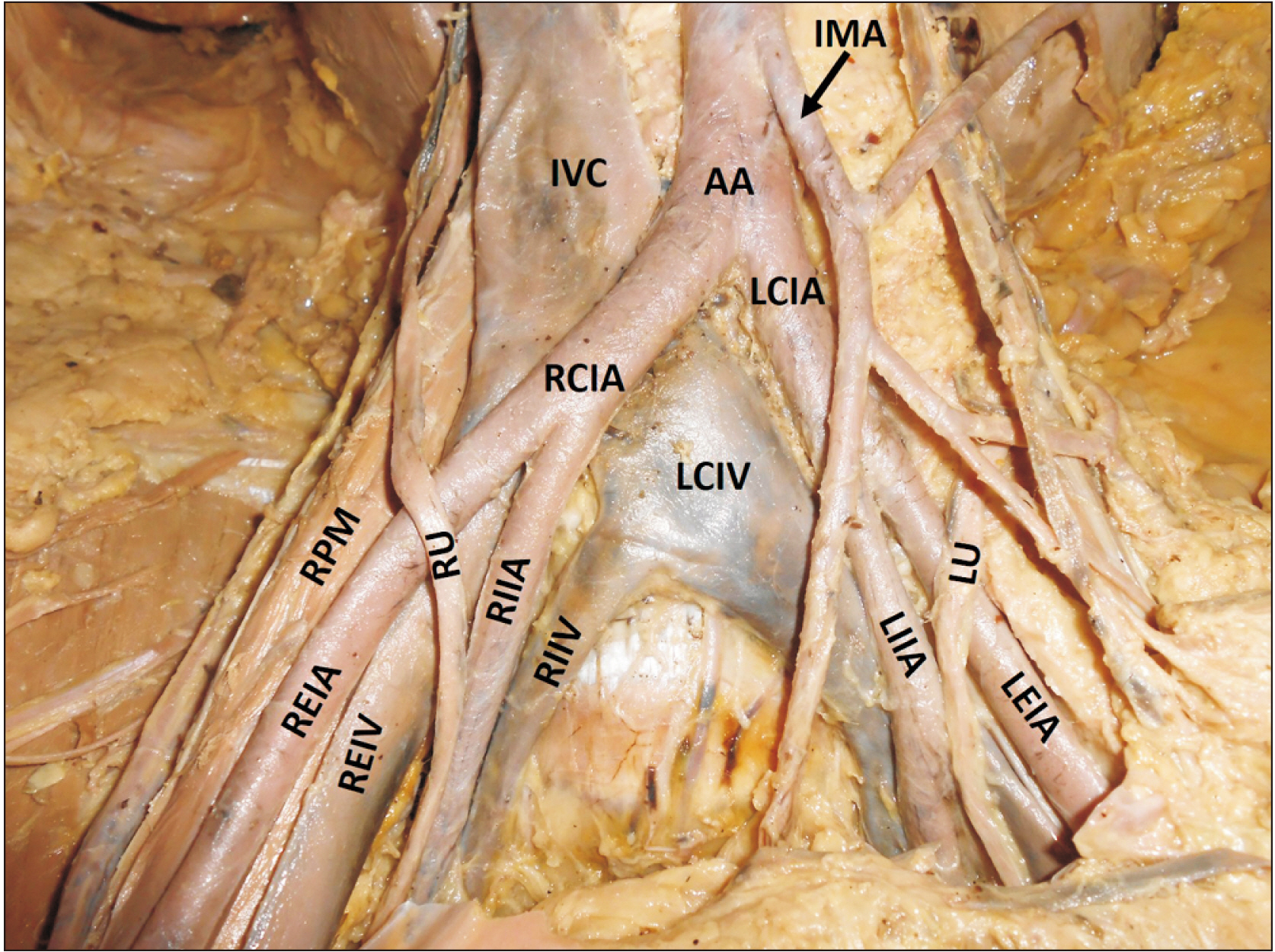
Fig. 1 Dissection of the abdominopelvic junction showing the blood vessels. IVC, inferior vena cava; LCIV, left common iliac vein; RIIV, right internal iliac vein; REIV, right external iliac vein; AA, abdominal aorta; LCIA, left common iliac artery; LEIA, left external iliac artery; LIIA, left internal iliac artery; RCIA, right common iliac artery; REIA, right external iliac artery; RIIA, right internal iliac artery; IMA, inferior mesenteric artery; RPM, right psoas major muscle; RU, right ureter; LU, left ureter.
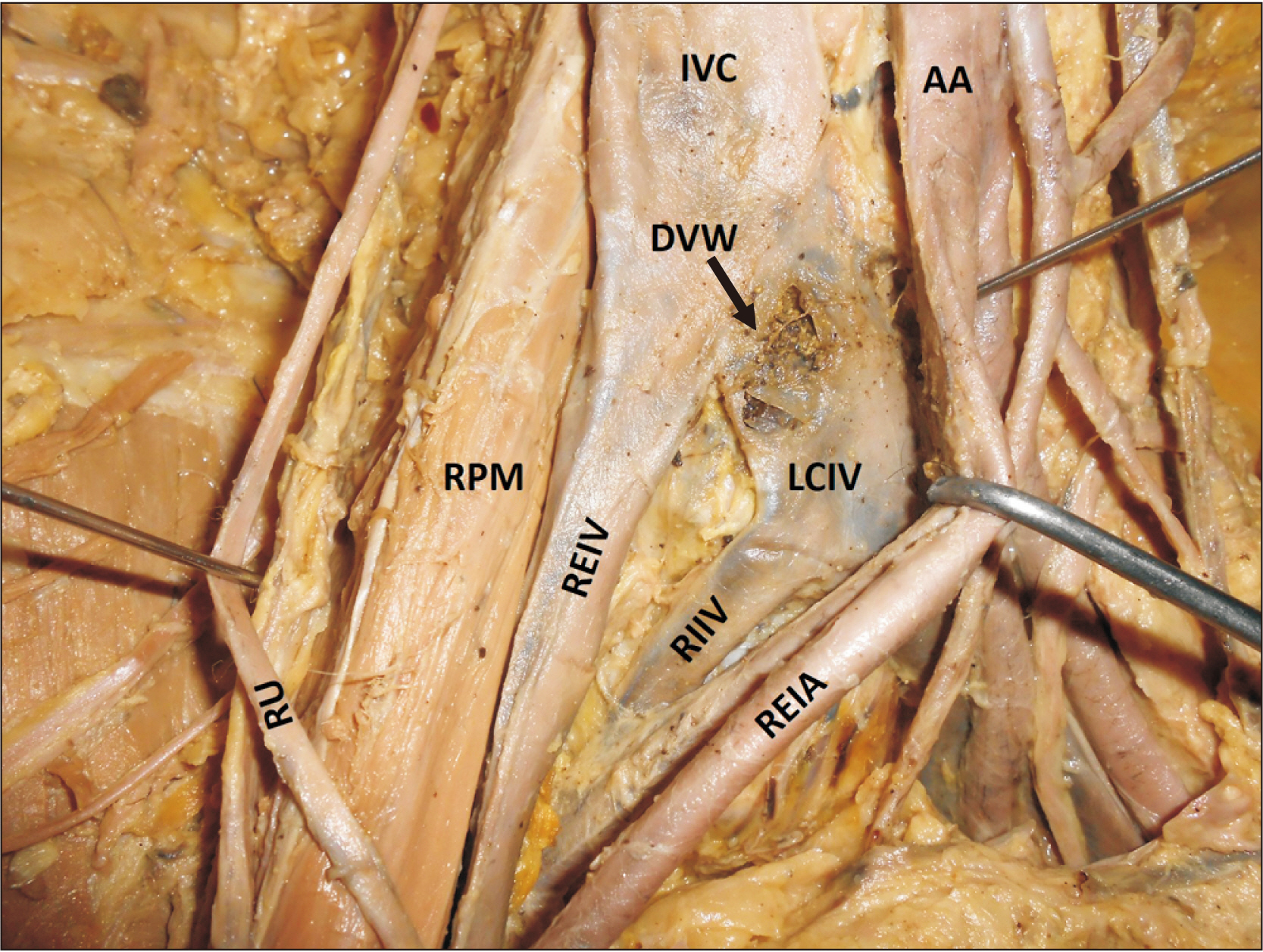
Fig. 2 Closer view of the variant blood vessels. The right external iliac artery has been pulled to the left to show the damaged wall of the left common iliac vein. IVC, inferior vena cava; LCIV, left common iliac vein; RIIV, right internal iliac vein; REIV, right external iliac vein; AA, abdominal aorta; REIA, right external iliac artery; RPM, right psoas major muscle; RU, right ureter; DVW, damaged wall of the left common iliac vein.
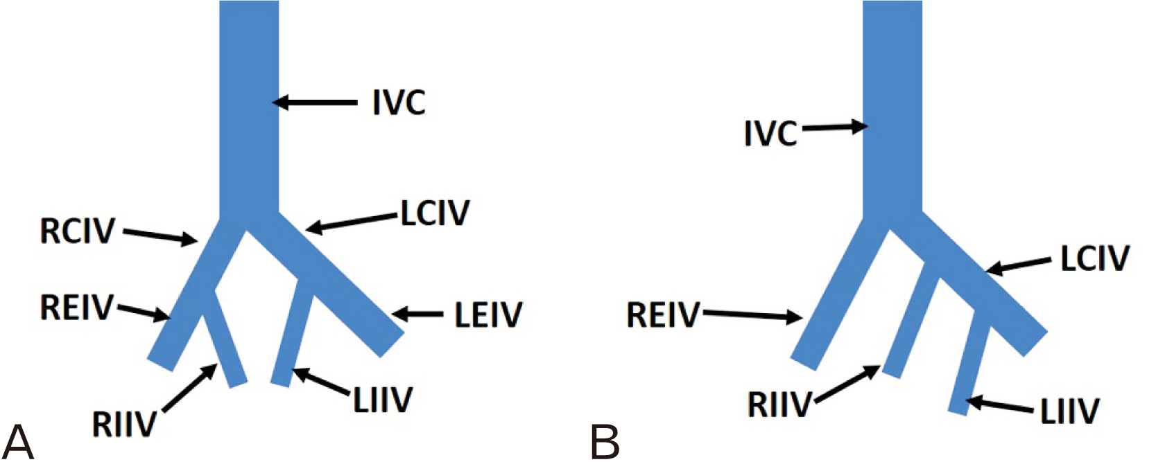
Fig. 3 A simplified, schematic diagram of the veins at the abdominopelvic junction. (A) The normal pattern of veins and (B) the variation observed. IVC, inferior vena cava; RCIV, right common iliac vein; LCIV, left common iliac vein; REIV, right external iliac vein; LEIV, left external iliac vein; RIIV, right internal iliac vein; LIIV, left internal iliac vein.
Reference
-
References
1. Nayak SB, Vasudeva SK. 2021; Jun. 16. Variant formation of left common iliac vein by the confluence of four veins. Morphologie. [Epub]. https://doi.org/10.1016/j.morpho.2021.05.116. DOI: 10.1016/j.morpho.2021.05.116. PMID: 34147368.
Article2. Nayak SB, Surendran S, Nelluri VM, Shetty P. 2019; Morphological and histological study of an 'iliac venous ladder' associated with very short common iliac arteries. J Morphol Sci. 36:255–60. DOI: 10.1055/s-0039-1697009.
Article3. Bass JE, Redwine MD, Kramer LA, Huynh PT, Harris JH Jr. 2000; Spectrum of congenital anomalies of the inferior vena cava: cross-sectional imaging findings. Radiographics. 20:639–52. DOI: 10.1148/radiographics.20.3.g00ma09639. PMID: 10835118.
Article4. Babaian RJ, Johnson DE. 1979; Major venous anomalies complicating retroperitoneal surgery. South Med J. 72:1254–8. DOI: 10.1097/00007611-197910000-00012. PMID: 482980.
Article5. Baldridge ED Jr, Canos AJ. 1987; Venous anomalies encountered in aortoiliac surgery. Arch Surg. 122:1184–8. DOI: 10.1001/archsurg.1987.01400220094018. PMID: 3662801.
Article6. Keating EM, Ritter MA, Faris PM. 1990; Structures at risk from medially placed acetabular screws. J Bone Joint Surg Am. 72:509–11. DOI: 10.2106/00004623-199072040-00006. PMID: 2324136.
Article7. Mirkovic S, Abitbol JJ, Steinman J, Edwards CC, Schaffler M, Massie J, Garfin SR. 1991; Anatomic consideration for sacral screw placement. Spine (Phila Pa 1976). 16(6 Suppl):S289–94. DOI: 10.1097/00007632-199106001-00022. PMID: 1862427.
Article8. Edwards WR. 1996; Hysterectomy, massive transfusion and packing to control haemorrhage from pelvic veins in the course of bilateral oophorectomy. Aust N Z J Obstet Gynaecol. 36:82–4. DOI: 10.1111/j.1479-828X.1996.tb02931.x. PMID: 8775260.
Article9. Oto A, Akpinar E, Sürücü HS, Denk CC, Celik HH. 2003; Right internal iliac vein joining the left common iliac vein: case report demonstrated by CT angiography. Surg Radiol Anat. 25:339–41. DOI: 10.1007/s00276-003-0123-0. PMID: 12910379.
Article10. Caggiati A, Amore M, Sedati P. 2016; Confluence of the right internal iliac vein into a compressed left common iliac vein. Phlebology. 31:145–6. DOI: 10.1177/0268355515586098. PMID: 25956550.
Article
- Full Text Links
-
- Actions
-
Cited
- CITED
-
- Close
- Share
-
- Similar articles
-
- Multiple Vascular Variations in Posterior Abdominal Region: A Case Report
- Inadvertent Arterial Catheterization of Central Venous Catheter: A Case Report
- Duplication of the ovarian vein: comprehensive review and case illustration
- Anatomical Variations Encountered during Adrenal Venous Sampling: A Report of Three Case Series and Review of Literature
- Incidental Musculoskeletal Lesions Detected on Abdominopelvic CT Scans: A Pictorial Essay
