Anat Cell Biol.
2022 Sep;55(3):380-383. 10.5115/acb.22.016.
A large sublingual glandular branch of the lingual nerve: a rare case report
- Albuck A
 1
1 - Haikata Y2,3
- Watanabe K2
- Tubbs RS4,5,6,7,8,
- Iwanaga J2,3,4,5
- Affiliations
-
- 1Tulane University School of Medicine, New Orleans, LA, USA.
- 2Division of Gross and Clinical Anatomy, Department of Anatomy, Kurume University School of Medicine, Kurume, Fukuoka, Japan.
- 3Dental and Oral Medical Center, Kurume University School of Medicine, Kurume, Fukuoka, Japan.
- 4Department of Neurosurgery, Tulane Center for Clinical Neurosciences, Tulane University School of Medicine, New Orleans, LA, USA.
- 5Department of Neurology, Tulane Center for Clinical Neurosciences, Tulane University School of Medicine, New Orleans, LA, USA.
- 6Department of Anatomical Sciences, St. George's University, St. George's, Grenada, West Indies.
- 7Department of Structural & Cellular Biology, Tulane University School of Medicine, New Orleans, LA, USA.
- 8Department of Neurosurgery and Ochsner Neuroscience Institute, Ochsner Health System, New Orleans, LA, USA.
- 9Department of Surgery, Tulane University School of Medicine, New Orleans, LA, USA.
- 10University of Queensland, Brisbane, Australia.
- KMID: 2533365
- DOI: http://doi.org/10.5115/acb.22.016
Abstract
- While the route, location, and pathology of the lingual nerve has been detailed extensively in reports in the literature, its terminal branch to the sublingual gland is often overlooked. It is known, via both gross and histological observation, that the sublingual glandular branch terminates at the posterior aspect of the sublingual gland. Upon routine cadaveric dissection of a male cadaver, one of the lingual nerve branches was found to terminate at the anteroinferior portion of a herniated sublingual gland. This specific course has not previously been discussed or reported via gross or histological observation. Therefore, a timely review of the lingual nerve’s terminal sublingual glandular branch’s anatomy and clinical significance pertaining to this case is warranted. Surgeons who treat patients with submental masses should be aware of the anatomy of this nerve and the potential variance described here in order to avoid postprocedural complications.
Keyword
Figure
-
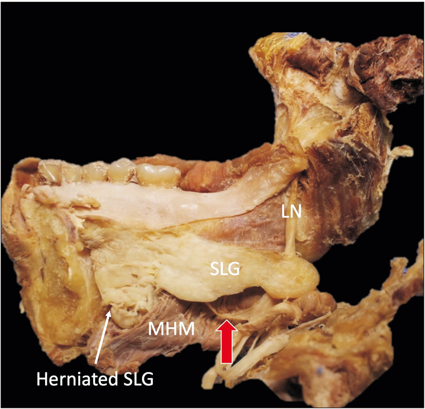
Fig. 1 A large branch (red arrow) of the LN running lateral to the SLG. LN, lingual nerve; MHM, mylohyoid muscle; SLG, sublingual gland.

Fig. 2 A branch (red arrows) of the lingual nerve (LN) entering the herniated part of the sublingual gland (SLG). Note the connection of the branch to the herniated part of the SLG in the defect of the mylohyoid muscle (MHM). (A) SLG being turned medially. (B) SLG being turned superiorly.

Fig. 3 Histological observation with H&E staining showing the fibrous tissue (harvested from the rectangular area in the left bottom image) was nerve tissue. Scale bar: 200 μm.
Reference
-
References
1. Standring S. 2020. Gray's anatomy. 42nd ed. Elsevier;London:2. Kikuta S, Iwanaga J, Kusukawa J, Tubbs RS. 2019; An anatomical study of the lingual nerve in the lower third molar area. Anat Cell Biol. 52:140–2. DOI: 10.5115/acb.2019.52.2.140. PMID: 31338230. PMCID: PMC6624336.
Article3. Iwanaga J, Cleveland MK, Wada J, Tubbs RS. 2020; How to avoid iatrogenic lingual nerve injury in the retromolar area: an anatomical study of retromolar pad and lingual nerve. Surg Radiol Anat. 42:523–8. DOI: 10.1007/s00276-020-02422-w. PMID: 31989215.
Article4. Iwanaga J, Nakamura K, Alonso F, Kirkpatrick C, Oskouian RJ, Watanabe K, Tubbs RS. 2018; Anatomical study of the so-called "retromolar gland": distinguishing normal anatomy from oral cavity pathology. Clin Anat. 31:462–5. DOI: 10.1002/ca.23047. PMID: 29349817.
Article5. Graff-Radford SB, Evans RW. 2003; Lingual nerve injury. Headache. 43:975–83. DOI: 10.1046/j.1526-4610.2003.03189.x. PMID: 14511274.
Article6. Ryumon S, Hage D, Ibaragi S, Okui T, Tubbs RS, Iwanaga J. 2021; Dual innervation of the submandibular gland by nerve to mylohyoid and chorda tympani. Morphologie. 105:316–8. DOI: 10.1016/j.morpho.2020.11.004. PMID: 33288421.
Article7. Sittitavornwong S, Babston M, Denson D, Zehren S, Friend J. 2017; Clinical anatomy of the lingual nerve: a review. J Oral Maxillofac Surg. 75:926.e1–926.e9. DOI: 10.1016/j.joms.2017.01.009. PMID: 28189657.
Article8. Holmberg KV, Hoffman MP. 2014; Anatomy, biogenesis and regeneration of salivary glands. Monogr Oral Sci. 24:1–13. DOI: 10.1159/000358776. PMID: 24862590. PMCID: PMC4048853.
Article9. Otonari-Yamamoto M, Nakajima K, Tsuji Y, Otonari T, Curtin HD, Okano T, Sano T. 2010; Imaging of the mylohyoid muscle: separation of submandibular and sublingual spaces. AJR Am J Roentgenol. 194:W431–8. DOI: 10.2214/AJR.09.3516. PMID: 20410390.
Article10. White DK, Davidson HC, Harnsberger HR, Haller J, Kamya A. 2001; Accessory salivary tissue in the mylohyoid boutonnière: a clinical and radiologic pseudolesion of the oral cavity. AJNR Am J Neuroradiol. 22:406–12. PMID: 11156791. PMCID: PMC7973941.11. Engel JD, Harn SD, Cohen DM. 1987; Mylohyoid herniation: gross and histologic evaluation with clinical correlation. Oral Surg Oral Med Oral Pathol. 63:55–9. DOI: 10.1016/0030-4220(87)90340-9. PMID: 3468466.
Article12. Yang HC, Kim SY, Kim SK, Oh CS, Chung IH, Nam KI. 2016; A cadaveric study on mylohyoid herniation of the sublingual gland. Eur Arch Otorhinolaryngol. 273:4413–6. DOI: 10.1007/s00405-016-4095-1. PMID: 27180250.
Article13. Jain P, Jain R. 2014; Types of sublingual gland herniation observed during sonography of plunging ranulas. J Ultrasound Med. 33:1491–7. DOI: 10.7863/ultra.33.8.1491. PMID: 25063415.
Article14. Shakeel M, Nair S, Ahmad Z. 2016; Painless, reproducible, reversible floor of mouth herniation: what's in there? J Otolaryngol ENT Res. 4:00118. DOI: 10.15406/joentr.2016.04.00118.
Article15. Attie JN, Friedman E, Rothberg MS. 1984; Submandibular and axillary schwannomas not associated with von Recklinghausen's disease. J Oral Maxillofac Surg. 42:391–4. DOI: 10.1016/S0278-2391(84)80012-9. PMID: 6425471.
Article
