Distribution of frontal sinus pattern amongst Malaysian population: a skull radiograph study
- Iwani Zulkiflee ND
 1
1 - Chainchel Singh MK2,3
- Alias A4,5,6
- Hadi Pritam HM7
- Chung E8
- Sakaran R9
- Zaidun NH1
- Woon CK1
- Affiliations
-
- 1Department of Anatomy, Faculty of Medicine, Universiti Teknologi MARA, Selangor, Malaysia.
- 2Institute of Pathology, Laboratory and Forensic Medicine (I-PPerForM), Faculty of Medicine, Universiti Teknologi MARA, Selangor, Malaysia.
- 3Department of Radiology, Faculty of Medicine, Universiti Teknologi MARA, Selangor, Malaysia.
- 4Department of Basic Sciences and Oral Biology, Faculty of Dentistry, Universiti Sains Islam Malaysia, Kuala Lumpur, Malaysia.
- 5Forensic Odontology Unit, Department of Imaging & Pathology, KU Leuven, Leuven, Belgium.
- 6Department of Forensic Odontology, Faculty of Dental Medicine, Universitas Airlangga, Surabaya, Indonesia.
- 7Forensic Unit, School of Health Sciences, Universiti Sains Malaysia, Kelantan, Malaysia.
- 8Department of Biomedical Imaging, University Malaya Medical Centre, Kuala Lumpur, Malaysia.
- 9Department of Anatomy, Asian Institute of Medicine, Science and Technology (AIMST), Kedah, Malaysia.
- KMID: 2533354
- DOI: http://doi.org/10.5115/acb.22.075
Abstract
- Frontal sinus has unique anatomical features that are distinct to every population. However, the distribution of frontal sinus patterns has yet to be explored in the Malaysian population. This study aimed to describe the distribution of frontal sinus patterns among adult Malaysians. 409 adult Malaysian posteroanterior skull radiographs, consisting of 200 males and 209 females of Malay, Chinese, and Indian races aged between 20–69 years old, were included in the study. The frontal sinus patterns were classified according to total and percentage of presence or absence of frontal sinus, symmetry or asymmetrical (right or left dominant), unilateral absence (right or left), bilateral absence, and lobulation. The findings showed that bilateral presence of frontal sinus is common, in 95.4% of individuals and bilateral absence was noted in 2.7% individuals. Unilateral absence was found in 2.0% of individuals. Asymmetrical frontal sinus was observed in 54.5% of population meanwhile 40.8% showed symmetrical frontal sinus. The majority of individuals, regardless of sex, race, and age, possessed 1 to 3 lobes on both sides of the frontal sinus. The findings suggest that the frontal sinus is highly asymmetric, and the absence of the frontal sinus is rare. This morphological variation provides an insight into the landmarking placement for measurement during forensic application and assists neurosurgeons in surgical procedure to avoid breaching of the frontal sinus.
Keyword
Figure
-
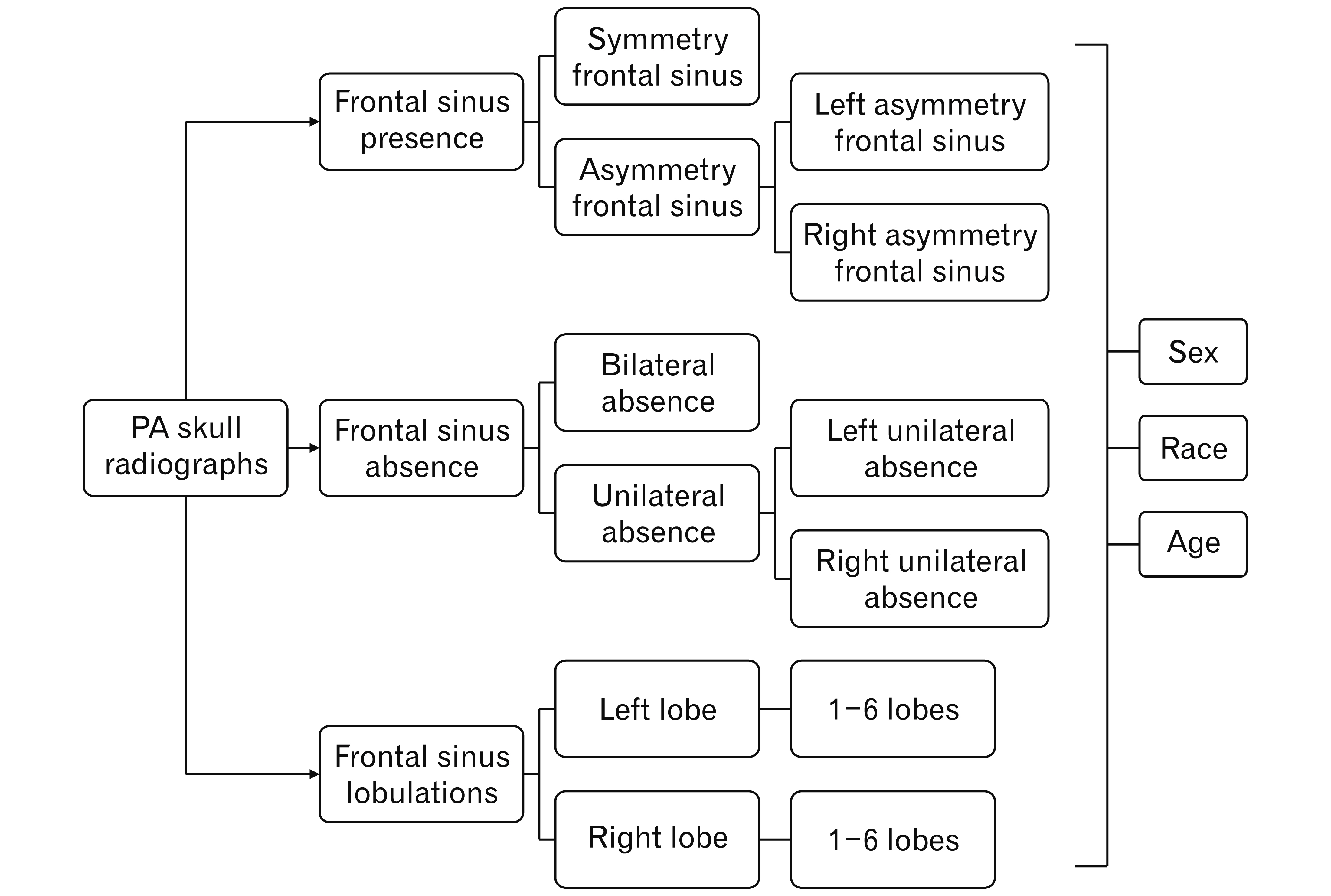
Fig. 1 Classification of frontal sinus pattern.
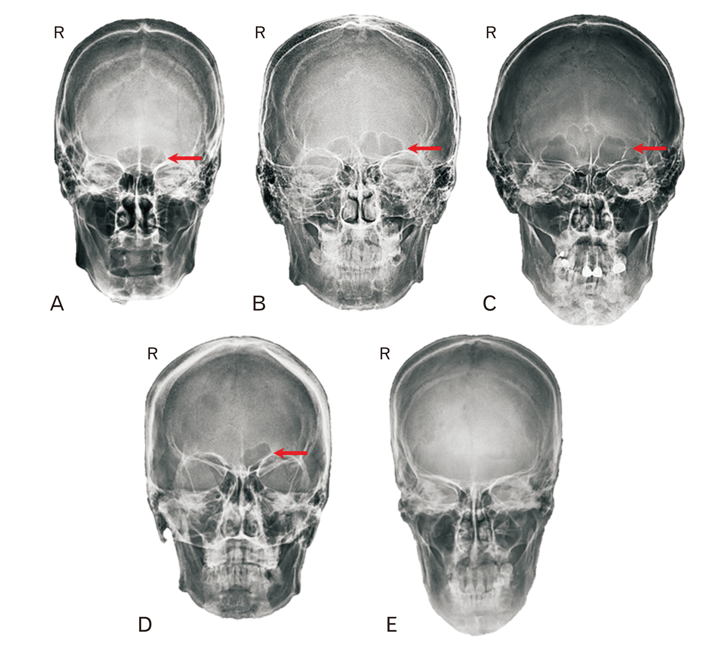
Fig. 2 Various frontal sinus patterns: (A) symmetrical, (B) left asymmetry, (C) right asymmetry, (D) unilateral absence, and (E) bilateral absence. Red arrows shows frontal sinus. R, right side.
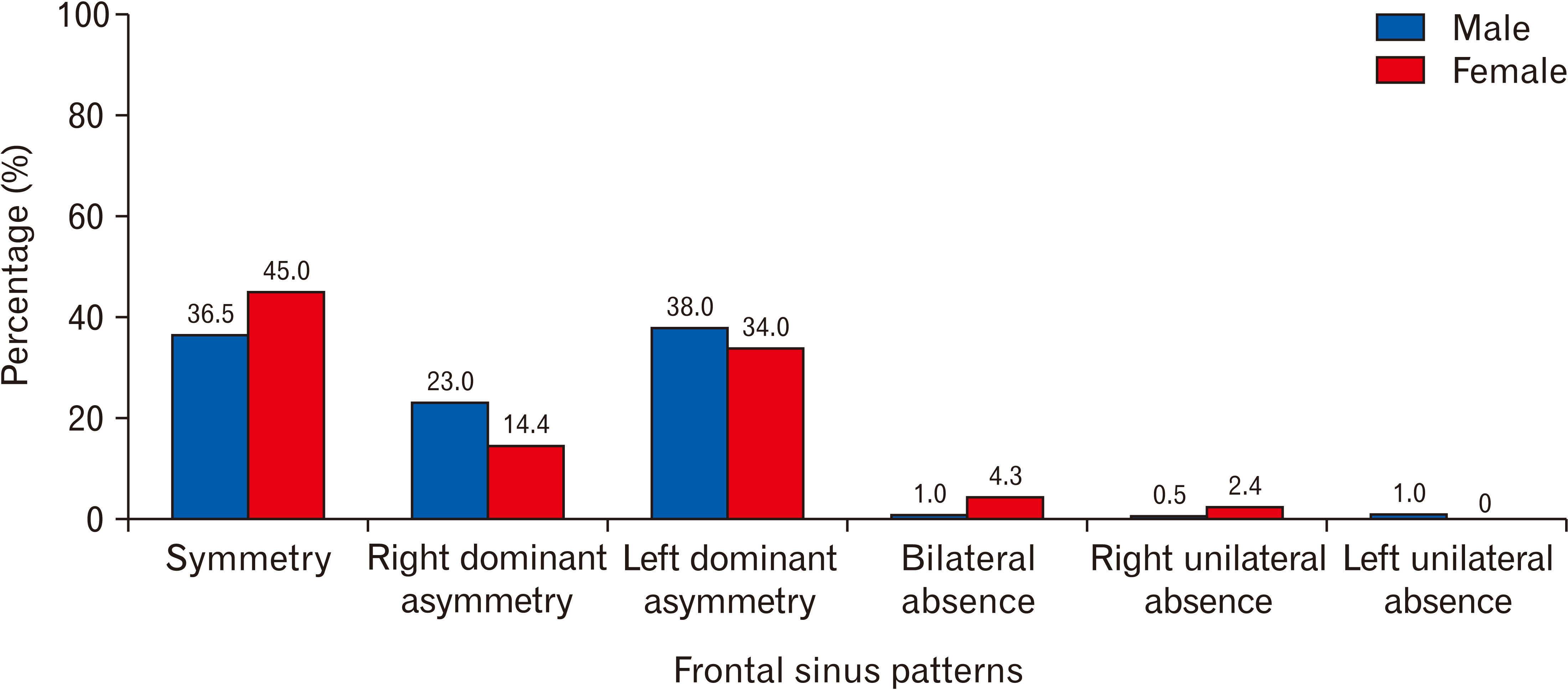
Fig. 3 Bar graph of frontal sinus patterns distribution between sex groups, males (n=200) and females (n=209).
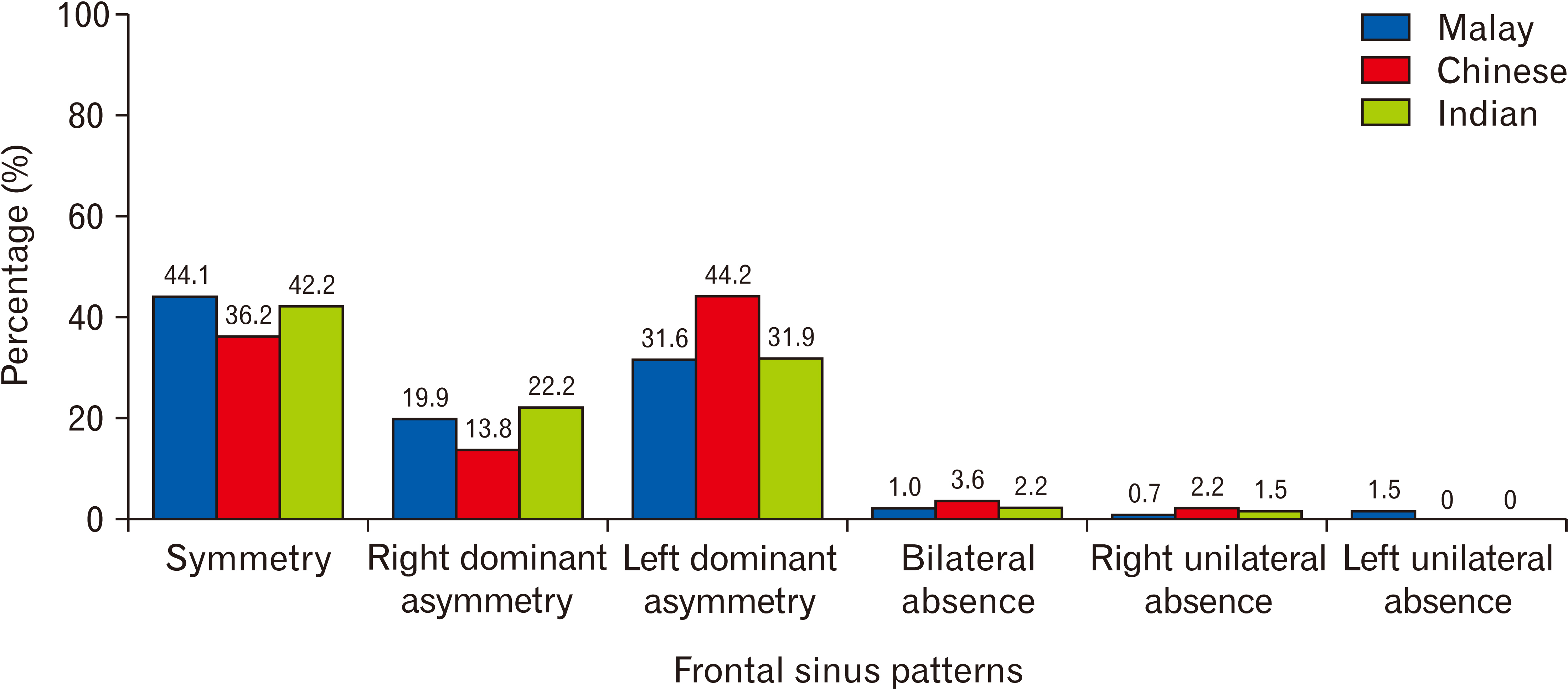
Fig. 4 Bar graph of frontal sinus patterns distribution between race groups, Malays (n=136), Chinese (n=138), and Indians (n=135).

Fig. 5 Bar graph of frontal sinus patterns distribution between age groups, group I (n=82), group II (n=79), group III (n=83), group IV (n=82), and group V (n=83). Age range: group I, 20–29 yr; group II, 30–39 yr; group III, 40–49 yr; group IV, 50–59 yr; group V, 60–69 yr.
Reference
-
References
1. Gadekar NB, Kotrashetti VS, Hosmani J, Nayak R. 2019; Forensic application of frontal sinus measurement among the Indian population. J Oral Maxillofac Pathol. 23:147–51. DOI: 10.4103/jomfp.JOMFP_214_18. PMID: 31110433. PMCID: PMC6503804.2. Crosta E. 2016. Sexual determination from frontal sinus analysis in a subadult population using archival radiographic records [thesis]. University of Nevada, Las Vegas;Las Vegas:3. Čechová M, Dupej J, Brůžek J, Bejdová Š, Horák M, Velemínská J. 2019; Sex estimation using external morphology of the frontal bone and frontal sinuses in a contemporary Czech population. Int J Legal Med. 133:1285–94. DOI: 10.1007/s00414-019-02063-8. PMID: 30982130.
Article4. Verma P, Verma KG, Khosa R, Kumar S, Basavaraju S, Patwardhan N. 2015; Combined use of frontal sinus and nasal septum patterns as an aid in forensics: a digital radiographic study. N Am J Med Sci. 7:47–52. DOI: 10.4103/1947-2714.152078. PMID: 25789248. PMCID: PMC4358048.
Article5. Christensen AM. 2003. An empirical examination of frontal sinus outline variability using elliptic fourier analysis: implications for identification, standardization, and legal admissibility [PhD dissertation]. University of Tennessee;Knoxville:6. Tatlisumak E, Asirdizer M, Bora A, Hekimoglu Y, Etli Y, Gumus O, Keskin S. 2017; The effects of gender and age on forensic personal identification from frontal sinus in a Turkish population. Saudi Med J. 38:41–7. DOI: 10.15537/smj.2017.1.16218. PMID: 28042629. PMCID: PMC5278064.
Article7. Ozgursoy OB, Comert A, Yorulmaz I, Tekdemir I, Elhan A, Kucuk B. 2010; Hidden unilateral agenesis of the frontal sinus: human cadaver study of a potential surgical pitfall. Am J Otolaryngol. 31:231–4. DOI: 10.1016/j.amjoto.2009.02.010. PMID: 20015751.
Article8. Amine A, Habashy KJ, Najem E, Abbas R, Moussalem C, Bsat S, Hourany R, Darwish H. 2021; Frontal sinus morphometry in relation to surgically relevant landmarks in the Middle East population: can we globalize? World Neurosurg. 148:e87–93. DOI: 10.1016/j.wneu.2020.12.018. PMID: 33309894.
Article9. Kim DI, Lee UY, Park SO, Kwak DS, Han SH. 2013; Identification using frontal sinus by three-dimensional reconstruction from computed tomography. J Forensic Sci. 58:5–12. DOI: 10.1111/j.1556-4029.2012.02185.x. PMID: 22563883.
Article10. Besana JL, Rogers TL. 2010; Personal identification using the frontal sinus. J Forensic Sci. 55:584–9. DOI: 10.1111/j.1556-4029.2009.01281.x. PMID: 20102475.
Article11. Patil N, Karjodkar FR, Sontakke S, Sansare K, Salvi R. 2012; Uniqueness of radiographic patterns of the frontal sinus for personal identification. Imaging Sci Dent. 42:213–7. DOI: 10.5624/isd.2012.42.4.213. PMID: 23301206. PMCID: PMC3534174.
Article12. Cox M, Malcolm M, Fairgrieve SI. 2009; A new digital method for the objective comparison of frontal sinuses for identification. J Forensic Sci. 54:761–72. DOI: 10.1111/j.1556-4029.2009.01075.x. PMID: 19486246.
Article13. Garhia P, Saxena S, Gupta A. 2019; Frontal sinus variability as a tool in forensic identification- a pilot study using radiographic images and software analysis. Int J Cur Res Rev. 11:8–12. DOI: 10.31782/IJCRR.2019.0812.
Article14. David MP, Saxena R. 2010; Use of frontal sinus and nasal septum patterns as an aid in personal identification: a digital radiographic pilot study. J Forensic Dent Sci. 2:77–80. DOI: 10.4103/0975-1475.81286. PMID: 21731344. PMCID: PMC3125957.
Article15. Shireen A, Goel S, Ahmed IM, Sabeh AM, Mahmoud W. 2019; Radiomorphometric evaluation of the frontal sinus in relation to age and gender in Saudi population. J Int Soc Prev Community Dent. 9:584–96. DOI: 10.4103/jispcd.jispcd_222_19. PMID: 32039079. PMCID: PMC6905308.16. Yoshino M, Miyasaka S, Sato H, Seta S. 1987; Classification system of frontal sinus patterns by radiography. Its application to identification of unknown skeletal remains. Forensic Sci Int. 34:289–99. DOI: 10.1016/0379-0738(87)90041-7. PMID: 3623370.
Article17. Taniguchi M, Sakoda S, Kano T, Zhu BL, Kamikodai Y, Fujita MQ, Maeda H. 2003; Possible use of nasal septum and frontal sinus patterns to radiographic identification of unknown human remains. Osaka City Med J. 49:31–8. PMID: 14703097.18. Koertvelyessy T. 1972; Relationships between the frontal sinus and climatic conditions: a skeletal approach to cold adaptation. Am J Phys Anthropol. 37:161–72. DOI: 10.1002/ajpa.1330370202. PMID: 5085494.
Article19. Vance HA. 2021. The influence of ancestry, sex, and age on the morphology of the frontal sinus in black and white individuals [thesis]. University of Montana;Missoula:20. Barros FD, Fernandes CMDS, Kuhnen B, Filho JS, Gonçalves M, da Costa Serra M. 2021; Paranasal sinuses and human identification. Res Soc Dev. 10:e48710918161. DOI: 10.33448/rsd-v10i9.18161.
Article21. MyCensus. 2020. Department of Statistics Malaysia Official Portal. Current population estimates, Malaysia; 2020 [Internet]. Department of Statistics Malaysia;Putrajaya: Available from: https://www.dosm.gov.my/v1/index.php?r=column2FcthemeByCat&cat=155&bul_id=OVByWjg5YkQ3MWFZRTN5bDJiaEVhZz09&menu_id=L0pheU43NWJwRWVSZklWdzQ4TlhUUT09#:~:text=The%20growth%20rate%20of%20Citizens,to%2029.7%20million%20in%202020.&text=Overall%2C%20there%20were%20more%20males,and%2015.9%20million%20(females). cited 2021 Nov 10.
- Full Text Links
-
- Actions
-
Cited
- CITED
-
- Close
- Share
-
- Similar articles
-
- Clinical Experience in Treatment of the Giant Frontal Sinus Osteoma using Cranialization
- A Reconstructive Transbasal Approach to Tumors Involving the Anterior Skull Base
- Morphological Analysis using Simple Skull AP of Frontal Sinus in Korea
- Frontal Sinus Stenting in Endoscopic Sinus Surgery
- Frontal Sinus Mucocele with Massive Skull Destruction
