A plunging ranula in a child with holoprosencephaly: a case of unique pathophysiology and difficult airway management
- Affiliations
-
- 1Department of Oral and Maxillofacial Surgery, Graduate School of Medicine, Kyoto University, Kyoto, Japan
- 2Department of Pediatrics, Graduate School of Medicine, Kyoto University, Kyoto, Japan
- 3Department of Anesthesia, Kyoto University Hospital, Kyoto, Japan
- KMID: 2532828
- DOI: http://doi.org/10.5125/jkaoms.2022.48.4.232
Abstract
- A ranula is a pseudocyst that originates from the sublingual gland after trauma. Acute cases of ranulas that progress rapidly and cause respiratory distress are rare. Holoprosencephaly is a complex brain malformation caused by incomplete cleavage of the prosencephalon. Children with holoprosencephaly may experience upper airway obstruction due to the associated dentoalveolar malformations and oromotor dysfunctions. We present the case of an eight-year-old female patient with holoprosencephaly and a plunging ranula that manifested as an acute course due to difficult airway management. She required gastrostomy for oromotor dysfunctions related to feeding and swallowing and difficulty managing oral secretions. The sublingual gland and ranula were removed under general anesthesia. Postoperatively, urgent reintubation and close monitoring in the intensive care unit were required due to upper airway obstruction. We successfully managed the patient with close cooperation of a pediatrician and an anesthetist, and no recurrence was observed at the one-year follow-up. A ranula can be caused by trauma to the floor of the mouth in association with lingually inclined mandibular teeth, a type of dentoalveolar compensation seen in maxillary hypoplasia associated with holoprosencephaly. Careful consideration is needed in such cases since airway management can be difficult due to postoperative swelling and oromotor dysfunctions.
Figure
-
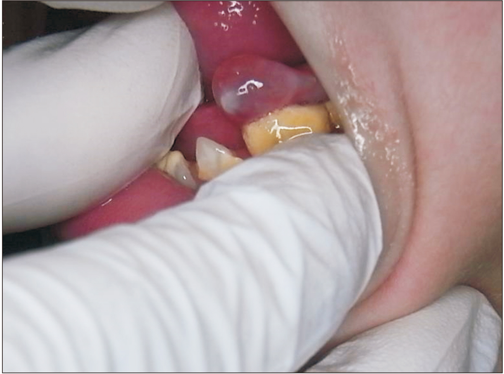
Fig. 1 A soft, fluctuant, and translucent mass with a diameter of 7 mm located on the left side of the floor of the mouth is seen.
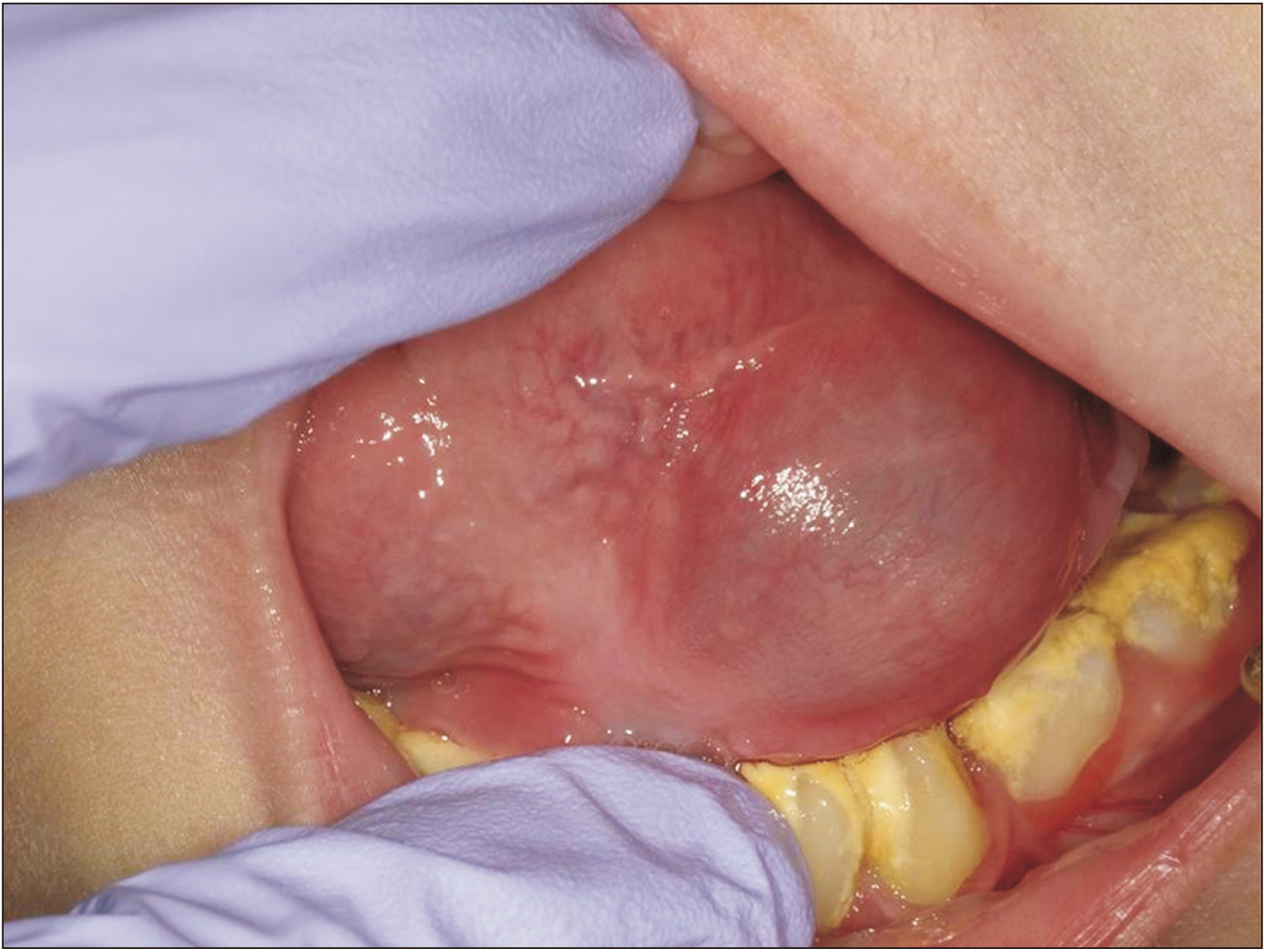
Fig. 2 A fluctuant, translucent, and dome-shaped swelling with a diameter of approximately 30 mm located on the left side of the floor of the mouth is shown.
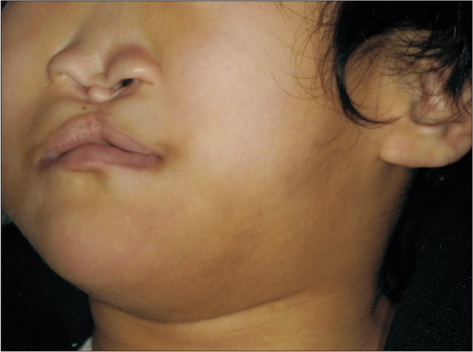
Fig. 3 A swelling is located in the left upper neck, and the overlying skin appears normal. The flat nose suggests nasal pyriform aperture stenosis.
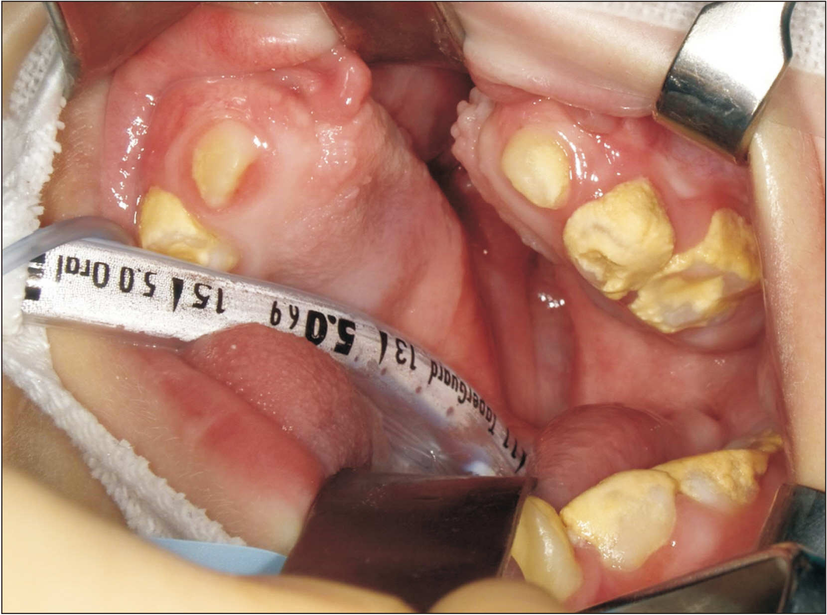
Fig. 4 The intraoperative view after orotracheal intubation, showing a cleft palate.
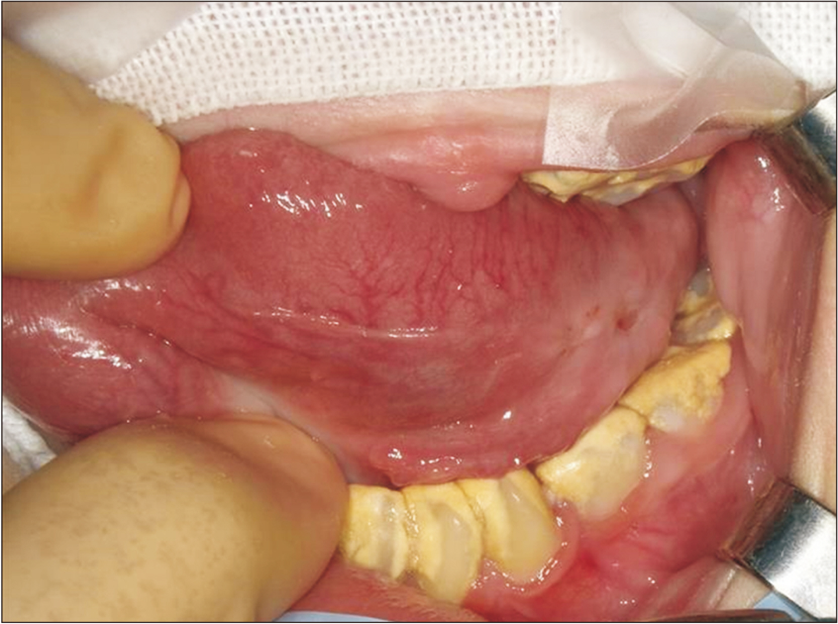
Fig. 5 The intraoperative view before incising, showing a mass with reduced tension and size and mandibular teeth with lingual inclination.
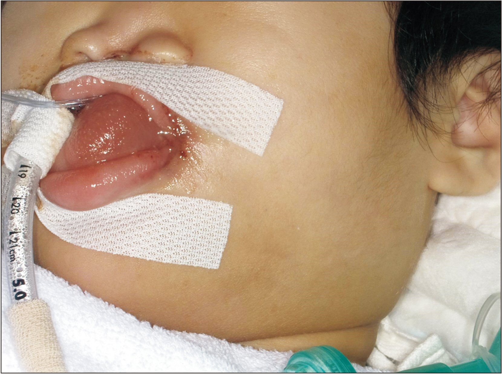
Fig. 6 On the day after surgery, the patient showed remarkable facial swelling and remained intubated.
Reference
-
References
1. Pandit RT, Park AH. 2002; Management of pediatric ranula. Otolaryngol Head Neck Surg. 127:115–8. https://doi.org/10.1067/mhn.2002.126590. DOI: 10.1067/mhn.2002.126590. PMID: 12161740.
Article2. Sathanantham DK, Shah GB, Ulüalp S. 2015; Rapid development of a simple ranula in a child. Ann Otol Rhinol Laryngol. 124:322–5. https://doi.org/10.1177/0003489414553653. DOI: 10.1177/0003489414553653. PMID: 25277701.
Article3. Bradley PJ. 2018; Sublingual gland. Oper Tech Otolaryngol Head Neck Surg. 29:168–76. https://doi.org/10.1016/j.otot.2018.06.006. DOI: 10.1016/j.otot.2018.06.006. PMID: 33526317.
Article4. Zhi K, Wen Y, Zhou H. 2009; Management of the pediatric plunging ranula: results of 15 years' clinical experience. Oral Surg Oral Med Oral Pathol Oral Radiol Endod. 107:499–502. https://doi.org/10.1016/j.tripleo.2008.09.023. DOI: 10.1016/j.tripleo.2008.09.023. PMID: 19071033.
Article5. Effat KG. 2012; Acute presentation of a plunging ranula causing respiratory distress: case report. J Laryngol Otol. 126:861–3. https://doi.org/10.1017/S0022215112000862. DOI: 10.1017/S0022215112000862. PMID: 22583866.
Article6. Granato L, Pinto CF, de Castro NP Jr, Rocha A, Bellido Rios OA. 2005; Holoprosencephaly--report of two cases. Int J Pediatr Otorhinolaryngol. 69:1563–8. https://doi.org/10.1016/j.ijporl.2005.04.031. DOI: 10.1016/j.ijporl.2005.04.031. PMID: 16083973.
Article7. Levey EB, Stashinko E, Clegg NJ, Delgado MR. 2010; Management of children with holoprosencephaly. Am J Med Genet C Semin Med Genet. 154C:183–90. https://doi.org/10.1002/ajmg.c.30254. DOI: 10.1002/ajmg.c.30254. PMID: 20104615.
Article8. Than JK, Rosenberg TL, Anand G, Sitton M. 2020; The importance of sublingual gland removal in treatment of ranulas: a large retrospective study. Am J Otolaryngol. 41:102418. https://doi.org/10.1016/j.amjoto.2020.102418. DOI: 10.1016/j.amjoto.2020.102418. PMID: 32087991.
Article9. Harrison JD. 2010; Modern management and pathophysiology of ranula: literature review. Head Neck. 32:1310–20. https://doi.org/10.1002/hed.21326. DOI: 10.1002/hed.21326. PMID: 20054853.
Article10. Dubourg C, Bendavid C, Pasquier L, Henry C, Odent S, David V. 2007; Holoprosencephaly. Orphanet J Rare Dis. 2:8. https://doi.org/10.1186/1750-1172-2-8. DOI: 10.1186/1750-1172-2-8. PMID: 17274816. PMCID: PMC1802747.
Article
- Full Text Links
-
- Actions
-
Cited
- CITED
-
- Close
- Share
-
- Similar articles
-
- PLUNGING RANULA IN A 4-YEAR-OLD CHILD: REPORT OF A CASE
- A Case of Bilateral Plunging Ranula
- Transoral Drainage of Plunging Ranula using Intraoperative Ultrasonography after Sublingual Gland Resection: A Case Report
- Case report of the management of the ranula
- A case report of Plunging ranula with metastatic adenocarcinoma
