Cerebral Metastases in Appendiceal Cancer: Comprehensive Review and Report of Rare Medullary Carcinoma Histology
- Affiliations
-
- 1Departments of Neurosurgery Beth Israel Deaconess Medical Center and Harvard Medical School, Boston, MA, USA
- 2Departments of Pathology, Beth Israel Deaconess Medical Center and Harvard Medical School, Boston, MA, USA
- KMID: 2532245
- DOI: http://doi.org/10.14791/btrt.2022.0019
Abstract
- Appendiceal cancer is an extremely rare malignancy, and its metastatic spread to the brain is even more unusual. We describe a 47-year-old female who presented with a rare cerebral appendiceal carcinoma metastasis, a case that is further remarkable for representing the first histologic diagnosis of primary medullary carcinoma in the appendix. Based on a comprehensive review of the English literature using PubMed, Embase, and Google Scholar, only six other cases of cerebral appendiceal metastases have been described.
Figure
-
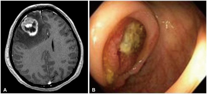
Fig. 1 Images of appendiceal carcinoma with cerebral metastasis. A: Axial T1-gadolinum enhanced MRI of the brain demonstrating a heterogeneously enhancing right frontal mass with extensive vasogenic edema prior to resection. B: Appendiceal mass identified on colonoscopy with stigmata of recent bleeding.
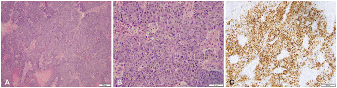
Fig. 2 Photomicrographs of the right frontal mass showing metastatic poorly differentiated carcinoma. A: Low power (H&E, ×4) shows a poorly differentiated neoplasm with prominent sheet-like growth and necrosis (bottom). B: Higher power (H&E, ×20) highlights poorly differentiated tumor cells with prominent nucleoli and mitotic activity. C: CDX-2 is positive in tumor cells, suggesting gastrointestinal origin. Scale bars = 200 µm (A), 50 µm (B), 100 µm (C).
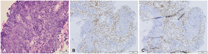
Fig. 3 Photomicrographs of the appendix tumor biopsy, poor differentiated carcinoma, favoring medullary carcinoma. A: High power (H&E, ×20) highlights a poorly differentiated tumor with sheet-like growth, prominent nucleoli, and mitotic activity, similar to the brain lesion. B: Immunohistochemistry for mismatch repair protein MLH-1 shows loss in tumor cells with retention in adjacent non-neoplastic cells. C: Immunohistochemistry for mismatch repair protein PMS-2 shows loss in tumor cells with retention in adjacent non-neoplastic cells. Scale bars = 50 µm (A), 100 µm (B and C).
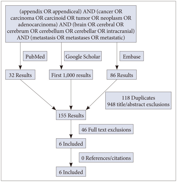
Fig. 4 Flow chart of search results for cerebral appendiceal metastases performed May 2022 using the databases of PubMed, Embase, and Google Scholar.
Reference
-
1. Siegel RL, Miller KD, Goding Sauer A, Fedewa SA, Butterly LF, Anderson JC, et al. Colorectal cancer statistics, 2020. CA Cancer J Clin. 2020; 70:145–164. PMID: 32133645.2. Minhas A, Hendrickson J, Minhas SA. Frequency and risk factors for metastasis in newly diagnosed appendiceal carcinoma. Cureus. 2021; 13:e16341. PMID: 34395124.3. Thompson E, Banerjee S, Thompson S, Silva R, Muse A, Arif-Tiwari H, et al. Incidence and predictors of brain metastasis in colorectal cancer patients. Int J Colorectal Dis. 2022; 37:153–159. PMID: 34596736.4. Habbous S, Forster K, Darling G, Jerzak K, Holloway CMB, Sahgal A, et al. Incidence and real-world burden of brain metastases from solid tumors and hematologic malignancies in Ontario: a population-based study. Neurooncol Adv. 2021; 3:vdaa178. PMID: 33585818.5. Biroli A, Cecchi PC, Pragal S, Hanspeter E, Schwarz A. Cerebral metastasis from a previously undiagnosed appendiceal adenocarcinoma. Case Rep Oncol Med. 2012; 2012:192807. PMID: 23198200.6. Foster A, Lofters J, Durham S, Jhawer M. A rare case of brain metastases from appendiceal carcinoma: a case report. J Case Rep Images Oncology. 2021; 7:100080Z10AF2021.7. Ruoff C, Hanna L, Zhi W, Shahzad G, Gotlieb V, Saif MW. Cancers of the appendix: review of the literatures. ISRN Oncol. 2011; 2011:728579. PMID: 22084738.8. Washington MK, Nagtegaal ID. Tumours of the appendix: Introduction. WHO Classification of Tumours Editorial Board. WHO classification of tumours. Digestive system tumours. 5th ed. Lyon: IARC Press;2019. p. 135–156.9. Roy SP, Al Zhahrani N, Barat S, Morris DL. Case series on high grade appendiceal cancer with peritoneal and liver carcinomatosis undergoing cytoreductive surgery and hyperthermic intraperitoneal chemotherapy (HIPEC). Int J Surg Case Rep. 2022; 94:107027. PMID: 35398783.10. Nonaka D, Papaxoinis G, Lamarca A, Fulford P, Valle J, Chakrabarty B. A study of appendiceal crypt cell adenocarcinoma (so-called goblet cell carcinoid and its related adenocarcinoma). Hum Pathol. 2018; 72:18–27. PMID: 28823572.11. Dietrich CS 3rd, Desimone CP, Modesitt SC, Depriest PD, Ueland FR, Pavlik EJ, et al. Primary appendiceal cancer: gynecologic manifestations and treatment options. Gynecol Oncol. 2007; 104:602–606. PMID: 17055559.12. Spann JL, Lowbeer L, VanWormer DE. Adenocarcinoma of the appendix vermiformis. J Surg Oncol. 1971; 3:185–196. PMID: 5094277.13. Christensen TD, Spindler KL, Palshof JA, Nielsen DL. Systematic review: brain metastases from colorectal cancer—incidence and patient characteristics. BMC Cancer. 2016; 16:260. PMID: 27037031.14. Jung M, Ahn JB, Chang JH, Suh CO, Hong S, Roh JK, et al. Brain metastases from colorectal carcinoma: prognostic factors and outcome. J Neurooncol. 2011; 101:49–55. PMID: 20467783.15. Fiehn AM, Grauslund M, Glenthøj A, Melchior LC, Vainer B, Willemoe GL. Medullary carcinoma of the colon: can the undifferentiated be differentiated? Virchows Arch. 2015; 466:13–20. PMID: 25339302.16. Lee LH, Yantiss RK, Sadot E, Ren B, Calvacanti MS, Hechtman JF, et al. Diagnosing colorectal medullary carcinoma: interobserver variability and clinicopathological implications. Hum Pathol. 2017; 62:74–82. PMID: 28034727.17. Winn B, Tavares R, Fanion J, Noble L, Gao J, Sabo E, et al. Differentiating the undifferentiated: immunohistochemical profile of medullary carcinoma of the colon with an emphasis on intestinal differentiation. Hum Pathol. 2009; 40:398–404. PMID: 18992917.18. Knox RD, Luey N, Sioson L, Kedziora A, Clarkson A, Watson N, et al. Medullary colorectal carcinoma revisited: a clinical and pathological study of 102 cases. Ann Surg Oncol. 2015; 22:2988–2996. PMID: 25572685.19. Ye J, Zhou Y, Weiser MR, Gönen M, Zhang L, Samdani T, et al. Immunohistochemical detection of ARID1A in colorectal carcinoma: loss of staining is associated with sporadic microsatellite unstable tumors with medullary histology and high TNM stage. Hum Pathol. 2014; 45:2430–2436. PMID: 25311944.
- Full Text Links
-
- Actions
-
Cited
- CITED
-
- Close
- Share
-
- Similar articles
-
- Appendiceal Mucocele Developed by Appendiceal Mucosal Prolapse: A Rare Case Report with Review of Literature
- Concurrent Medullay and Papillary Carcinoma of the Thyroid
- Fine Needle Aspiration Cytology of Medullary Carcinoma of the Breast: A Case Report
- A Case of Medullary Thyroid Carcinoma in which the Skin Metastasis was Concurrently Present and Response Occurred to Chemotherapy
- Concurrent Papillary and Medullary Carcinoma of the Thyroid Gland
