Utility of transoral and transcranial ultrasonography in the diagnosis of internal carotid dissection: a case report
- Affiliations
-
- 1Department of Critical Care Medicine, Sanatorio de los Arcos, Autonomous City of Buenos Aires, Argentina
- 2Argentinian Critical Care Ultrasonography Association (ASARUC), Autonomous City of Buenos Aires, Argentina
- KMID: 2532000
- DOI: http://doi.org/10.18700/jnc.210033
Abstract
- Background
Internal carotid artery (ICA) dissection is one of the most common causes of stroke in young patients. Ultrasonographic assessment of the carotid artery is the method of choice for early detection. The use of the conventional technique for diagnosis is limited since dissection frequently occurs in the distal portion of the ICA.
Case Report
We describe the case of a 48-year-old Argentinian woman with malignant cerebral infarction secondary to ICA dissection. We diagnosed her with neck vessels using conventional ultrasonography and a transoral ultrasonographic approach combined with transcranial ultrasonography.
Conclusion
We recommend the transoral assessment of ICA combined with ultrasonography as a non-invasive method for the diagnosis of ICA dissection.
Keyword
Figure
-
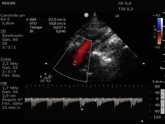
Fig. 1. Internal carotid artery conventional ultrasonography.

Fig. 2. Transoral ultrasonography. Yellow arrow; homogeneous hypoechoic image with absence of Doppler signal compatible with false lumen thrombosis and refinement of the true lumen.
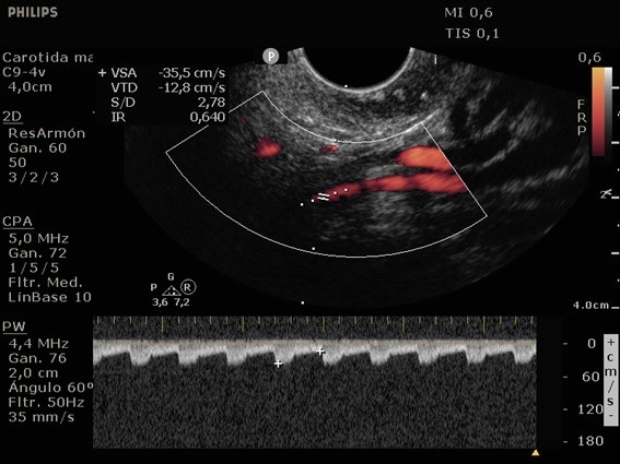
Fig. 3. Transoral ultrasonography using pulsed wave Doppler.

Fig. 4. Transcranial ultrasonography of the anterior cerebral artery.
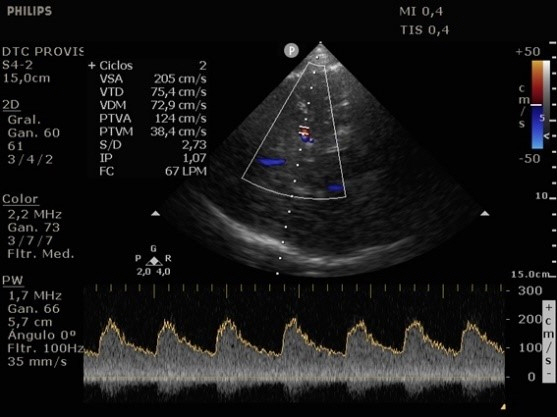
Fig. 5. Transcranial ultrasonography of the middle cerebral artery.
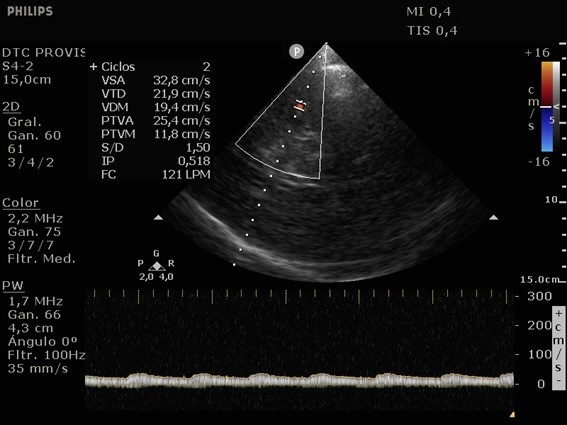
Fig. 6. Transcranial ultrasonography of the distal middle cerebral artery.
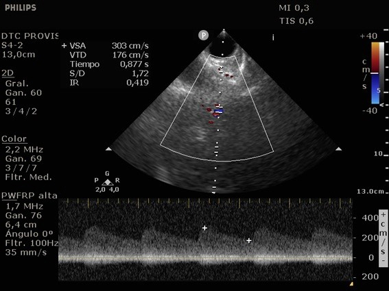
Fig. 7. Transorbital ultrasonography.
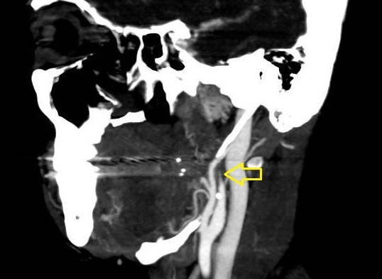
Fig. 8. Computed tomography angiography. Yellow arrow: postbulbar region of the left internal carotid artery (ICA) showing progressive reduction of the lumen until it becomes filiform and even undetectable compatible with ICA dissection.
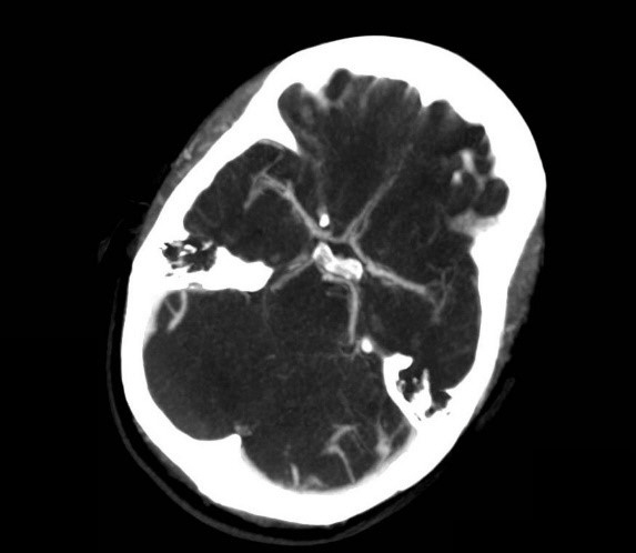
Fig. 9. Computed tomography angiography of the intracranial arteries.
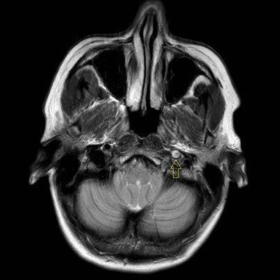
Fig. 10. Magnetic resonance imaging. Yellow arrow: absence of emptied flow at the level of the petrous and cavernous portion of the left internal carotid artery.
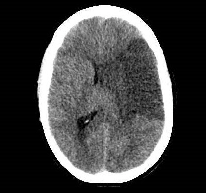
Fig. 11. Brain computed tomography of the patient in a coma.
Reference
-
1. Kim YK, Schulman S. Cervical artery dissection: pathology, epidemiology and management. Thromb Res. 2009; 123:810–21.
Article2. Lee VH, Brown RD Jr, Mandrekar JN, Mokri B. Incidence and outcome of cervical artery dissection: a population-based study. Neurology. 2006; 67:1809–12.
Article3. Rodallec MH, Marteau V, Gerber S, Desmottes L, Zins M. Craniocervical arterial dissection: spectrum of imaging findings and differential diagnosis. Radiographics. 2008; 28:1711–28.
Article4. Boßelmann C, Poli S. Sonographic features of carotid artery dissection due to extension of aortic dissection: a case report. Ultrasound J. 2019; 11:32.
Article5. Agarwala MK, Asad A, Gummadi N, Chidambaram S, Venkateswaralu J. Bilateral spontaneous internal carotid artery dissection managed with endovascular stenting: a case report. Indian Heart J. 2016; 68 Suppl 2(Suppl 2):S69–71.6. Yasaka M, Kimura K, Otsubo R, Isa K, Wada K, Nagatsuka K, et al. Transoral carotid ultrasonography. Stroke. 1998; 29:1383–8.
Article7. Suzuki R, Koga M, Toyoda K, Uemura M, Nagasawa H, Yakushiji Y, et al. Identification of internal carotid artery dissection by transoral carotid ultrasonography. Cerebrovasc Dis. 2012; 33:369–77.
Article8. Benninger DH, Georgiadis D, Gandjour J, Baumgartner RW. Accuracy of color duplex ultrasound diagnosis of spontaneous carotid dissection causing ischemia. Stroke. 2006; 37:377–81.
Article9. Wang H, Fei L, Xia H, Zhang Q, Huang Y. Diagnostic significance of transcranial doppler combined with carotid ultrasound in patients with cerebral ischemic stroke. Am J Transl Res. 2021; 13:6980–6.
- Full Text Links
-
- Actions
-
Cited
- CITED
-
- Close
- Share
-
- Similar articles
-
- Traumatic intracranial internal carotid artery dissection and pseudoaneurysm presenting as oculomotor nerve palsy
- Internal Carotid Artery Dissection Presenting as Isolated Unilateral Hypoglossal Nerve Palsy
- Vasospasm of Proximal Internal Carotid Artery Following Transcranial Removal of a Pituitary Adenoma
- Treatment of Internal Carotid Artery Dissections with Endovascular Stent Placement: Report of Two Cases
- Diagnostic vascular ultrasonography with the help of color Doppler and contrast-enhanced ultrasonography
