J Cerebrovasc Endovasc Neurosurg.
2022 Jun;24(2):154-159. 10.7461/jcen.2021.E2021.06.011.
A Ruptured lenticulostriate artery aneurysm in moyamoya disease treated with Onyx embolization
- Affiliations
-
- 1Department of Neurological Surgery, Asan Medical Center, University of Ulsan College of Medicine, Seoul, Korea
- 2Department of Neurological Surgery, Gangneung Asan Hospital, University of Ulsan College of Medicine, Gangneung, Korea
- KMID: 2531658
- DOI: http://doi.org/10.7461/jcen.2021.E2021.06.011
Abstract
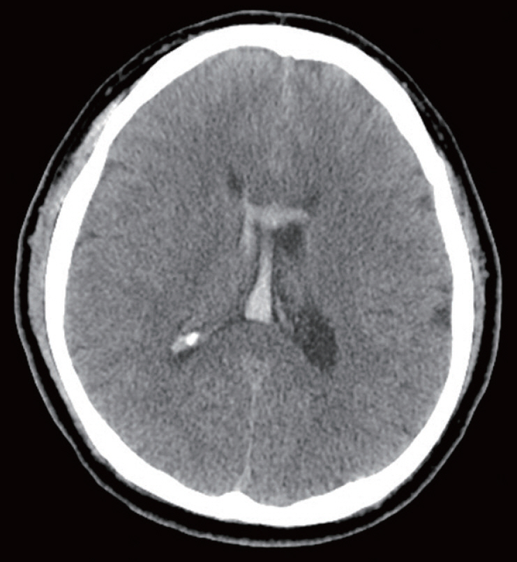
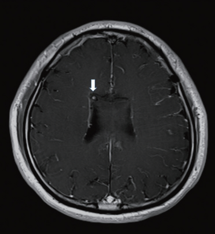
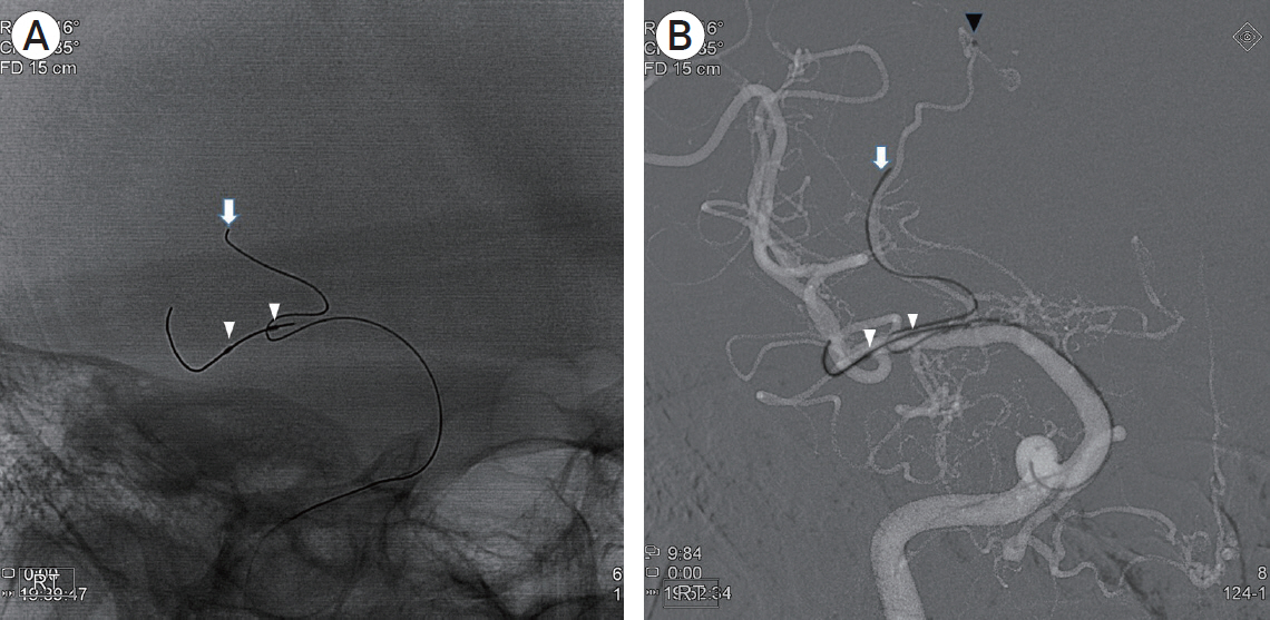
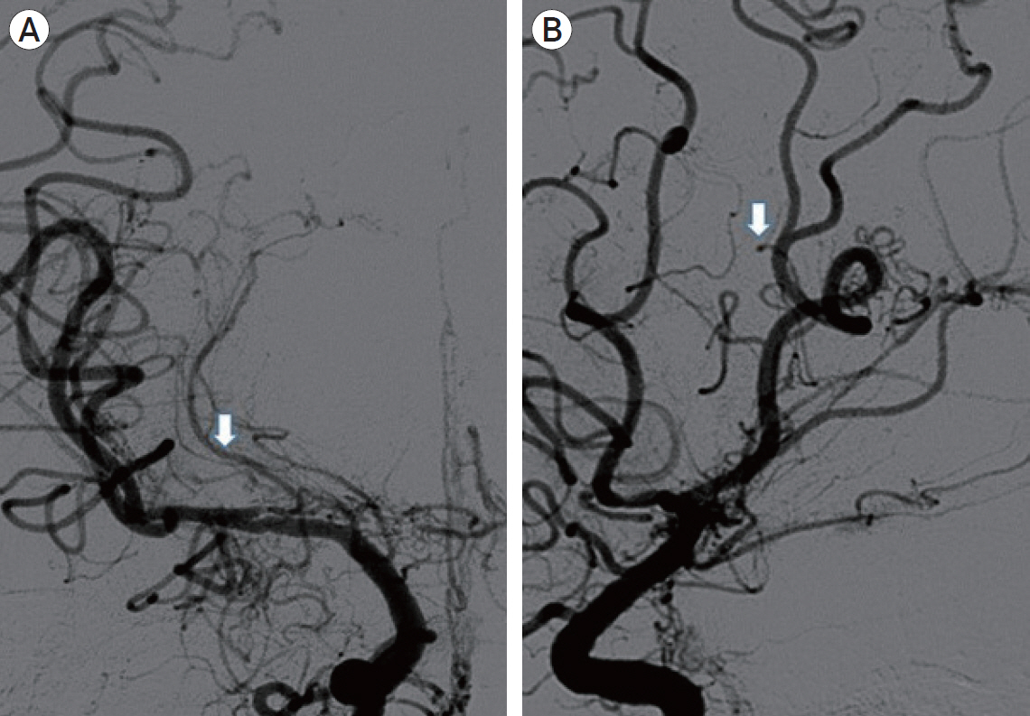
- Lenticulostriate artery (LSA) aneurysms are uncommon. Here, we report one case of ruptured LSA aneurysm which is related to Moyamoya disease (MMD). Surgical treatment of this aneurysm is challenging because of its deep location and complex neural structures around the LSA. We report one case treated with endovascular Onyx embolization, successfully and review LSA aneurysm associated with MMD.
Figure
Cited by 1 articles
-
Ruptured Peripheral Cerebral Aneurysms Associated With Moyamoya Disease: A Systematic Review
Zheng Feng, Yongquan Chang, Chao Fu
J Stroke. 2024;26(3):360-370. doi: 10.5853/jos.2024.02061.
Reference
-
1. David E H-G, Ana SF, Hector RM, Juan SF, Enrique C-O, Jose A F-S. Aneurysms of the Lenticulostriate Artery: A Systematic Review. World Neurosurg. 2021; Jan. 145:471–9.
Article2. Halpin SF, Britton JA, Byrne JV, Clifton A, Hart G, Moore A. Prospective evaluation of cerebral angiography and computed tomography in cerebral haematoma. Journal of neurology, neurosurgery, and psychiatry. 1994; Oct. 57(10):1180–6.
Article3. Kawaguchi S, Sakaki T, Morimoto T, Kakizaki T, Kamada K. Characteristics of intracranial aneurysms associated with moyamoya disease. A review of 111 cases. Acta neurochirurgica. 1996; 138(11):1287–94.4. Lei Z, Kan X, Yandong Z, Xin W, Jinlu Y. Treatment Strategies for Aneurysms Associated with Moyamoya Disease. International Journal of Medical Sciences. 2015; 12(3):234–42.
Article5. Nohra C, Stavropoula T, L. Fernando G, Aaron SD, Qaisar S, David G, Robert R, Pascal J. Onyx Embolization of a Ruptured Lenticulostriate Artery Aneurysm in a Patient with Moyamoya Disease. World Neurosurg. 2013; Sep-Oct. 80(3-4):436.e7–10.6. Takahashi JC, Miyamoto S. Moyamoya disease: recent progress and outlook. Neurologia medico-chirurgica. 2010; 50(9):824–32.
Article7. Yoshida Y, Yoshimoto T, Shirane R, Sakurai Y. Clinical course, surgical management, and long-term outcome of moyamoya patients with rebleeding after an episode of intracerebral hemorrhage: An extensive follow-Up study. Stroke; a journal of cerebral circulation. 1999; Nov. 30(11):2272–6.
- Full Text Links
- Actions
-
Cited
- CITED
-
- Close
- Share
- Similar articles
-
- Endovascular Embolization of a Ruptured Distal Lenticulostriate Artery Aneurysm in Patients with Moyamoya Disease
- Ruptured Distal Lenticulostriate Artery Aneurysm Associated with Ipsilateral Middle Cerebral Artery Occlusion : Case Report
- Successful Endovascular Treatment of Ruptured Superior Cerebellar Artery Aneurysm Associated with Moyamoya Disease : A Case Report and Review of the Literature
- Ruptured Persistent Trigeminal Artery Aneurysm Associated with Moyamoya Disease
- Coil Embolization for Distal Middle Cerebral Artery Aneurysm


