Clinicopathologic features and survival outcomes of ocular melanoma: a series of 31 cases from a tertiary university hospital
- Affiliations
-
- 1Department of Pathology, Faculty of Medicine, Gazi University, Ankara, Turkey
- KMID: 2531610
- DOI: http://doi.org/10.4132/jptm.2022.03.10
Abstract
- Background
We aimed to determine the effect of clinicopathologic features on overall survival among Caucasian ocular melanoma patients in the Central Anatolia region of Turkey.
Methods
This single-center study included conjunctival (n = 12) and uveal (n = 19) melanoma patients diagnosed between January 2008 and March 2020. Clinicopathologic features and outcomes were reviewed retrospectively. Five cases were tested for BRAF V600 mutations with real-time polymerase chain reaction, and one case was tested with nextgeneration sequencing. Survival was calculated using the Kaplan-Meier method.
Results
Thirty-one patients had a mean initial age of 58.32 years (median, 61 years; range 25 to 78 years). There were 13 male and 18 female patients. The median follow-up time was 43.5 months (range, 6 to 155 months) for conjunctival melanoma and 35 months (range, 8 to 151 months) for uveal melanoma. When this study ended, eight of the 12 conjunctival melanoma patients (66.7%) and nine of the 19 uveal melanoma patients (47.4%) had died. The presence of tumor-infiltrating lymphocytes was related to improved overall survival in conjunctival melanoma (p = .014), whereas the presence of ulceration (p = .030), lymphovascular invasion (p = .051), tumor in the left eye (p = .012), tumor thickness of > 2 mm (p = .012), and mitotic count of >1/mm² (p = .012) reduced the overall survival in conjunctival melanoma. Uveal melanoma tumors with the largest diameter of 9.1–15 mm led to the lowest overall survival among subgroups (p = .035). Involvement of the conjunctiva (p=.005) and lens (p = .003) diminished overall survival in uveal melanoma. BRAF V600 mutation was present in one case of conjunctival melanoma, GNAQ R183Q mutation was present in one case of uveal melanoma. Patients with uveal melanoma presented with an advanced pathological tumor stage compared to those with conjunctival melanoma (p = .019).
Conclusions
This study confirmed the presence of tumor-infiltrating lymphocytes as a favorable factor in conjunctival melanoma and conjunctival and lens involvement as unfavorable prognostic factors in uveal melanoma for overall survival, respectively.
Figure
-
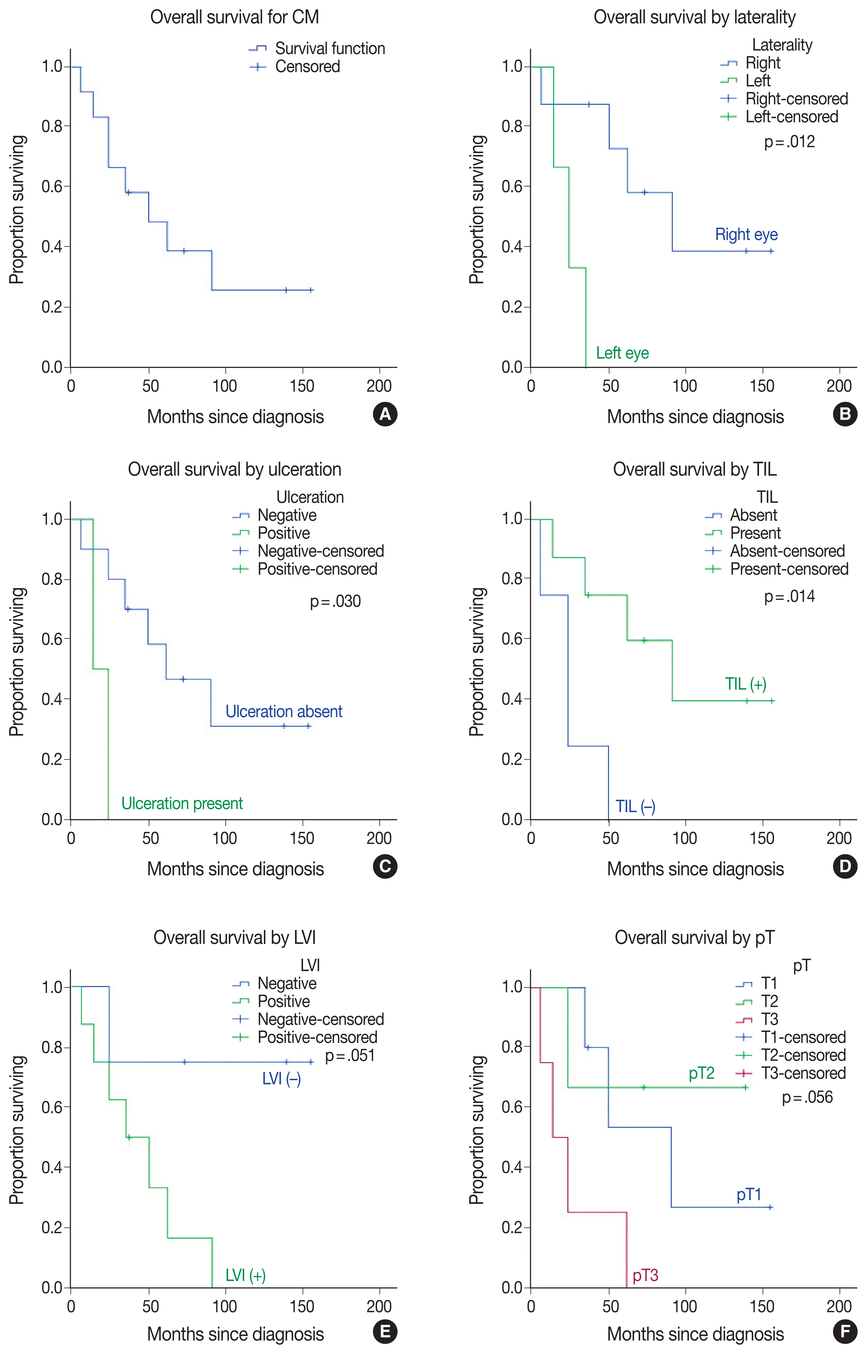
Fig. 1 Kaplan-Meier overall survival curves in conjunctival melanoma (CM) (A) patients are compared for tumor variables by laterality (B), ulceration (C), tumor-infiltrating lymphocytes (TILs) (D), lymphovascular invasion (LVI) (E), and pathological tumor (pT) staging (F).

Fig. 2 (A) A conjunctival melanoma with nodular and well-circumscribed appearance is located in the left-eye nasal side bulbar conjunctiva in a 73-year-old male patient. (B, C) A subepithelial portion of melanoma nodule shows increased vascularization and a moderate amount of tumor-infiltrating lymphocytes. In addition, junctional involvement of the conjunctival epithelium (C, D) and prominent pagetoid spreading (D) are present at the nodule periphery. (D) Monotonous-appearing melanocytes have invaded the stroma in a nested growth pattern and exhibit slight, scattered pigmentation. (E) Beneath the intraepithelial melanocytic proliferation, the conjunctival stroma is invaded by epithelioid melanocytes with large eosinophilic cytoplasm (F) with rare intranuclear eosinophilic pseudo-inclusion (arrow), and prominent nucleoli dominate in this BRAF V600 mutant conjunctival melanoma.
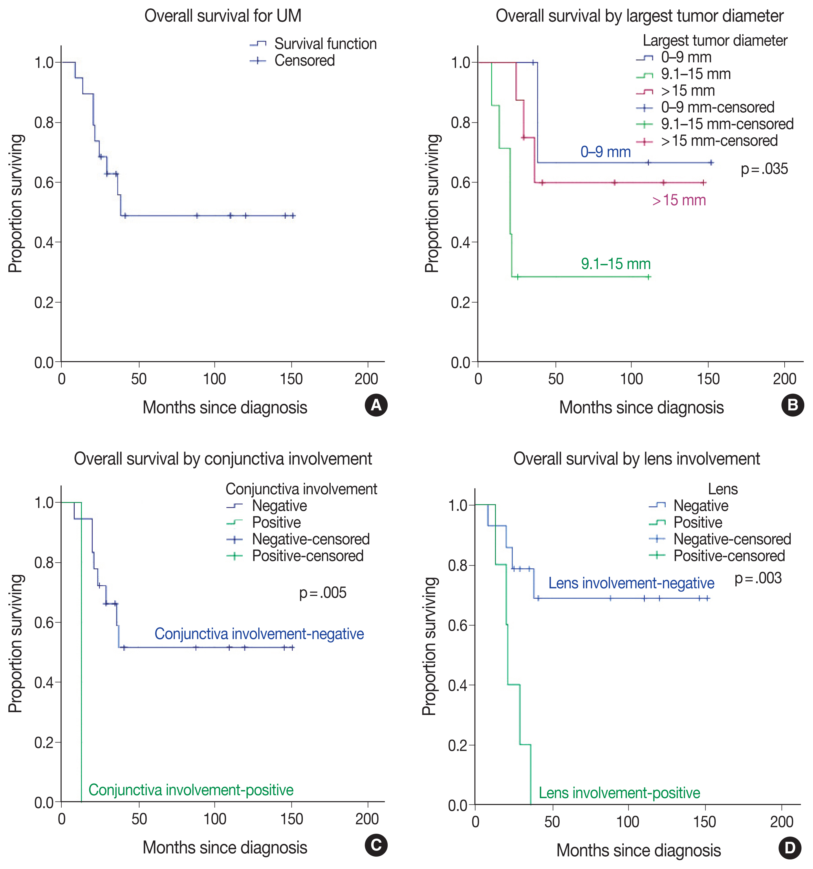
Fig. 3 Kaplan-Meier overall survival curves in uveal melanoma (UM) patients (A) compared by largest tumor diameter (B), conjunctival involvement (C), and lens involvement (D).

Fig. 4 Right enucleation from a 25-year-old female patient revealed a densely pigmented, dome-shaped posterior choroidal melanoma with a basal diameter of 16 mm and a tumor thickness of 3.3 mm (A–C). Effacement of the overlying retinal layer by infiltrating melanocytes (D) (arrow). Densely pigmented atypical melanocytes are arranged around the necrosis reminiscent of pseudo-palisading necrosis (E). A closer look highlights the atypical epithelioid melanocytes with large nuclei and prominent nucleoli (F). This case is wild-type for BRAF V600 mutation.
Reference
-
References
1. Elder DE, Massi D, Scolyer RA, Willemze R. WHO classification of skin tumours. 4th ed. Lyon: IARC Press;2018.2. Singh AD, Turell ME, Topham AK. Uveal melanoma: trends in incidence, treatment, and survival. Ophthalmology. 2011; 118:1881–5.
Article3. Kastelan S, Gverovic Antunica A, Beketic Oreskovic L, Salopek Rabatic J, Kasun B, Bakija I. Conjunctival melanoma: epidemiological trends and features. Pathol Oncol Res. 2018; 24:787–96.4. Kastelan S, Antunica AG, Oreskovic LB, Pelcic G, Kasun E, Hat K. Immunotherapy for uveal melanoma: current knowledge and perspectives. Curr Med Chem. 2020; 27:1350–66.5. Shields CL, Kaliki S, Cohen MN, Shields PW, Furuta M, Shields JA. Prognosis of uveal melanoma based on race in 8100 patients: The 2015 Doyne Lecture. Eye (Lond). 2015; 29:1027–35.
Article6. Jovanovic P, Mihajlovic M, Djordjevic-Jocic J, Vlajkovic S, Cekic S, Stefanovic V. Ocular melanoma: an overview of the current status. Int J Clin Exp Pathol. 2013; 6:1230–44.7. Li Y, Fan X, Jia R. Conjunctival melanoma: update on management. Int Ophthalmol Clin. 2019; 59:27–35.
Article8. Yu GP, Hu DN, McCormick S, Finger PT. Conjunctival melanoma: is it increasing in the United States? Am J Ophthalmol. 2003; 135:800–6.
Article9. Vora GK, Demirci H, Marr B, Mruthyunjaya P. Advances in the management of conjunctival melanoma. Surv Ophthalmol. 2017; 62:26–42.
Article10. Tan LLY, Hong J, Goh WL, et al. Clinical features and survival outcomes of ocular melanoma in a multi-ethnic Asian cohort. Sci Rep. 2020; 10:16367.
Article11. Jain P, Finger PT, Damato B, et al. Multicenter, international assessment of the eighth edition of the American Joint Committee on Cancer cancer staging manual for conjunctival melanoma. JAMA Ophthalmol. 2019; 137:905–11.
Article12. Sagiv O, Thakar SD, Kandl TJ, et al. Immunotherapy with programmed cell death 1 inhibitors for 5 patients with conjunctival melanoma. JAMA Ophthalmol. 2018; 136:1236–41.
Article13. Kaliki S, Shields CL. Uveal melanoma: relatively rare but deadly cancer. Eye (Lond). 2017; 31:241–57.
Article14. Amin MB, Edge S, Greene FL, et al. AJCC cancer staging manual. 8th ed. New York: Springer;2017.15. Esmaeli B, Roberts D, Ross M, et al. Histologic features of conjunctival melanoma predictive of metastasis and death (an American Ophthalmological thesis). Trans Am Ophthalmol Soc. 2012; 110:64–73.16. Protocol for the examination of specimens from patients with uveal melanoma [Internet]. Northfield: College of American Pathologists;2017. [cited 2021 May 10]. Available from: https://documents.cap.org/protocols/cp-uveal-melanoma-17protocol-4000.pdf .17. Jager MJ, Shields CL, Cebulla CM, et al. Uveal melanoma. Nat Rev Dis Primers. 2020; 6:24.
Article18. Shields CL, Shields JA, De Potter P, Cater J, Tardio D, Barrett J. Diffuse choroidal melanoma: clinical features predictive of metastasis. Arch Ophthalmol. 1996; 114:956–63.19. McLean IW, Foster WD, Zimmerman LE, Gamel JW. Modifications of Callender’s classification of uveal melanoma at the Armed Forces Institute of Pathology. Am J Ophthalmol. 1983; 96:502–9.
Article20. Koc I, Kiratli H. Current management of conjunctival melanoma part 1: clinical features, diagnosis and histopathology. Turk J Ophthalmol. 2020; 50:293–303.
Article21. Berta-Antalics AI, Kruse FE, Holbach L. Pathology and prognostic factors of conjunctival melanoma. Ophthalmologe. 2015; 112:892894–8.22. Wolf J, Auw-Haedrich C, Schlecht A, et al. Transcriptional characterization of conjunctival melanoma identifies the cellular tumor microenvironment and prognostic gene signatures. Sci Rep. 2020; 10:17022.
Article23. Tarlan B, Kiratli H. Uveal melanoma: current trends in diagnosis and management. Turk J Ophthalmol. 2016; 46:123–37.24. Kujala E, Makitie T, Kivela T. Very long-term prognosis of patients with malignant uveal melanoma. Invest Ophthalmol Vis Sci. 2003; 44:4651–9.
Article25. Alkatan HM, Al Qahtani AA, Maktabi AM. Enucleated globes with choroidal melanoma: a retrospective histopathological study and correlation with cytogenetic profile in 2 eye centers. Ann Med Surg (Lond). 2020; 55:227–33.
Article26. Shields CL, Kaliki S, Furuta M, Mashayekhi A, Shields JA. Clinical spectrum and prognosis of uveal melanoma based on age at presentation in 8,033 cases. Retina. 2012; 32:1363–72.
Article27. Kaliki S, Shields CL, Mashayekhi A, Ganesh A, Furuta M, Shields JA. Influence of age on prognosis of young patients with uveal melanoma: a matched retrospective cohort study. Eur J Ophthalmol. 2013; 23:208–16.
Article28. Esmaeli B, Rubin ML, Xu S, et al. Greater tumor thickness, ulceration, and positive sentinel lymph node are associated with worse prognosis in patients with conjunctival melanoma: implications for future AJCC classifications. Am J Surg Pathol. 2019; 43:1701–10.29. Tuomaala S, Toivonen P, Al-Jamal R, Kivela T. Prognostic significance of histopathology of primary conjunctival melanoma in Caucasians. Curr Eye Res. 2007; 32:939–52.
Article30. Damato B, Coupland SE. A reappraisal of the significance of largest basal diameter of posterior uveal melanoma. Eye (Lond). 2009; 23:2152–60.
Article31. Kaliki S, Shields CL, Shields JA. Uveal melanoma: estimating prognosis. Indian J Ophthalmol. 2015; 63:93–102.
Article32. Cao J, Brouwer NJ, Richards KE, et al. PD-L1/PD-1 expression and tumor-infiltrating lymphocytes in conjunctival melanoma. Oncotarget. 2017; 8:54722–34.
Article33. Anastassiou G, Esser M, Bader E, Steuhl KP, Bornfeld N. Expression of cell adhesion molecules and tumour infiltrating leucocytes in conjunctival melanoma. Melanoma Res. 2004; 14:381–5.
Article34. Brouwer NJ, Verdijk RM, Heegaard S, Marinkovic M, Esmaeli B, Jager MJ. Conjunctival melanoma: New insights in tumour genetics and immunology, leading to new therapeutic options. Prog Retin Eye Res. 2022; 86:100971.
Article35. Crawford JB. Conjunctival melanomas: prognostic factors a review and an analysis of a series. Trans Am Ophthalmol Soc. 1980; 78:467–502.36. Folberg R, McLean IW, Zimmerman LE. Malignant melanoma of the conjunctiva. Hum Pathol. 1985; 16:136–43.
Article37. Bobic-Radovanovic A, Latkovic Z, Marinkovic J, Radovanovic Z. Predictors of survival in malignant melanoma of the conjunctiva: a clinico-pathological and follow-up study. Eur J Ophthalmol. 1998; 8:4–7.
Article38. Vodencarevic AN, Jusufovic V, Terzic S, Burgic M, Halibasic M, Sinanovic M. Epidemiological analysis of ocular melanoma in university clinic center in Tuzla, Bosnia and Herzegovina. Mater Sociomed. 2016; 28:314–5.
Article39. Papastefanou VP, Cohen VM. Uveal melanoma. J Skin Cancer. 2011; 2011:573974.
Article40. Demirci H, Shields CL, Shields JA, Honavar SG, Eagle RC Jr. Ring melanoma of the ciliary body: report on twenty-three patients. Retina. 2002; 22:698–706.41. Shildkrot Y, Wilson MW. Conjunctival melanoma: pitfalls and dilemmas in management. Curr Opin Ophthalmol. 2010; 21:380–6.
Article42. Missotten GS, Keijser S, De Keizer RJ, De Wolff-Rouendaal D. Conjunctival melanoma in the Netherlands: a nationwide study. Invest Ophthalmol Vis Sci. 2005; 46:75–82.
Article43. Ortega MA, Fraile-Martinez O, Garcia-Honduvilla N, et al. Update on uveal melanoma: translational research from biology to clinical practice (Review). Int J Oncol. 2020; 57:1262–79.
Article44. Toomey CB, Fraser K, Thorson JA, Goldbaum MH, Lin JH. GNAQ and PMS1 mutations associated with uveal melanoma, ocular surface melanosis, and nevus of ota. Ocul Oncol Pathol. 2019; 5:267–72.
Article45. Rodriguez A, Duenas-Gonzalez A, Delgado-Pelayo S. Clinical presentation and management of uveal melanoma. Mol Clin Oncol. 2016; 5:675–7.
Article
- Full Text Links
-
- Actions
-
Cited
- CITED
-
- Close
- Share
-
- Similar articles
-
- Prognostic Significance of Epidermal Growth Factor Receptor Expression in Distant Metastatic Melanoma from Primary Cutaneous Melanoma
- Clinical Study of Malignant Melanoma for Recent 14 Years
- A Clinicopathologic Study of Melanonychia
- Ki67 Antigen as a Predictive Factor for Prognosis of Sinonasal Mucosal Melanoma
- A Case of Metastatic Malignant Melanoma of the Liver Resulting from Choroidal Melanoma
