Neurointervention.
2022 Jul;17(2):121-125. 10.5469/neuroint.2022.00101.
Delayed Rupture of an Anterior Communicating Artery Pseudoaneurysm Caused by Distal Occlusion Thrombectomy Using a Stent Retriever: A Case Report and Mechanism of Injury
- Affiliations
-
- 1Department of Neurology, Daegu Catholic University Medical Center, Daegu, Korea
- 2Department of Radiology, Daegu Catholic University Medical Center, Daegu, Korea
- KMID: 2531569
- DOI: http://doi.org/10.5469/neuroint.2022.00101
Abstract
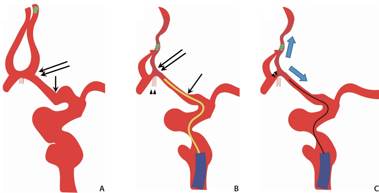
- We report a case of delayed rupture of an anterior communicating artery (Acom) pseudoaneurysm following mechanical thrombectomy (MT) of a distal artery occlusion using a stent retriever. An elderly patient with right hemiparesis showed left proximal internal cerebral artery and middle cerebral artery occlusions. During MT, a fragmented thrombus moved to the anterior cerebral artery (ACA). A stent retriever was deployed to the occluded ACA, and the Acom and proximal ACA segment were significantly straightened. Additionally, we attempted a blind exchange mini-pinning (BEMP) technique, but a subarachnoid hemorrhage (SAH) occurred. Bleeding was almost entirely absorbed 9 days after the procedure, but the SAH recurred at 20 days, and computed tomography angiography revealed a new pseudoaneurysm formation in the Acom. We suggest that the proposed mechanism of pseudoaneurysm formation was likely due to the dislocation and avulsion of the Acom perforators when the ipsilateral ACA was pushed and pulled during MT.
Figure
Reference
-
1. Palaniswami M, Yan B. Mechanical thrombectomy is now the gold standard for acute ischemic stroke: implications for routine clinical practice. Interv Neurol. 2015; 4:18–29.
Article2. Lee H, Qureshi AM, Mueller-Kronast NH, Zaidat OO, Froehler MT, Liebeskind DS, et al. Subarachnoid hemorrhage in mechanical thrombectomy for acute ischemic stroke: analysis of the STRATIS registry, systematic review, and meta-analysis. Front Neurol. 2021; 12:663058.
Article3. Jeong EO, Kwon HJ, Choi SW, Koh HS. Pseudoaneurysm formation after repetitive suction thrombectomy using a penumbra suction catheter. J Cerebrovasc Endovasc Neurosurg. 2016; 18:296–301.
Article4. Imahori T, Okamura Y, Sakata J, Shose H, Yamanishi S, Kohmura E. Delayed rebleeding from pseudoaneurysm after mechanical thrombectomy using stent retriever due to small artery avulsion confirmed by open surgery. World Neurosurg. 2020; 133:150–154.
Article5. Misaki K, Uchiyama N, Mohri M, Kamide T, Tsutsui T, Kanamori N, et al. Pseudoaneurysm formation caused by the withdrawal of a Trevo ProVue stent at a tortuous cerebral vessel: a case report. Acta Neurochir (Wien). 2016; 158:2085–2088.
Article6. Munich SA, Vakharia K, Levy EI. Overview of mechanical thrombectomy techniques. Neurosurgery. 2019; 85(Suppl 1):S60–S67.
Article7. Haussen DC, Al-Bayati AR, Eby B, Ravindran K, Rodrigues GM, Frankel MR, et al. Blind exchange with mini-pinning technique for distal occlusion thrombectomy. J Neurointerv Surg. 2020; 12:392–395.
Article8. Pérez-García C, Moreu M, Rosati S, Simal P, Egido JA, Gomez-Escalonilla C, et al. Mechanical thrombectomy in medium vessel occlusions: blind exchange with mini-pinning technique versus mini stent retriever alone. Stroke. 2020; 51:3224–3231.
Article9. Yoshimoto T, Tanaka K, Koge J, Shiozawa M, Yamagami H, Inoue M, et al. Blind exchange with mini-pinning technique using the tron stent retriever for middle cerebral artery M2 occlusion thrombectomy in acute ischemic stroke. Front Neurol. 2021; 12:667835.
Article10. Kim BM, Kim DJ, Kim DI. Stent application for the treatment of cerebral aneurysms. Neurointervention. 2011; 6:53–70.
Article11. Chapot R, Nordmeyer H, Heddier M, Velasco A, Schooss P, Stauder M, et al. The sheeping technique or how to avoid exchange maneuvers. Neuroradiology. 2013; 55:989–992.
Article12. Koge J, Kato S, Hashimoto T, Nakamura Y, Kawajiri M, Yamada T. Vessel wall injury after stent retriever thrombectomy for internal carotid artery occlusion with duplicated middle cerebral artery. World Neurosurg. 2019; 123:54–58.
Article13. Lescher S, Zimmermann M, Konczalla J, Deller T, Porto L, Seifert V, et al. Evaluation of the perforators of the anterior communicating artery (AComA) using routine cerebral 3D rotational angiography. J Neurointerv Surg. 2016; 8:1061–1066.
Article
- Full Text Links
- Actions
-
Cited
- CITED
-
- Close
- Share
- Similar articles
-
- Successful Cross-circulation Stent-Retriever Embolectomy Through Posterior Communicating Artery for Acute MCA Occlusion by Using Trevo XP ProVue
- Suction thrombectomy of distal medium vessel occlusion using microcatheter during mechanical thrombectomy for acute ischemic stroke: A case series
- Balloon Tamponade Treatment of a Stent-graft Related Rupture with a Splenic Artery Pseudoaneurysm: A Case Report
- Effectiveness of Anchoring with Balloon Guide Catheter and Stent Retriever in Difficult Mechanical Thrombectomy for Large Vessel Occlusion
- Posterior Thigh Compartment Syndrome as a Result of Pseudoaneurysm of the Popliteal Artery in the Distal Femoral Fracture: A Case Report


