J Korean Assoc Oral Maxillofac Surg.
2022 Jun;48(3):159-166. 10.5125/jkaoms.2022.48.3.159.
Marginal bone loss around crestal or subcrestal dental implants: prospective clinical study
- Affiliations
-
- 1Dental Research Center, School of Dentistry, Mashhad University of Medical Sciences, Mashhad, Iran
- 2Department of Oral and Maxillofacial Radiology, School of Dentistry and Dental Research Center, Mashhad University of Medical Sciences, Mashhad, Iran
- 3School of Dentistry, Mashhad University of Medical Sciences, Mashhad, Iran
- 4Department of Periodontics, School of Dentistry, Mashhad University of Medical Sciences, Mashhad, Iran
- KMID: 2531052
- DOI: http://doi.org/10.5125/jkaoms.2022.48.3.159
Abstract
Objectives
The stability of crestal bone has been reported as a major factor in the success of dental implants. Implants can be placed in an equicrestal (crestal) or subcrestal position. The aim of this study was to evaluate the effect of implant depth placement on marginal bone loss.
Materials and Methods
The study was created in a split-mouth design. Immediately after implant surgery, digital parallel radiographs were prepared and levels of bone were measured where marginal bone loss and bone level changes occurred. These measurements were repeated at 3-month and 6-month follow-up periods.
Results
In this interventional study, 49 implants were evaluated in 18 patients. Primary bone height was not significant between the intervention and control groups in both mesial and distal aspects at 3 months and 6 months from the baseline. The mean marginal bone loss on the mesial side was 1.03 mm in the subcrestal group and 0.83 mm in the crestal group. In addition, mean marginal bone loss on the distal side was 0.88 mm and 0.81 mm in the subcrestal and crestal groups, respectively. Marginal bone loss was not significantly different between sexes, the maxilla or mandible, and in the anterior or posterior regions as well as between different lengths and diameters of implants.
Conclusion
Based on the results of this study, there was no significant difference in terms of marginal bone loss between crestal and subcrestal implants.
Keyword
Figure
-
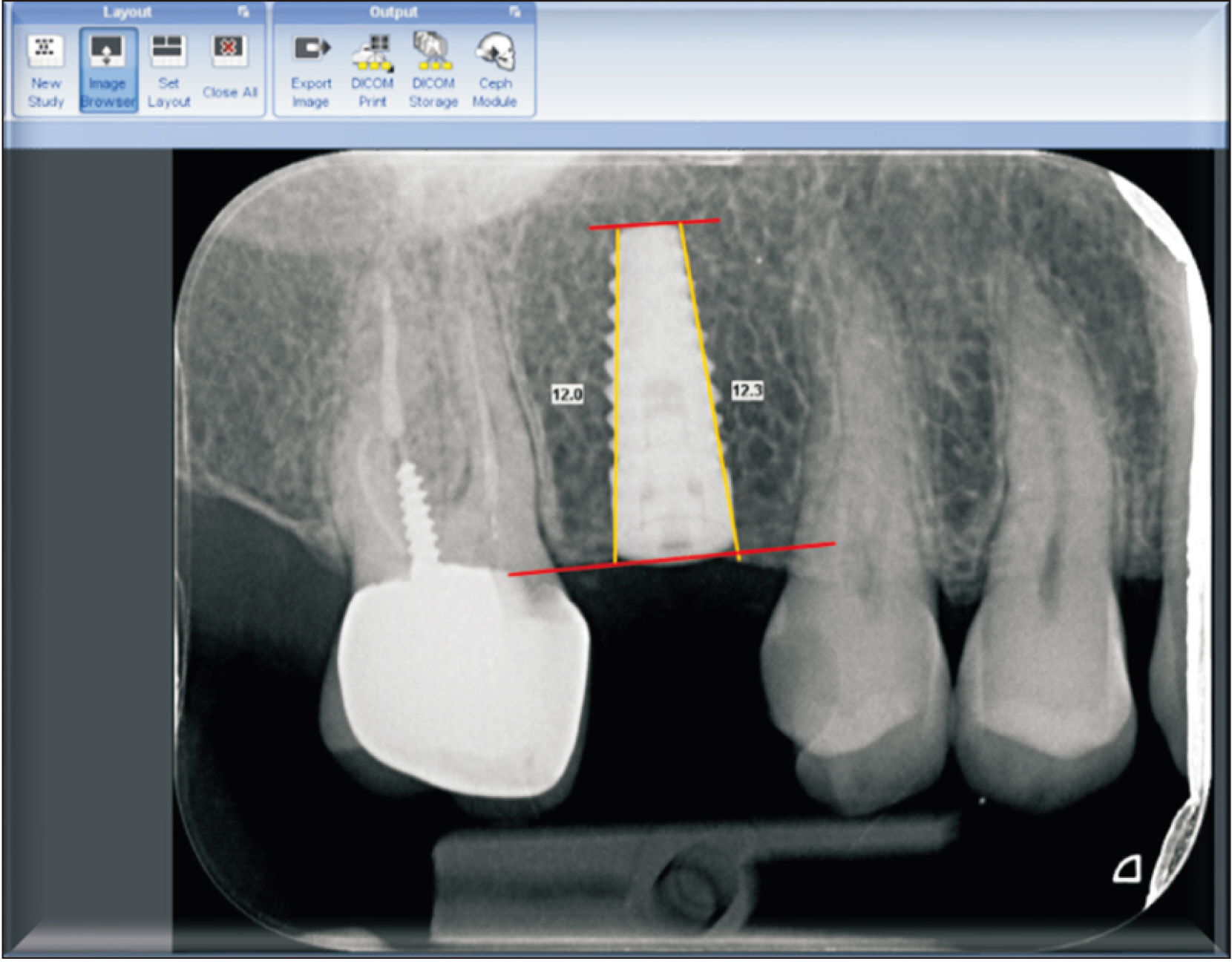
Fig. 1 Measurement of the bone crest to the apex of the fixture in both mesial and distal aspects. Baseline radiographic image.
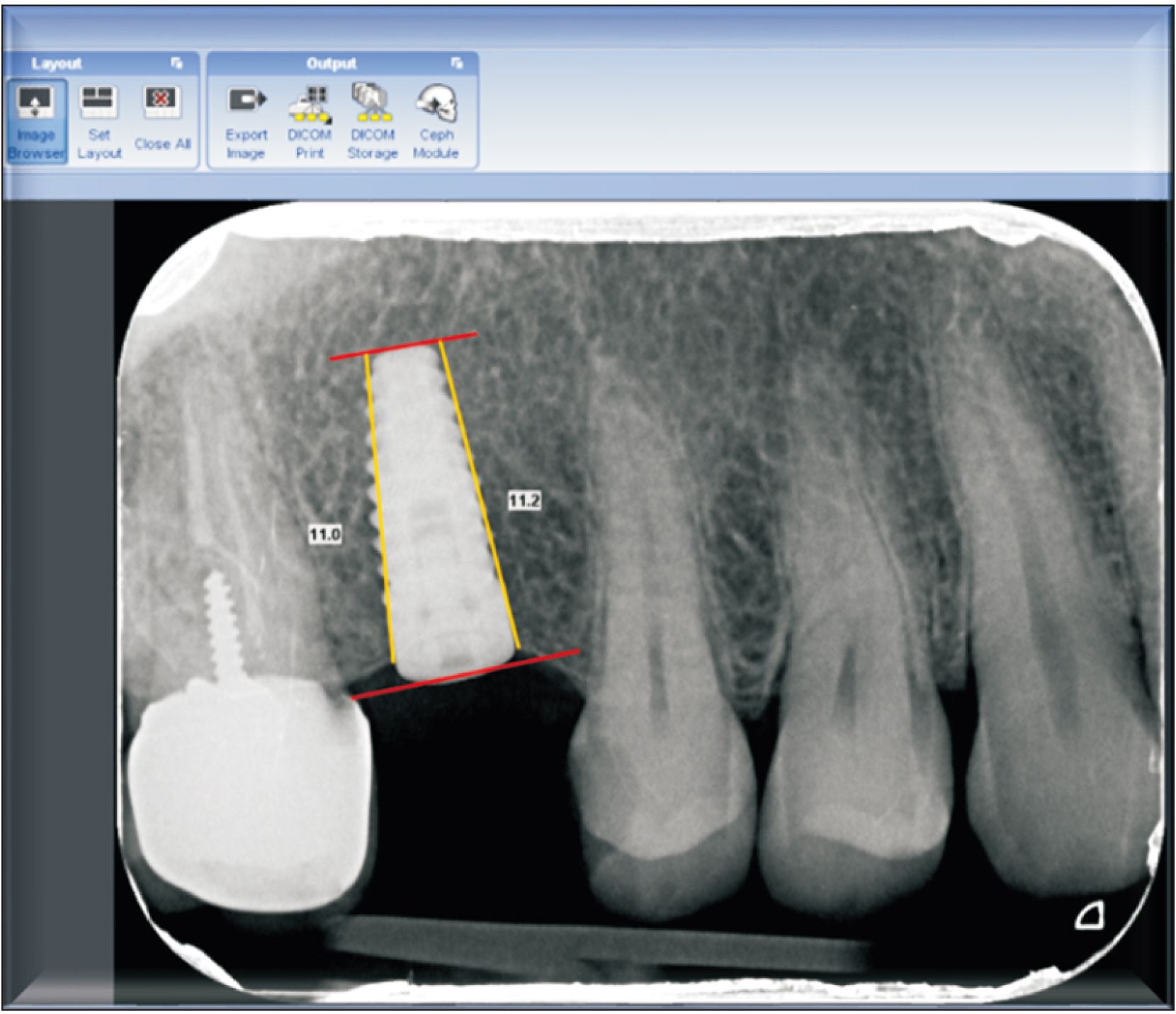
Fig. 2 Measurement of the bone crest to the apex of the fixture at both mesial and distal aspects. Three-month radiographic image.

Fig. 3 Mesial bone level change.
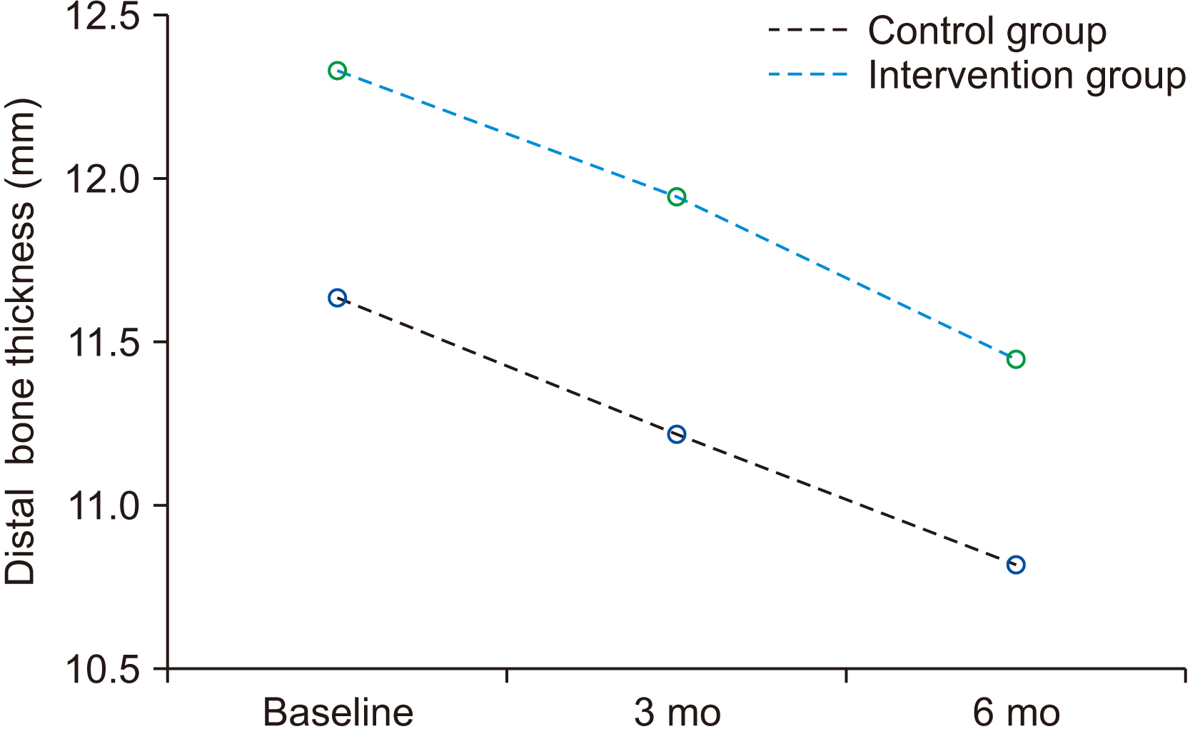
Fig. 4 Distal bone level change.
Reference
-
References
1. Borie E, Orsi IA, de Araujo CP. 2015; The influence of the connection, length and diameter of an implant on bone biomechanics. Acta Odontol Scand. 73:321–9. https://doi.org/10.3109/00016357.2014.961957. DOI: 10.3109/00016357.2014.961957. PMID: 25598357.
Article2. von Wilmowsky C, Moest T, Nkenke E, Stelzle F, Schlegel KA. 2014; Implants in bone: part II. Research on implant osseointegration: material testing, mechanical testing, imaging and histoanalytical methods. Oral Maxillofac Surg. 18:355–72. https://doi.org/10.1007/s10006-013-0397-2. DOI: 10.1007/s10006-013-0397-2. PMID: 23430020.
Article3. Chrcanovic BR, Albrektsson T, Wennerberg A. 2014; Reasons for failures of oral implants. J Oral Rehabil. 41:443–76. https://doi.org/10.1111/joor.12157. DOI: 10.1111/joor.12157. PMID: 24612346.
Article4. Pellicer-Chover H, Díaz-Sanchez M, Soto-Peñaloza D, Peñarrocha-Diago MA, Canullo L, Peñarrocha-Oltra D. 2019; Impact of crestal and subcrestal implant placement upon changes in marginal peri-implant bone level. A systematic review. Med Oral Patol Oral Cir Bucal. 24:e673–83. https://doi.org/10.4317/medoral.23006. DOI: 10.4317/medoral.23006. PMID: 31433391. PMCID: PMC6764703.
Article5. Kowalski J, Lapinska B, Nissan J, Lukomska-Szymanska M. 2021; Factors influencing marginal bone loss around dental implants: a narrative review. Coatings. 11:865. https://doi.org/10.3390/coatings11070865. DOI: 10.3390/coatings11070865.
Article6. Kütan E, Bolukbasi N, Yildirim-Ondur E, Ozdemir T. 2015; Clinical and radiographic evaluation of marginal bone changes around platform-switching implants placed in crestal or subcrestal positions: a randomized controlled clinical trial. Clin Implant Dent Relat Res. 17 Suppl 2:e364–75. https://doi.org/10.1111/cid.12248. DOI: 10.1111/cid.12248. PMID: 25041252.
Article7. Palacios-Garzón N, Velasco-Ortega E, López-López J. 2019; Bone loss in implants placed at subcrestal and crestal level: a systematic review and meta-analysis. Materials (Basel). 12:154. https://doi.org/10.3390/ma12010154. DOI: 10.3390/ma12010154. PMID: 30621286. PMCID: PMC6337530.
Article8. Puisys A, Linkevicius T. 2015; The influence of mucosal tissue thickening on crestal bone stability around bone-level implants. A prospective controlled clinical trial. Clin Oral Implants Res. 26:123–9. https://doi.org/10.1111/clr.12301. DOI: 10.1111/clr.12301. PMID: 24313250.
Article9. Romanos GE. 2015; Wound healing in immediately loaded implants. Periodontol 2000. 68:153–67. https://doi.org/10.1111/prd.12058. DOI: 10.1111/prd.12058. PMID: 25867985.
Article10. Hämmerle CH, Brägger U, Bürgin W, Lang NP. 1996; The effect of subcrestal placement of the polished surface of ITI implants on marginal soft and hard tissues. Clin Oral Implants Res. 7:111–9. https://doi.org/10.1034/j.1600-0501.1996.070204.x. DOI: 10.1034/j.1600-0501.1996.070204.x. PMID: 9002829.
Article11. Gatti C, Gatti F, Silvestri M, Mintrone F, Rossi R, Tridondani G, et al. 2018; A prospective multicenter study on radiographic crestal bone changes around dental implants placed at crestal or subcrestal level: one-year findings. Int J Oral Maxillofac Implants. 33:913–8. https://doi.org/10.11607/jomi.6509. DOI: 10.11607/jomi.6509. PMID: 30025009.
Article12. Veis A, Parissis N, Tsirlis A, Papadeli C, Marinis G, Zogakis A. 2010; Evaluation of peri-implant marginal bone loss using modified abutment connections at various crestal level placements. Int J Periodontics Restorative Dent. 30:609–17. PMID: 20967307.13. Romanos GE, Aydin E, Gaertner K, Nentwig GH. 2015; Long-term results after subcrestal or crestal placement of delayed loaded implants. Clin Implant Dent Relat Res. 17:133–41. https://doi.org/10.1111/cid.12084. DOI: 10.1111/cid.12084. PMID: 23675969.
Article14. Al Amri MD, Al-Johany SS, Al Baker AM, Al Rifaiy MQ, Abduljabbar TS, Al-Kheraif AA. 2017; Soft tissue changes and crestal bone loss around platform-switched implants placed at crestal and subcrestal levels: 36-month results from a prospective split-mouth clinical trial. Clin Oral Implants Res. 28:1342–7. https://doi.org/10.1111/clr.12990. DOI: 10.1111/clr.12990. PMID: 27743396.
Article15. de Siqueira RAC, Fontão FNGK, Sartori IAM, Santos PGF, Bernardes SR, Tiossi R. 2017; Effect of different implant placement depths on crestal bone levels and soft tissue behavior: a randomized clinical trial. Clin Oral Implants Res. 28:1227–33. https://doi.org/10.1111/clr.12946. DOI: 10.1111/clr.12946. PMID: 27480573.
Article16. Ercoli C, Jammal G, Buyers M, Tsigarida AA, Chochlidakis KM, Feng C, et al. 2017; Influence of apico-coronal implant placement on post-surgical crestal bone loss in humans. J Periodontol. 88:762–70. https://doi.org/10.1902/jop.2017.160802. DOI: 10.1902/jop.2017.160802. PMID: 28387610.
Article17. Valles C, Rodríguez-Ciurana X, Clementini M, Baglivo M, Paniagua B, Nart J. 2018; Influence of subcrestal implant placement compared with equicrestal position on the peri-implant hard and soft tissues around platform-switched implants: a systematic review and meta-analysis. Clin Oral Investig. 22:555–70. https://doi.org/10.1007/s00784-017-2301-1. DOI: 10.1007/s00784-017-2301-1. PMID: 29313133.
Article18. Cruz RS, Lemos CAA, de Luna Gomes JM, Pellizzer EP, Verri FR. Fernandes e Oliveira HF. 2022; Clinical comparison between crestal and subcrestal dental implants: a systematic review and meta-analysis. J Prosthet Dent. 127:408–17. https://doi.org/10.1016/j.prosdent.2020.11.003. DOI: 10.1016/j.prosdent.2020.11.003. PMID: 33358610.
Article19. Degidi M, Perrotti V, Shibli JA, Novaes AB, Piattelli A, Iezzi G. 2011; Equicrestal and subcrestal dental implants: a histologic and histomorphometric evaluation of nine retrieved human implants. J Periodontol. 82:708–15. https://doi.org/10.1902/jop.2010.100450. DOI: 10.1902/jop.2010.100450. PMID: 21138355.
Article20. Jung RE, Jones AA, Higginbottom FL, Wilson TG, Schoolfield J, Buser D, et al. 2008; The influence of non-matching implant and abutment diameters on radiographic crestal bone levels in dogs. J Periodontol. 79:260–70. https://doi.org/10.1902/jop.2008.070132. DOI: 10.1902/jop.2008.070132. PMID: 18251640.
Article21. Yi JM, Lee JK, Um HS, Chang BS, Lee MK. 2010; Marginal bony changes in relation to different vertical positions of dental implants. J Periodontal Implant Sci. 40:244–8. https://doi.org/10.5051/jpis.2010.40.5.244. DOI: 10.5051/jpis.2010.40.5.244. PMID: 21072222. PMCID: PMC2967813.
Article22. Pontes AE, Ribeiro FS, da Silva VC, Margonar R, Piattelli A, Cirelli JA, et al. 2008; Clinical and radiographic changes around dental implants inserted in different levels in relation to the crestal bone, under different restoration protocols, in the dog model. J Periodontol. 79:486–94. https://doi.org/10.1902/jop.2008.070145. DOI: 10.1902/jop.2008.070145. PMID: 18315431.
Article23. Fetner M, Fetner A, Koutouzis T, Clozza E, Tovar N, Sarendranath A, et al. 2015; The effects of subcrestal implant placement on crestal bone levels and bone-to-abutment contact: a microcomputed tomographic and histologic study in dogs. Int J Oral Maxillofac Implants. 30:1068–75. https://doi.org/10.11607/jomi.4043. DOI: 10.11607/jomi.4043. PMID: 26394343.
Article24. Pedro RE, De Carli JP, Linden MS, Lima IF, Paranhos LR, Costa MD, et al. 2017; Influence of age on factors associated with peri-implant bone loss after prosthetic rehabilitation over osseointegrated implants. J Contemp Dent Pract. 18:3–10. https://doi.org/10.5005/jp-journals-10024-1979. DOI: 10.5005/jp-journals-10024-1979. PMID: 28050977.
Article25. Wagenberg B, Froum SJ. 2006; A retrospective study of 1925 consecutively placed immediate implants from 1988 to 2004. Int J Oral Maxillofac Implants. 21:71–80. PMID: 16519184.26. Jang HW, Kang JK, Lee K, Lee YS, Park PK. 2011; A retrospective study on related factors affecting the survival rate of dental implants. J Adv Prosthodont. 3:204–15. https://doi.org/10.4047/jap.2011.3.4.204. DOI: 10.4047/jap.2011.3.4.204. PMID: 22259704. PMCID: PMC3259446.
Article27. Güven SŞ, Cabbar F, Güler N. 2020; Local and systemic factors associated with marginal bone loss around dental implants: a retrospective clinical study. Quintessence Int. 51:128–41. https://doi.org/10.3290/j.qi.a42950. DOI: 10.3290/j.qi.a42950. PMID: 31942574.
Article28. Raikar S, Talukdar P, Kumari S, Panda SK, Oommen VM, Prasad A. 2017; Factors affecting the survival rate of dental implants: a retrospective study. J Int Soc Prev Community Dent. 7:351–5. https://doi.org/10.4103/jispcd.JISPCD_380_17. DOI: 10.4103/jispcd.JISPCD_380_17. PMID: 29387619. PMCID: PMC5774056.
Article
- Full Text Links
-
- Actions
-
Cited
- CITED
-
- Close
- Share
-
- Similar articles
-
- Influence of platform switching on crestal bone resorption
- Corrigendum: Marginal bone loss around crestal or subcrestal dental implants: prospective clinical study
- Radiographic evaluation of the proximal bone level between two implants: A 3-year comparative study between Branemark and ITI implants in the mandibular posterior region
- Influence of interimplant distance on bone resorption : A radiological and histological study in beagle dogs
- An 1 year prospective comparative study evaluating the effect of microthread on the maintenance of marginal bone level
