Surgical importance of the tympanic bone: multidetector computed tomography findings
- Affiliations
-
- 1Department of Radiology, Kütahya Health Sciences University, Kütahya, Turkey
- 2Department of Otorhinolaryngology, Kütahya Health Sciences University, Kütahya, Turkey
- KMID: 2531050
- DOI: http://doi.org/10.5125/jkaoms.2022.48.3.149
Abstract
Objectives
To measure tympanic bone thickness (anterior-superior, anterior-inferior, and inferior wall), external ear canal length, and tympanomandibular distance that can be useful in cases that undergo tympanic bone resection.
Materials and Methods
The temporal computed tomography (CT) images of 349 patients were retrospectively evaluated. The anterior-inferior, anterior-superior, and inferior wall thicknesses; tympanomandibular distance; and external auditory canal (EAC) bone canal length were measured from the narrowest part of the canal. The shapes of the EAC in the coronal and sagittal planes were also examined.
Results
The numbers of female and male patients were similar, and the mean age was 49.45±13.95 years. The anterior-superior, anterior-inferior, and inferior wall thicknesses were 1.92±0.60, 2.54±0.74, and 9.16±2.20 mm, respectively. The anterior-superior and anterior-inferior wall thicknesses and canal lengths were greater on the right side (P<0.001). All measurement values were higher in males, except right tympanomandibular distance (P<0.05). A non-significant negative correlation was found between the age of the participants and the left anterior-inferior wall and tympanomandibular distance on both sides. Intra-observer agreement was high for all measurements. We observed four main shapes in the external ear canal in the coronal plane: Type 3, Type 2, Type 1, and Type 4 in order of frequency on the right, and Type 2, Type 3, Type 1, and Type 4 on the left. In the sagittal plane, we detected three shapes: oval (74.4%), triangular (16.3%), and round (9.4%).
Conclusion
The anterior wall thicknesses and tympanomandibular distance should be measured on preoperative temporal bone CT to safely perform tympanic bone anterior resection, which is required in some otological procedures, and also to prevent temporomandibular joint damage.
Figure
-

Fig. 1 Coronal oblique bone window computed tomography image. The narrowest part of the canal is marked in the coronal oblique image.

Fig. 2 Sagittal oblique bone window computed tomography image. Anterior-superior (AS), anterior-inferior (AI), and inferior (I) wall thicknesses were consecutively measured twice, as shown in the figure.

Fig. 3 Sagittal oblique bone window computed tomography image. Measurements were made from the inner cortex to the outer cortex.

Fig. 4 Four main shapes were observed in the coronal plane. A. Type 1: The anterior wall shows convexity towards the external auditory canal, the posterior wall is straight. B. Type 2: The anterior wall and posterior wall are straight, while the posterior wall is angled upwards at the canal entrance. C. Type 3: The anterior and posterior walls are straight. D. Type 4: While the anterior wall and posterior wall are straight, the anterior wall is angled upwards at the canal entrance.
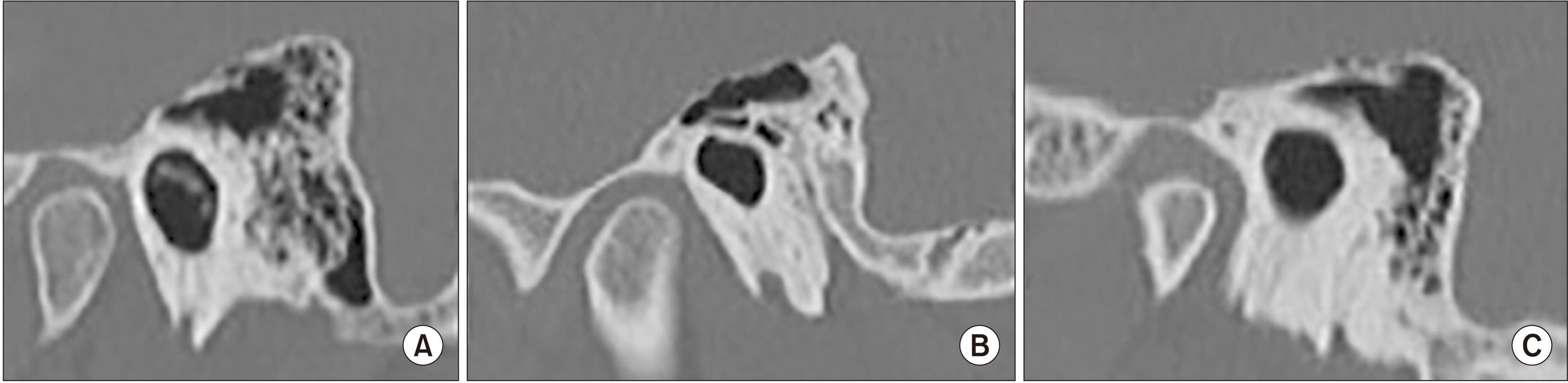
Fig. 5 Three main shapes were observed in the sagittal plane: oval (A), round (B), and triangular (C).
Reference
-
References
1. Isaacson B. 2018; Anatomy and surgical approach of the ear and temporal bone. Head Neck Pathol. 12:321–7. https://doi.org/10.1007/s12105-018-0926-2. DOI: 10.1007/s12105-018-0926-2. PMID: 30069845. PMCID: PMC6081290.
Article2. Lavy J, Fagan P. 2001; Canalplasty: review of 100 cases. J Laryngol Otol. 115:270–3. https://doi.org/10.1258/0022215011907424. DOI: 10.1258/0022215011907424. PMID: 11276326.
Article3. Selesnick SH, Carew JF, DiBartolomeo JR. 1995; Herniation of the temporomandibular joint into the external auditory canal: a complication of otologic surgery. Am J Otol. 16:751–7. PMID: 8572137.4. Kim CW. 2015; Osteomyelitis of the temporomandibular joint following canal wall down mastoidectomy. J Craniofac Surg. 26:e351–3. https://doi.org/10.1097/SCS.0000000000001821. DOI: 10.1097/SCS.0000000000001821. PMID: 26080261.
Article5. Litton WB, Krause CJ, Anson BA, Cohen WN. 1969; The relationship of the facial canal to the annular sulcus. Laryngoscope. 79:1584–604. DOI: 10.1002/lary.5540790905. PMID: 5821135.
Article6. Williams BJ. 1988; The relationship of the facial nerve to the tympanic annulus and external auditory canal. J Otolaryngol Soc Aust. 6:95–6.7. Adad B, Rasgon BM, Ackerson L. 1999; Relationship of the facial nerve to the tympanic annulus: a direct anatomic examination. Laryngoscope. 109:1189–92. https://doi.org/10.1097/00005537-199908000-00002. DOI: 10.1097/00005537-199908000-00002. PMID: 10443818.
Article8. Rodrigues S, Fagan P, Doust B, Moffat K. 2003; A radiologic study of the tympanic bone: anatomy and surgery. Otol Neurotol. 24:796–9. https://doi.org/10.1097/00129492-200309000-00017. DOI: 10.1097/00129492-200309000-00017. PMID: 14501458.
Article9. Eckerdal O, Ahlqvist J. 1980; External bony auditory canal and the tympanic bone. Morphologic properties and influences on the tomographic reproduction. Acta Radiol Diagn (Stockh). 21:425–31. https://doi.org/10.1177/028418518002100314. DOI: 10.1177/028418518002100314. PMID: 7435227.
Article10. Mancini F, Taibah AK, Falcioni M. 1999; Complications and their management in tympanomastoid surgery. Otolaryngol Clin North Am. 32:567–83. https://doi.org/10.1016/s0030-6665(05)70153-5. DOI: 10.1016/S0030-6665(05)70153-5. PMID: 10393787.
Article
- Full Text Links
-
- Actions
-
Cited
- CITED
-
- Close
- Share
-
- Similar articles
-
- Isolated tympanic plate fracture detected by cone-beam computed tomography: report of four cases with review of literature
- Opacification of tympanic membrane - as an anatomic landmark of otoradiology-
- Congenital Anomalies of the Aortic Arch: Evaluation with the Use of Multidetector Computed Tomography
- Clinical Application of Modified Technique of Temporal HRCT for Dehiscence of Tympanic Segment of Facial Nerve
- Orbital Rim Uptake on Bone Scans and Its Clinical Significance
