Photoacoustic Analysis Techniques for Thyroid Cancers: Principles and Applications
- Affiliations
-
- 1Department of Optics and Mechatronics Engineering, Pusan National University, Busan, Korea
- KMID: 2530122
- DOI: http://doi.org/10.11106/ijt.2022.15.1.23
Abstract
- Thyroid cancers are commonly diagnosed worldwide with a continuously increasing incident rate. The ultrasonography is the gold standard method for triaging thyroid nodules to biopsy. Although it has successfully triaged cancerous nodules, many benign nodules also have been triaged to be taken an invasive biopsy, which leads to the overdiagnosing issue. Photoacoustic imaging is an emerging biomedical imaging technique that can provide molecular functional information of biological tissues. Recently, there have been trials to investigate thyroid nodules by using clinically relevant photoacoustic imaging systems. In this review, the principles and applications of the photoacoustic imaging systems for analyzing thyroid nodules are overviewed. Although this technique still has a lot of ways to go for clinical translation, its initial results show great potential to be used for triaging thyroid nodules in vivo.
Keyword
Figure
-
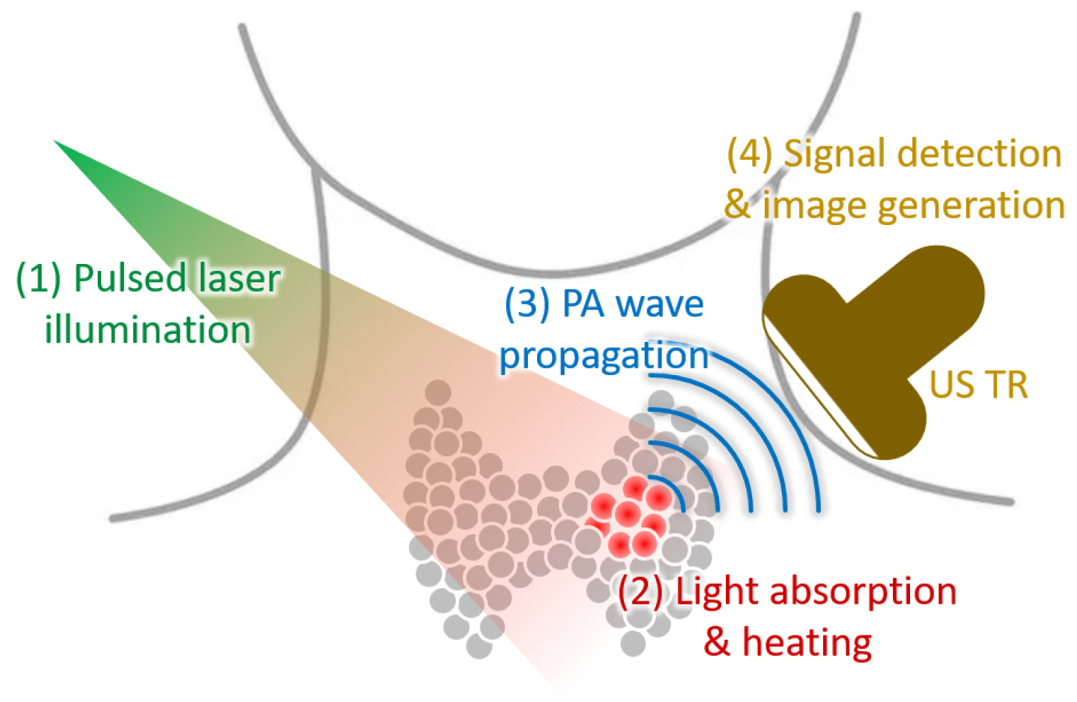
Fig. 1 Schematic illustration of PA imaging. PA: photoacoustic, US: ultrasound
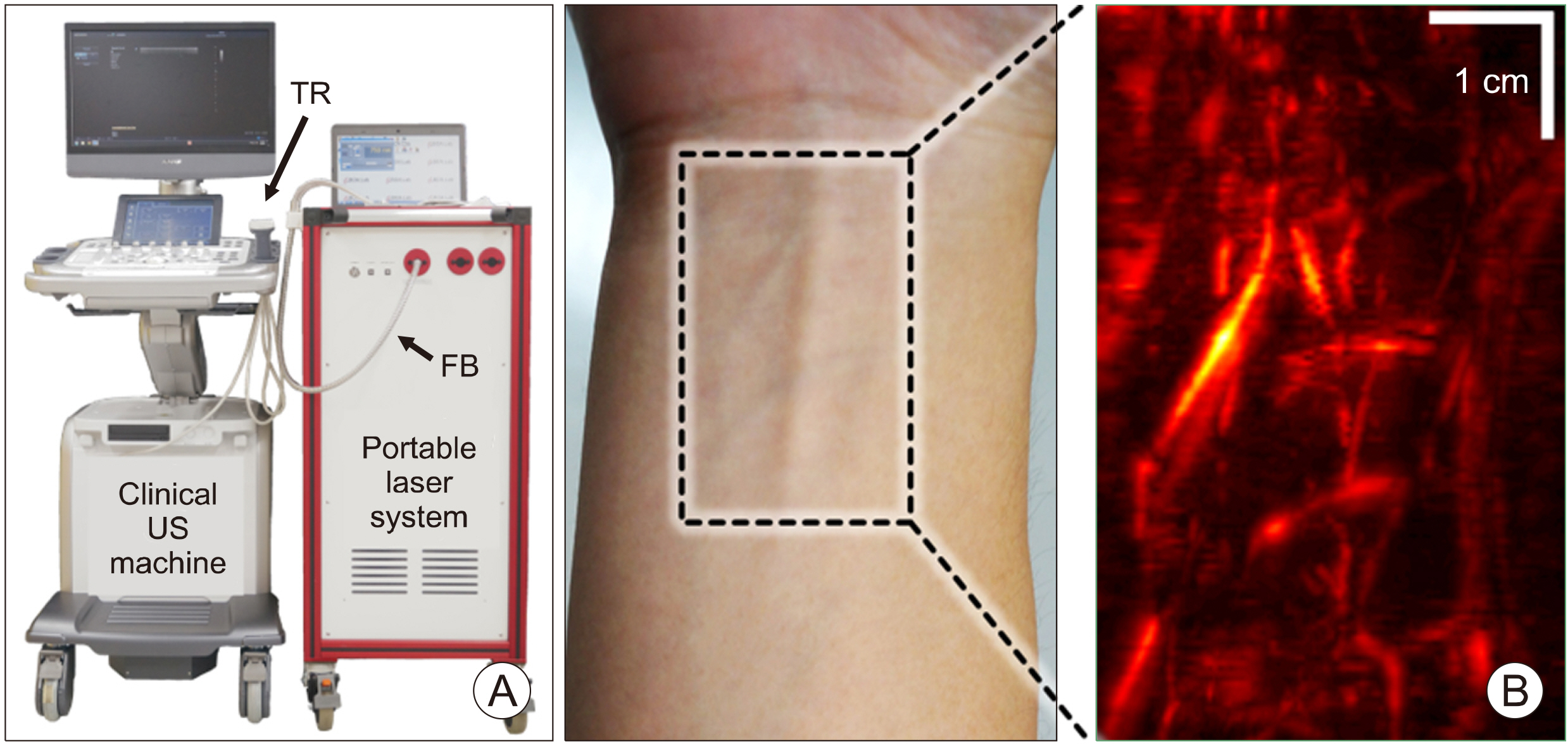
Fig. 2 (A) Photograph of the clinical PA and US imaging system. (B) PA images of blood vessel network in the human forearm. FB: fiber bundle, PA: photoacoustic, TR: transducer, US: ultrasound. The images are reproduced with permission from Ref. 26.
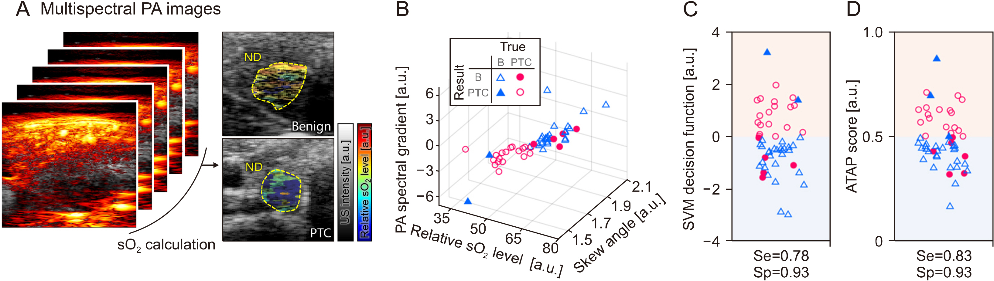
Fig. 3 (A) Multispectral PA analysis of human thyroid in vivo. (B) Three-dimensional illustrations of the multiparametric values with the classification results. (C) Classification results of the thyroid nodules based on the SVM decision function values. (D) Classification results of the thyroid nodules based on the ATAP score. ATAP: the American Thyroid Association and the photoacoustic probability of PTC, B: benign, ND: nodule, PA: photoacoustic, PTC: papillary thyroid cancer, Se: sensitivity, sO2: hemoglobin oxygen saturation, Sp: specificity, SVM: support vector machine, US: ultrasound. The images are reproduced with permission from Ref. 28.
Reference
-
References
1. Ferlay J, Colombet M, Soerjomataram I, Mathers C, Parkin DM, Pineros M, et al. Estimating the global cancer incidence and mortality in 2018: GLOBOCAN sources and methods. Int J Cancer. 2019; 144(8):1941–53. DOI: 10.1002/ijc.31937. PMID: 30350310.
Article2. Davies L, Morris LG, Haymart M, Chen AY, Goldenberg D, Morris J, et al. 2015; American Association of Clinical Endocrinologists and American College of Endocrinology disease state clinical review: the increasing incidence of thyroid cancer. Endocr Pract. 21(6):686–96. DOI: 10.4158/EP14466.DSCR. PMID: 26135963. PMCID: PMC4923940.
Article3. Horn-Ross PL, Lichtensztajn DY, Clarke CA, Dosiou C, Oakley-Girvan I, Reynolds P, et al. 2014; Continued rapid increase in thyroid cancer incidence in California: trends by patient, tumor, and neighborhood characteristics. Cancer Epidemiol Biomarkers Prev. 23(6):1067–79. DOI: 10.1158/1055-9965.EPI-13-1089. PMID: 24842625. PMCID: PMC4071298.
Article4. Siegel RL, Miller KD, Fuchs HE, Jemal A. 2022; Cancer statistics, 2022. CA Cancer J Clin. 72(1):7–33. DOI: 10.3322/caac.21708. PMID: 35020204.
Article5. Welch HG, Black WC. 2010; Overdiagnosis in cancer. J Natl Cancer Inst. 102(9):605–13. DOI: 10.1093/jnci/djq099. PMID: 20413742.
Article6. Vaccarella S, Franceschi S, Bray F, Wild CP, Plummer M, Dal Maso L. 2016; Worldwide thyroid-cancer epidemic? The increasing impact of overdiagnosis. N Engl J Med. 375(7):614–7. DOI: 10.1056/NEJMp1604412. PMID: 27532827.
Article7. Moon WJ, Jung SL, Lee JH, Na DG, Baek JH, Lee YH, et al. 2008; Benign and malignant thyroid nodules: US differentiation-- multicenter retrospective study. Radiology. 247(3):762–70. DOI: 10.1148/radiol.2473070944. PMID: 18403624.
Article8. Frates MC, Benson CB, Charboneau JW, Cibas ES, Clark OH, Coleman BG, et al. 2005; Management of thyroid nodules detected at US: Society of Radiologists in Ultrasound consensus conference statement. Radiology. 237(3):794–800. DOI: 10.1148/radiol.2373050220. PMID: 16304103.
Article9. Gharib H, Goellner JR. 1993; Fine-needle aspiration biopsy of the thyroid: an appraisal. Ann Intern Med. 118(4):282–9. DOI: 10.7326/0003-4819-118-4-199302150-00007. PMID: 8420446.
Article10. Haugen BR, Alexander EK, Bible KC, Doherty GM, Mandel SJ, Nikiforov YE, et al. 2016; 2015 American Thyroid Association Management guidelines for adult patients with thyroid nodules and differentiated thyroid cancer: the American Thyroid Association guidelines task force on thyroid nodules and differentiated thyroid cancer. Thyroid. 26(1):1–133. DOI: 10.1089/thy.2015.0020. PMID: 26462967. PMCID: PMC4739132.
Article11. Perros P, Boelaert K, Colley S, Evans C, Evans RM, Gerrard Ba G, et al. 2014; Guidelines for the management of thyroid cancer. Clin Endocrinol (Oxf). 81 Suppl 1:1–122. DOI: 10.1111/cen.12515. PMID: 24989897.
Article12. Kwak JY, Han KH, Yoon JH, Moon HJ, Son EJ, Park SH, et al. 2011; Thyroid imaging reporting and data system for US features of nodules: a step in establishing better stratification of cancer risk. Radiology. 260(3):892–9. DOI: 10.1148/radiol.11110206. PMID: 21771959.
Article13. Chng CL, Tan HC, Too CW, Lim WY, Chiam PPS, Zhu L, et al. 2018; Diagnostic performance of ATA, BTA and TIRADS sonographic patterns in the prediction of malignancy in histo-logically proven thyroid nodules. Singapore Med J. 59(11):578–83. DOI: 10.11622/smedj.2018062. PMID: 29774361. PMCID: PMC6250759.
Article14. Kim C, Favazza C, Wang LV. 2010; In vivo photoacoustic tomography of chemicals: high-resolution functional and molecular optical imaging at new depths. Chem Rev. 110(5):2756–82. DOI: 10.1021/cr900266s. PMID: 20210338. PMCID: PMC2872199.
Article15. Choi W, Park EY, Jeon S, Kim C. 2018; Clinical photoacoustic imaging platforms. Biomed Eng Lett. 8(2):139–55. DOI: 10.1007/s13534-018-0062-7. PMID: 30603199. PMCID: PMC6208525.
Article16. Steinberg I, Huland DM, Vermesh O, Frostig HE, Tummers WS, Gambhir SS. 2019; Photoacoustic clinical imaging. Photoacoustics. 14:77–98. DOI: 10.1016/j.pacs.2019.05.001. PMID: 31293884. PMCID: PMC6595011.
Article17. Kim C, Erpelding TN, Jankovic L, Pashley MD, Wang LV. 2010; Deeply penetrating in vivo photoacoustic imaging using a clinical ultrasound array system. Biomed Opt Express. 1(1):278–84. DOI: 10.1364/BOE.1.000278. PMID: 21258465. PMCID: PMC3005157.
Article18. Wang LV, Hu S. 2012; Photoacoustic tomography: in vivo imaging from organelles to organs. Science. 335(6075):1458–62. DOI: 10.1126/science.1216210. PMID: 22442475. PMCID: PMC3322413.
Article19. Kim J, Park S, Lee C, Kim JY, Kim C. 2015; Organic nanostructures for photoacoustic imaging. ChemNanoMat. 2(3):156–66. DOI: 10.1002/cnma.201500171.
Article20. Park EY, Lee H, Han S, Kim C, Kim J. 2022; Photoacoustic imaging systems based on clinical ultrasound platform. Exp Biol Med (Maywood). 247(7):551–60. DOI: 10.1177/15353702211073684. PMID: 35068228. PMCID: PMC9014524.
Article21. Bell AG. 1880; The photophone. Science. 1(11):130–4. DOI: 10.1126/science.os-1.11.130. PMID: 17834320.
Article22. Lee C, Kim J, Zhang Y, Jeon M, Liu C, Song L, et al. 2015; Dual-color photoacoustic lymph node imaging using nanofor-mulated naphthalocyanines. Biomaterials. 73:142–8. DOI: 10.1016/j.biomaterials.2015.09.023. PMID: 26408999.
Article23. Zhang Y, Jeon M, Rich LJ, Hong H, Geng J, Zhang Y, et al. 2014; Non-invasive multimodal functional imaging of the intestine with frozen micellar naphthalocyanines. Nat Nanotechnol. 9(8):631–8. DOI: 10.1038/nnano.2014.130. PMID: 24997526. PMCID: PMC4130353.
Article24. Park B, Lee KM, Park S, Yun M, Choi HJ, Kim J, et al. 2020; Deep tissue photoacoustic imaging of nickel(II) dithiolene- containing polymeric nanoparticles in the second near-infrared window. Theranostics. 10(6):2509–21. DOI: 10.7150/thno.39403. PMID: 32194816. PMCID: PMC7052900.
Article25. Zhang HF, Maslov K, Sivaramakrishnan M, Stoica G, Wang LV. 2007; Imaging of hemoglobin oxygen saturation variations in single vessels in vivo using photoacoustic microscopy. Appl Phys Lett. 90(5):053901. DOI: 10.1063/1.2435697.26. Kim J, Park S, Jung Y, Chang S, Park J, Zhang Y, et al. 2016; Programmable real-time clinical photoacoustic and ultrasound imaging system. Sci Rep. 6:35137. DOI: 10.1038/srep35137. PMID: 27731357. PMCID: PMC5059665.
Article27. Kim J, Park EY, Park B, Choi W, Lee KJ, Kim C. 2020; Towards clinical photoacoustic and ultrasound imaging: probe improvement and real-time graphical user interface. Exp Biol Med (Maywood). 245(4):321–9. DOI: 10.1177/1535370219889968. PMID: 31916849. PMCID: PMC7370595.
Article28. Dogra VS, Chinni BK, Valluru KS, Moalem J, Giampoli EJ, Evans K, et al. 2014; Preliminary results of ex vivo multispectral photoacoustic imaging in the management of thyroid cancer. AJR Am J Roentgenol. 202(6):W552–8. DOI: 10.2214/AJR.13.11433. PMID: 24848849.
Article29. Dima A, Ntziachristos V. 2016; In-vivo handheld optoacoustic tomo-graphy of the human thyroid. Photoacoustics. 4(2):65–9. DOI: 10.1016/j.pacs.2016.05.003. PMID: 27766210. PMCID: PMC5066088.
Article30. Roll W, Markwardt NA, Masthoff M, Helfen A, Claussen J, Eisenblatter M, et al. 2019; Multispectral optoacoustic tomography of benign and malignant thyroid disorders: a pilot study. J Nucl Med. 60(10):1461–6. DOI: 10.2967/jnumed.118.222174. PMID: 30850507. PMCID: PMC6785793.
Article31. Kim J, Park B, Ha J, Steinberg I, Hooper SM, Jeong C, et al. 2021; Multiparametric photoacoustic analysis of human thyroid cancers in vivo. Cancer Res. 81(18):4849–60. DOI: 10.1158/0008-5472.CAN-20-3334. PMID: 34185675.32. Mao Y, Xing M. 2016; Recent incidences and differential trends of thyroid cancer in the USA. Endocr Relat Cancer. 23(4):313–22. DOI: 10.1530/ERC-15-0445. PMID: 26917552. PMCID: PMC4891202.
Article
- Full Text Links
-
- Actions
-
Cited
- CITED
-
- Close
- Share
-
- Similar articles
-
- Clinical photoacoustic imaging of cancer
- Photoacoustic microscopy: principles and biomedical applications
- Review of Photoacoustic Imaging for Imaging-Guided Spinal Surgery
- Ultrasound elastography of the thyroid: principles and current status
- Photoacoustic imaging of occlusal incipient caries in the visible and near-infrared range
