Korean J Orthod.
2022 May;52(3):201-209. 10.4041/kjod21.217.
Influence of late removal after treatment on the removal torque of microimplants
- Affiliations
-
- 1Department of Orthodontics, School of Dentistry, Kyungpook National University, Daegu, Korea
- KMID: 2529943
- DOI: http://doi.org/10.4041/kjod21.217
Abstract
Objective
To compare the removal torque of microimplants upon post-use removal and post-retention removal and to assess the influencing factors.
Methods
The sample group included 241 patients (age, 30.25 ± 12.2 years) with 568 microimplants. They were divided into the post-use (microimplants removed immediately after use or treatment) and post-retention (microimplants removed during the retention period) removal groups. The removal torque in both groups was assessed according to sex, age, placement site and method, and microimplant size. Pearson correlation and multiple linear regression analyses were performed for evaluating variables influencing the removal torque.
Results
The mean period of total in-bone stay of microimplants in the postretention removal group (1,237 days) was approximately two times longer than that in the post-use removal group (656.28 days). The removal torques in the post-retention removal group (range, 4–5 N cm) were also higher than those in the post-use removal group. The mandible and pre-drilling groups demonstrated higher placement and removal torques than did the maxilla and no-drilling groups, respectively. In the no-drilling post-use removal group, the placement torque and microimplant length positively correlated with the removal torque. In the post-retention removal group, unloading in-bone stay period and microimplant diameter positively correlated with the removal torque in the no-drilling and pre-drilling methods, respectively.
Conclusions
The removal torques differed according to the orthodontic loading and removal time of microimplants. With prolonged retention of microimplants inserted using the no-drilling method, the removal torque was clinically acceptable and positively correlated with the unloading in-bone stay period.
Keyword
Figure
-
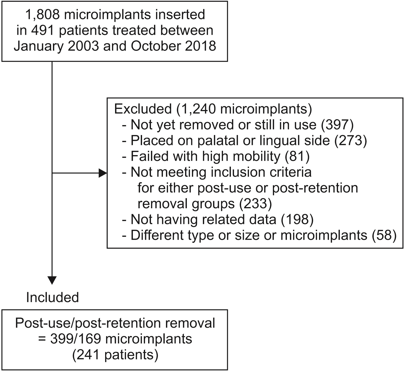
Figure 1 Flow chart of the sampling performed in the study.

Figure 2 Dimensions of the microimplants used in the study.

Figure 3 Measurement of the placement and removal torques by using a digital torque gauge.
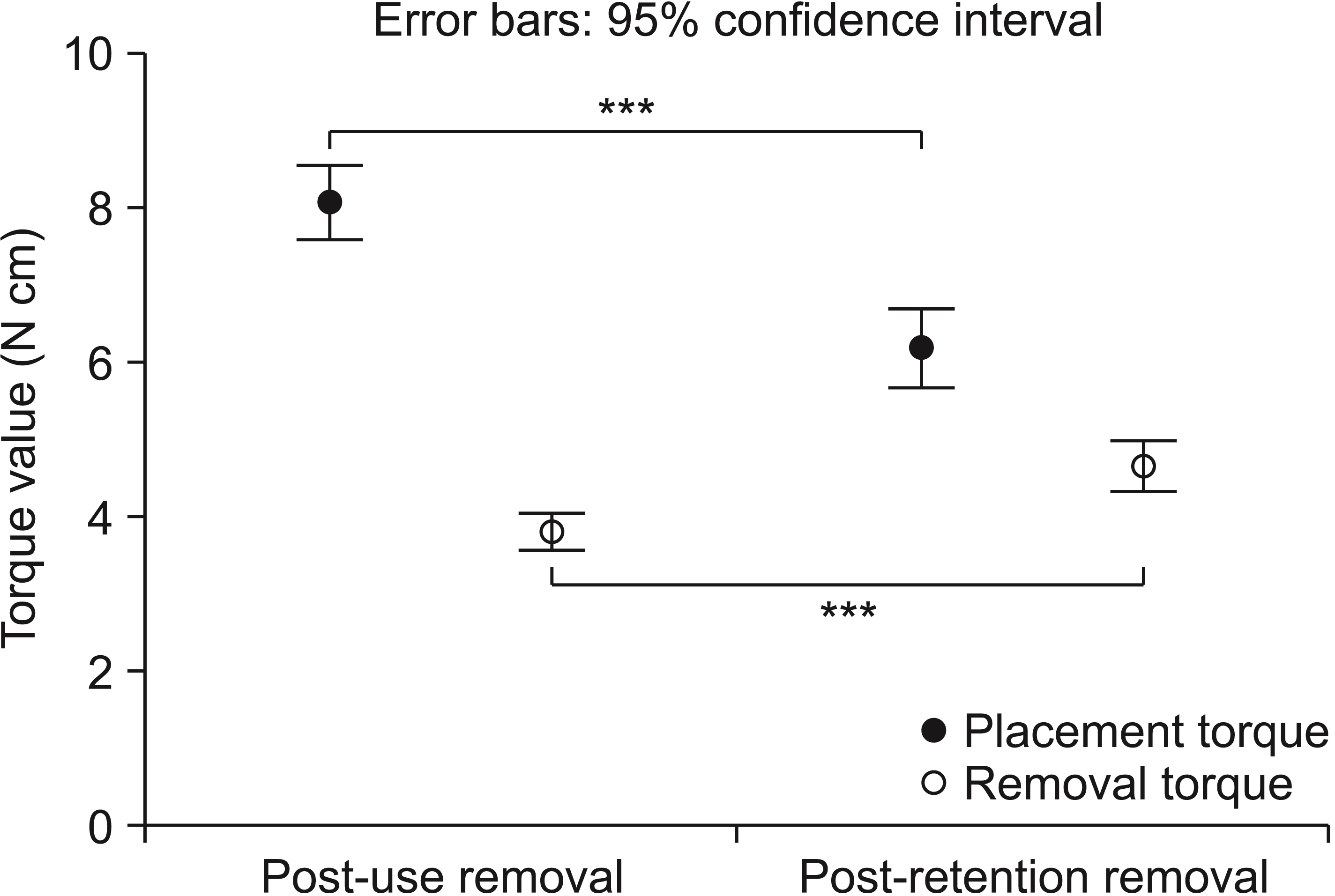
Figure 4 Error-bar plot of the overall placement and removal torques in the post-use and post-retention removal groups. ***p < 0.001.
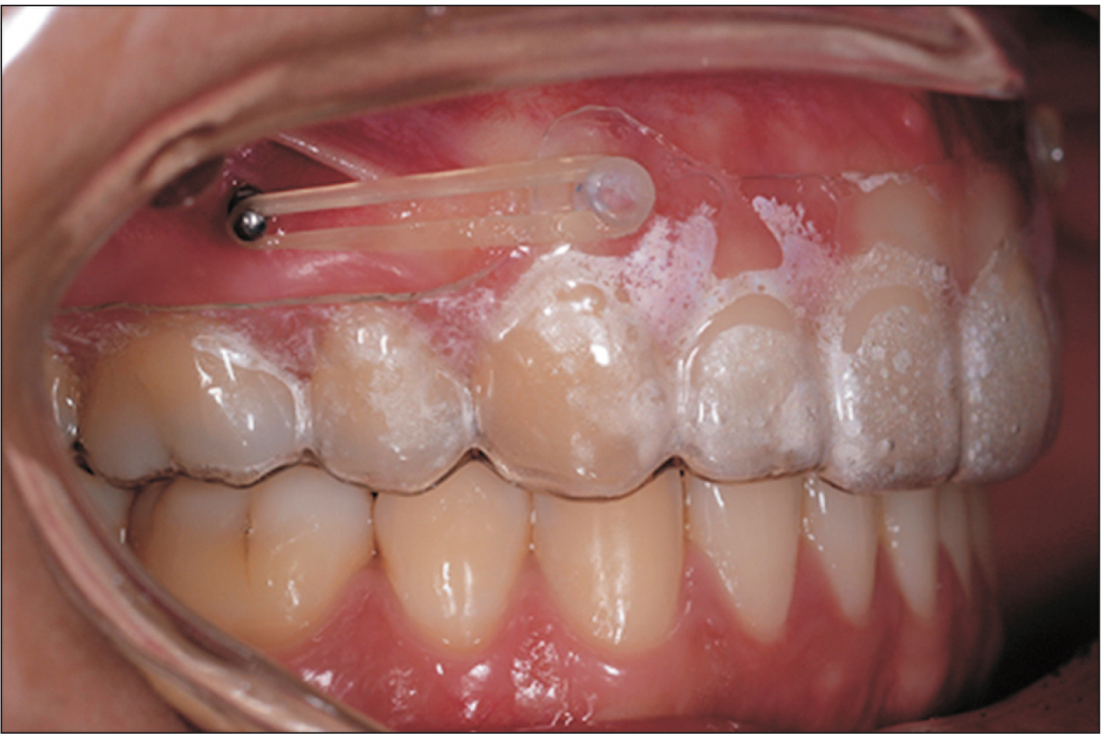
Figure 5 Orthodontic force application for additional correction or retention after treatment by using retained microimplants and elastics.
Reference
-
1. Huja SS, Litsky AS, Beck FM, Johnson KA, Larsen PE. 2005; Pull-out strength of monocortical screws placed in the maxillae and mandibles of dogs. Am J Orthod Dentofacial Orthop. 127:307–13. DOI: 10.1016/j.ajodo.2003.12.023. PMID: 15775945.
Article2. Motoyoshi M, Hirabayashi M, Uemura M, Shimizu N. 2006; Recommended placement torque when tightening an orthodontic mini-implant. Clin Oral Implants Res. 17:109–14. DOI: 10.1111/j.1600-0501.2005.01211.x. PMID: 16441792.
Article3. Suzuki EY, Suzuki B. 2011; Placement and removal torque values of orthodontic miniscrew implants. Am J Orthod Dentofacial Orthop. 139:669–78. DOI: 10.1016/j.ajodo.2010.11.017. PMID: 21536211.
Article4. Guler AU, Sumer M, Duran I, Sandikci EO, Telcioglu NT. 2013; Resonance frequency analysis of 208 Straumann dental implants during the healing period. J Oral Implantol. 39:161–7. DOI: 10.1563/AAID-JOI-D-11-00060. PMID: 22103915.
Article5. Watanabe T, Miyazawa K, Fujiwara T, Kawaguchi M, Tabuchi M, Goto S. 2017; Insertion torque and Periotest values are important factors predicting outcome after orthodontic miniscrew placement. Am J Orthod Dentofacial Orthop. 152:483–8. DOI: 10.1016/j.ajodo.2017.01.026. PMID: 28962732.
Article6. Büchter A, Wiechmann D, Gaertner C, Hendrik M, Vogeler M, Wiesmann HP, et al. 2006; Load-related bone modelling at the interface of orthodontic micro-implants. Clin Oral Implants Res. 17:714–22. DOI: 10.1111/j.1600-0501.2006.01233.x. PMID: 17092232.
Article7. Motoyoshi M, Uemura M, Ono A, Okazaki K, Shigeeda T, Shimizu N. 2010; Factors affecting the long-term stability of orthodontic mini-implants. Am J Orthod Dentofacial Orthop. 137:588.e1–5. discussion 588–9. DOI: 10.1016/j.ajodo.2009.05.019. PMID: 20451776.
Article8. Park HS, Yen S, Jeong SH. 2006; Histologic and biomechanical characteristics of orthodontic self-drilling and self-tapping microscrew implants. Korean J Orthod. 36:295–307.9. Ure DS, Oliver DR, Kim KB, Melo AC, Buschang PH. 2011; Stability changes of miniscrew implants over time. Angle Orthod. 81:994–1000. DOI: 10.2319/120810-711.1. PMID: 21612317. PMCID: PMC8903854.
Article10. Park HS, Kyung HM, Sung JH. McNamara JA, editor. 2008. The treatment of open bite with microimplant anchorage. Microimplants as temporary orthodontic anchorage. The University of Michigan;Ann Arbor: p. 111–4.11. Yadav S, Upadhyay M, Liu S, Roberts E, Neace WP, Nanda R. 2012; Microdamage of the cortical bone during mini-implant insertion with self-drilling and self-tapping techniques: a randomized controlled trial. Am J Orthod Dentofacial Orthop. 141:538–46. DOI: 10.1016/j.ajodo.2011.12.016. PMID: 22554747.
Article12. Roberts WE, Huja SS. Graber LW, Vanarsdall RL, Vig KWL, Huang GJ, editors. 2017. Bone physiology, metabolism, and biomechanics in orthodontic practice. Orthodontics: current principles and techniques. 6th ed. Elsevier;St. Louis: p. 99–153.13. Lakshmikantha HT, Ravichandran NK, Jeon M, Kim J, Park HS. 2019; 3-Dimensional characterization of cortical bone microdamage following placement of orthodontic microimplants using Optical Coherence Tomography. Sci Rep. 9:3242. DOI: 10.1038/s41598-019-39670-9. PMID: 30824805. PMCID: PMC6397251.
Article14. Park HS, Lee YJ, Jeong SH, Kwon TG. 2008; Density of the alveolar and basal bones of the maxilla and the mandible. Am J Orthod Dentofacial Orthop. 133:30–7. DOI: 10.1016/j.ajodo.2006.01.044. PMID: 18174068.
Article15. Park HS, Jeong SH, Kwon OW. 2006; Factors affecting the clinical success of screw implants used as orthodontic anchorage. Am J Orthod Dentofacial Orthop. 130:18–25. DOI: 10.1016/j.ajodo.2004.11.032. PMID: 16849067.
Article16. Frost HM. 2003; Bone's mechanostat: a 2003 update. Anat Rec A Discov Mol Cell Evol Biol. 275:1081–101. DOI: 10.1002/ar.a.10119. PMID: 14613308.
Article17. Degidi M, Scarano A, Piattelli M, Perrotti V, Piattelli A. 2005; Bone remodeling in immediately loaded and unloaded titanium dental implants: a histologic and histomorphometric study in humans. J Oral Implantol. 31:18–24. DOI: 10.1563/0-717.1. PMID: 15751384.
Article18. Misch CE, Bidez MW, Sharawy M. 2001; A bioengineered implant for a predetermined bone cellular response to loading forces. A literature review and case report. J Periodontol. 72:1276–86. DOI: 10.1902/jop.2000.72.9.1276. PMID: 11577963.
Article19. Ozdemir F, Tozlu M, Germec-Cakan D. 2013; Cortical bone thickness of the alveolar process measured with cone-beam computed tomography in patients with different facial types. Am J Orthod Dentofacial Orthop. 143:190–6. DOI: 10.1016/j.ajodo.2012.09.013. PMID: 23374925.
Article20. Miyawaki S, Koyama I, Inoue M, Mishima K, Sugahara T, Takano-Yamamoto T. 2003; Factors associated with the stability of titanium screws placed in the posterior region for orthodontic anchorage. Am J Orthod Dentofacial Orthop. 124:373–8. DOI: 10.1016/S0889-5406(03)00565-1. PMID: 14560266.
Article
- Full Text Links
-
- Actions
-
Cited
- CITED
-
- Close
- Share
-
- Similar articles
-
- Comparison of histologic observation and insertional and removal torque values between titanium grade 2 and 4 microimplants
- The influence of intentional mobilization of implant fixtures before osseointegration
- Influence of Tightening Torque on Implant-Abutment Screw Joint Stability
- Effect of cutting flute length and shape on insertion and removal torque of orthodontic mini-implants
- The influence of abutment screw tightening timing and DLC coating of conical connection implant system
