Brain Tumor Res Treat.
2022 Apr;10(2):117-122. 10.14791/btrt.2022.0005.
Rapid-Growing Intracranial Immature Teratoma Presenting Obstructive Hydrocephalus and Abducens Nerve Palsy: A Case Report and Literature Review
- Affiliations
-
- 1Departments of Neurosurgery, Dongsan Medical Center, Keimyung University School of Medicine, Daegu, Korea
- 2Departments of Pediatrics, Dongsan Medical Center, Keimyung University School of Medicine, Daegu, Korea
- 3Departments of Pathology, Dongsan Medical Center, Keimyung University School of Medicine, Daegu, Korea
- KMID: 2529444
- DOI: http://doi.org/10.14791/btrt.2022.0005
Abstract
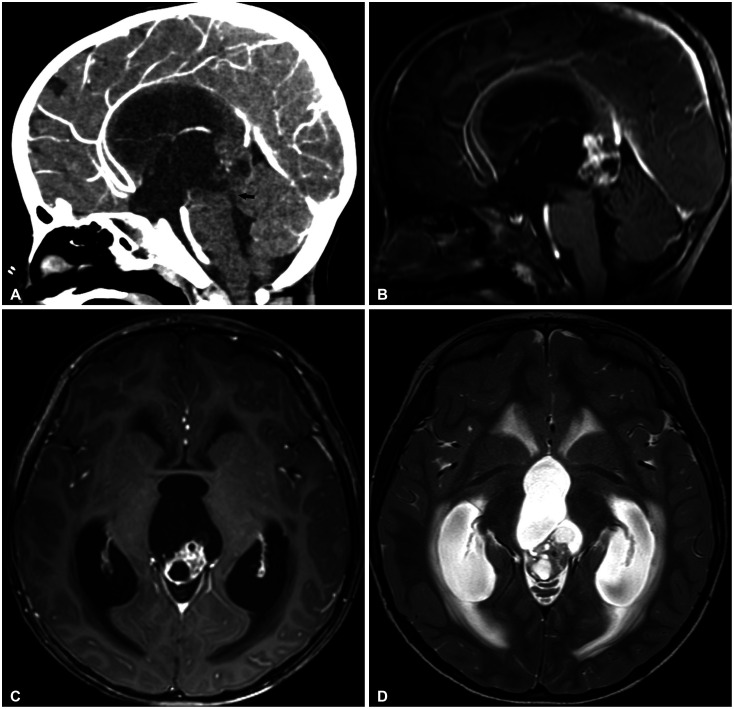
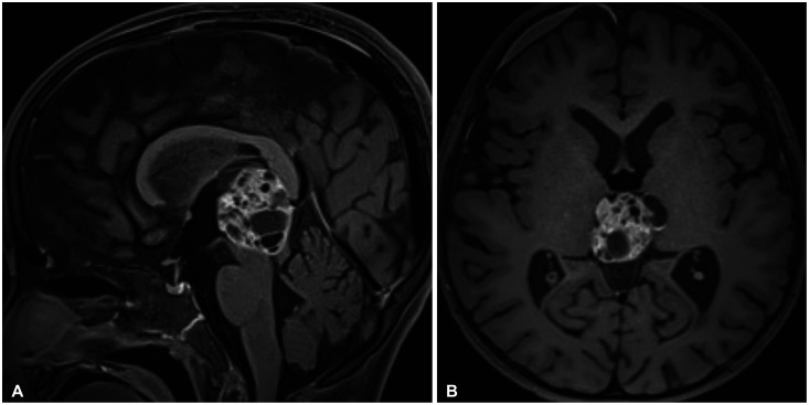
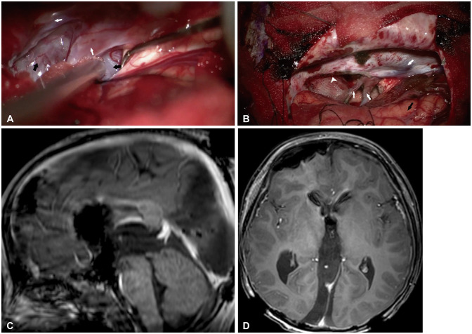
- Intracranial immature teratoma is an extremely rare disease with poor prognosis and requires complicated treatment. Owing to the deep midline location of the tumor, total surgical resection of the tumor is challenging. We present our experience with a fast-growing pineal gland immature teratoma in a 4-year-old boy, who presented with obstructive hydrocephalus and abducens nerve palsy, which was treated with total surgical resection of the tumor. In addition, we aimed to determine the appropriate treatment modality for intracranial immature teratomas by reviewing the literature and investigating the prognosis.
Keyword
Figure
Reference
-
1. Goyal N, Kakkar A, Singh PK, Sharma MC, Chandra PS, Mahapatra AK, et al. Intracranial teratomas in children: a clinicopathological study. Childs Nerv Syst. 2013; 29:2035–2042. PMID: 23568500.
Article2. Georgiu C, Opincariu I, Cebotaru CL, Mirescu ŞC, Stănoiu BP, Domşa TA, et al. Intracranial immature teratoma with a primitive neuroectodermal malignant transformation - case report and review of the literature. Rom J Morphol Embryol. 2016; 57:1389–1395. PMID: 28174809.3. Abdelmuhdi AS, Almazam AE, Dissi NA, Albastaki UM, Pierre-Jerome C. Intracranial teratoma: imaging, intraoperative, and pathologic features: AIRP best cases in radiologic-pathologic correlation. Radiographics. 2017; 37:1506–1511. PMID: 28898192.
Article4. Huang X, Zhang R, Zhou LF. Diagnosis and treatment of intracranial immature teratoma. Pediatr Neurosurg. 2009; 45:354–360. PMID: 19907199.
Article5. Lee YH, Park EK, Park YS, Shim KW, Choi JU, Kim DS. Treatment and outcomes of primary intracranial teratoma. Childs Nerv Syst. 2009; 25:1581–1587. PMID: 19693515.
Article6. Ogawa K, Toita T, Nakamura K, Uno T, Onishi H, Itami J, et al. Treatment and prognosis of patients with intracranial nongerminomatous malignant germ cell tumors: a multiinstitutional retrospective analysis of 41 patients. Cancer. 2003; 98:369–376. PMID: 12872359.
Article7. Ferraz ST, Valera ET, Brassesco MS, Santos de Oliveira R, Carlos dos Santos A, Saggioro FP, et al. Intracranial teratoma in children: the role of chromosome 21 trisomy. Neuropathology. 2014; 34:197–200. PMID: 24812702.
Article8. Liu Z, Lv X, Wang W, An J, Duan F, Feng X, et al. Imaging characteristics of primary intracranial teratoma. Acta Radiol. 2014; 55:874–881. PMID: 24103916.
Article9. Noudel R, Vinchon M, Dhellemmes P, Litré CF, Rousseaux P. Intracranial teratomas in children: the role and timing of surgical removal. J Neurosurg Pediatr. 2008; 2:331–338. PMID: 18976103.
Article10. Han JW, Koh KN, Kim JY, Baek HJ, Lee JW, Shim KW, et al. Current trends in management for central nervous system germ cell tumor. Clin Pediatr Hematol Oncol. 2016; 23:17–27.11. Zhang X, Wang H, Hong F, Xu T, Chen J. “Bones in the medulla oblongata?”— A case report of intracranial teratoma and review of the literature. Front Pediatr. 2021; 9:628265. PMID: 34026683.
Article12. Osborn AG, Preece MT. Intracranial cysts: radiologic-pathologic correlation and imaging approach. Radiology. 2006; 239:650–664. PMID: 16714456.
Article13. Beschorner R, Schittenhelm J, Bueltmann E, Ritz R, Meyermann R, Mittelbronn M. Mature cerebellar teratoma in adulthood. Neuropathology. 2009; 29:176–180. PMID: 18627482.14. Li Q, You C, Zan X, Chen N, Zhou L, Xu J. Mature cystic teratoma (dermoid cyst) in the sylvian fissure: a case report and review of the literature. J Child Neurol. 2012; 27:211–217. PMID: 22190504.
Article15. Hoffman HJ, Otsubo H, Hendrick EB, Humphreys RP, Drake JM, Becker LE, et al. Intracranial germ-cell tumors in children. J Neurosurg. 1991; 74:545–551. PMID: 1848284.16. Ziyal IM, Bozkurt G, Bilginer B, Gülsen S, Ozcan OE. Abducens nerve palsy in a patient with a parasagittal meningioma--case report. Neurol Med Chir (Tokyo). 2006; 46:98–100. PMID: 16498221.
- Full Text Links
- Actions
-
Cited
- CITED
-
- Close
- Share
- Similar articles
-
- A Case of Benign Abducens Nerve Palsy of Childhood
- A case of vaginally terminated fetus who had intracranial immature teratoma after transabdominal fetal cephalocentesis
- A Case of Traumatic Bilateral Abducens Nerve Palsy Associated with Skull Base Fracture
- A Case of Isolated Unilateral Abducens Nerve Palsy Caused by Clival Metastasis from Rectal Cancer
- A Case of Spontaneous Intracranial Hypotension presented as Bilateral Abducens Nerve Palsy without Postural Headache


