Prog Med Phys.
2022 Mar;33(1):1-10. 10.14316/pmp.2022.33.1.1.
Effective Volume of the Korea Research Institute of Standards and Science Free Air Chamber L1 for Low-Energy X-Ray Measurement
- Affiliations
-
- 1Ionizing Radiation Metrology Group, Daejeon, Korea
- 2Length Group, Korea Research Institute of Standards and Science (KRISS), Daejeon, Korea
- KMID: 2528099
- DOI: http://doi.org/10.14316/pmp.2022.33.1.1
Abstract
- Purpose
To evaluate the effective volume of the Korea Research Institute of Standards and Science free air chamber (KRISS FAC) L1 used for the primary standard device of the low-energy X-ray air kerma.
Methods
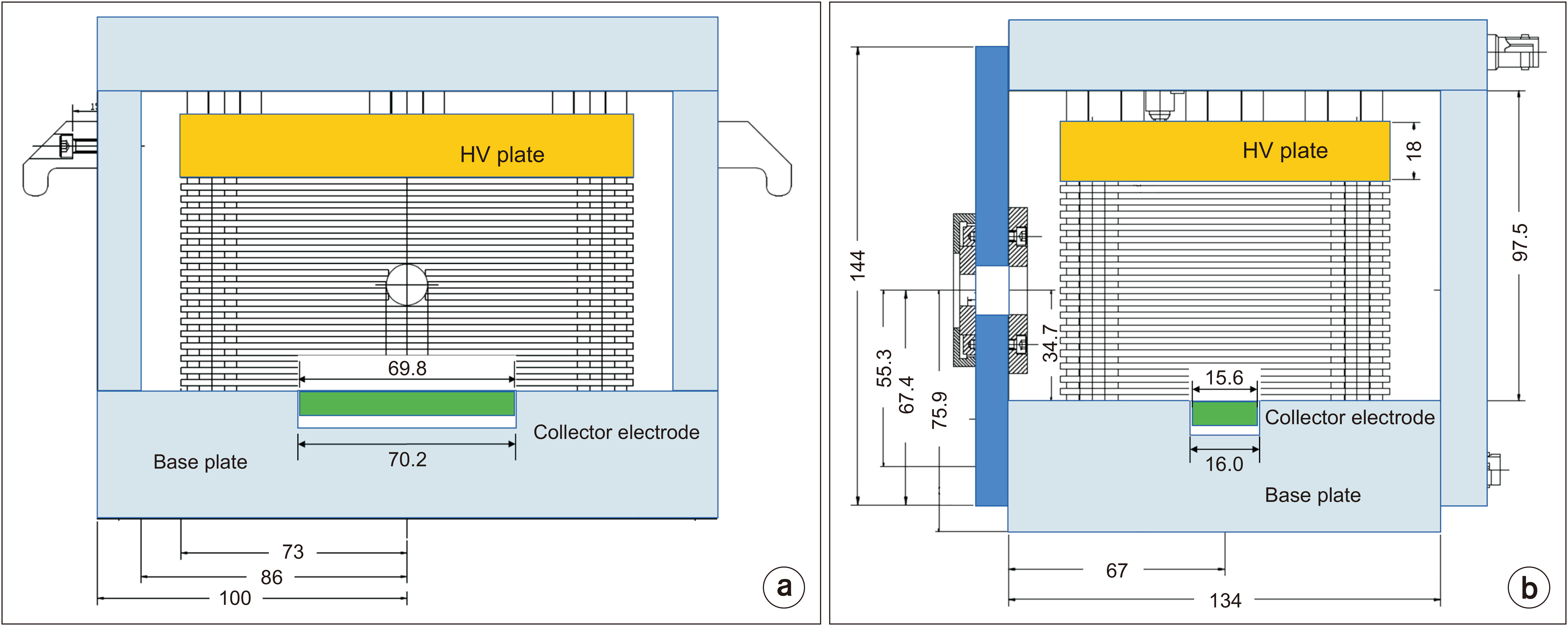
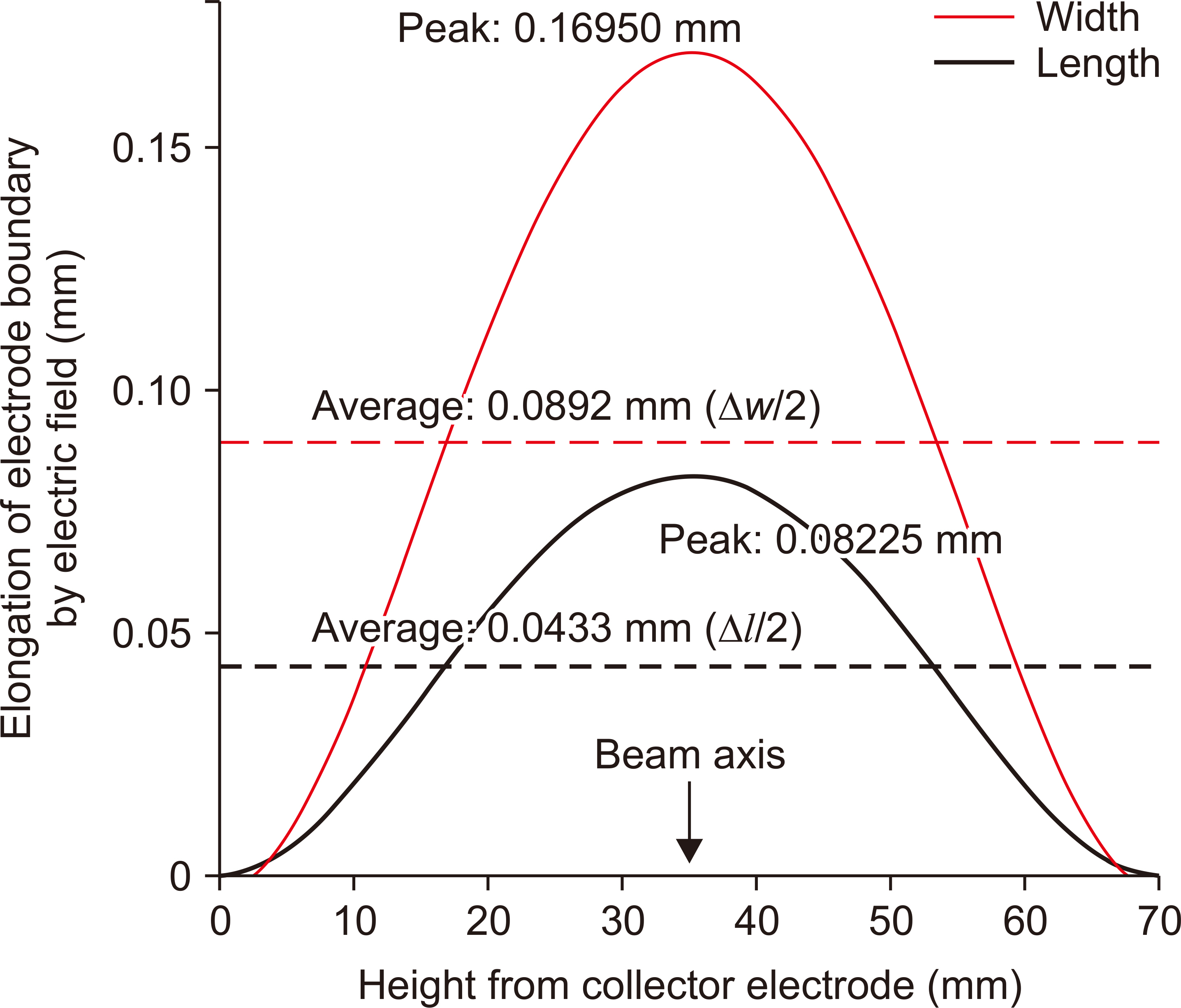
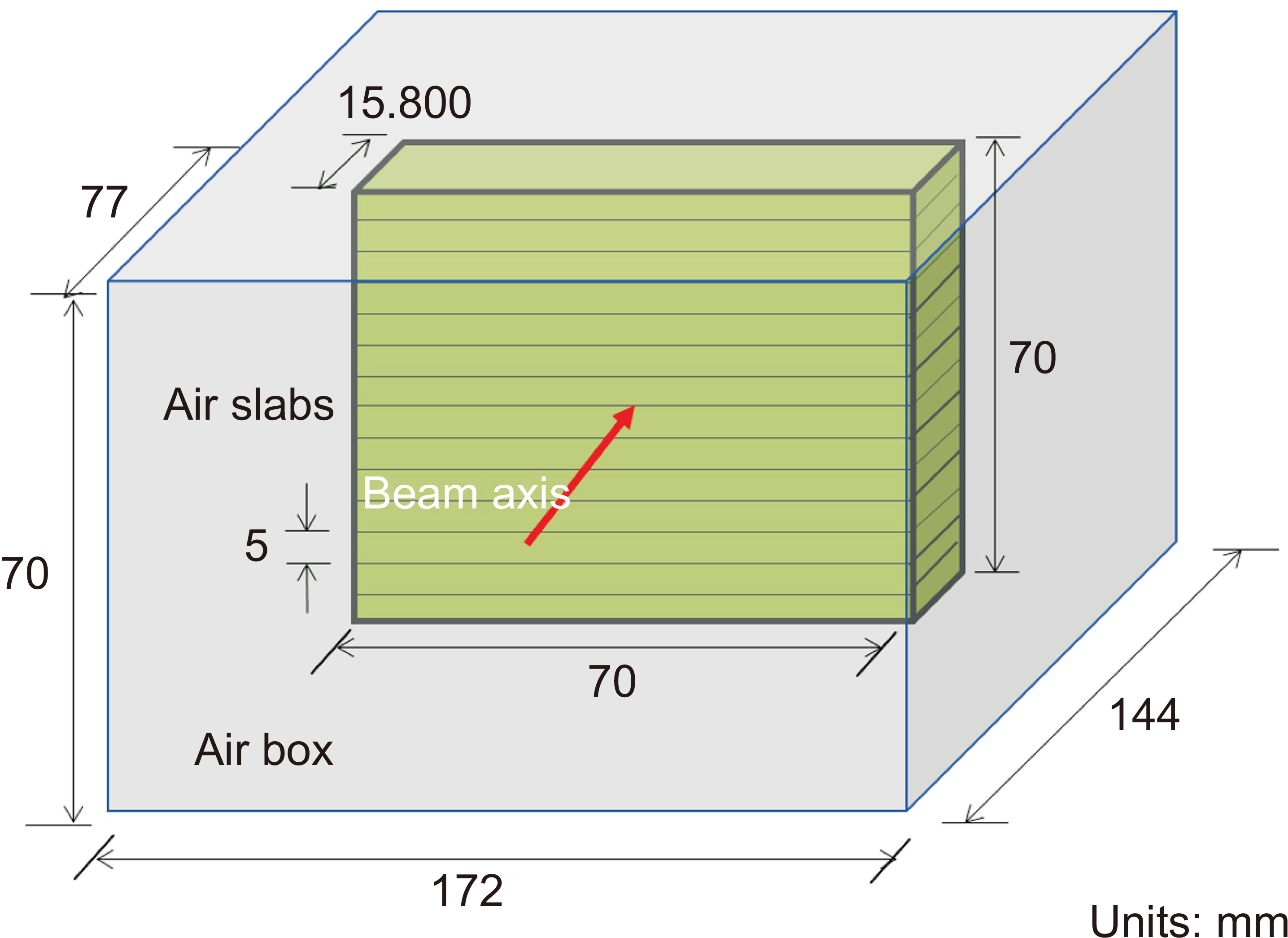
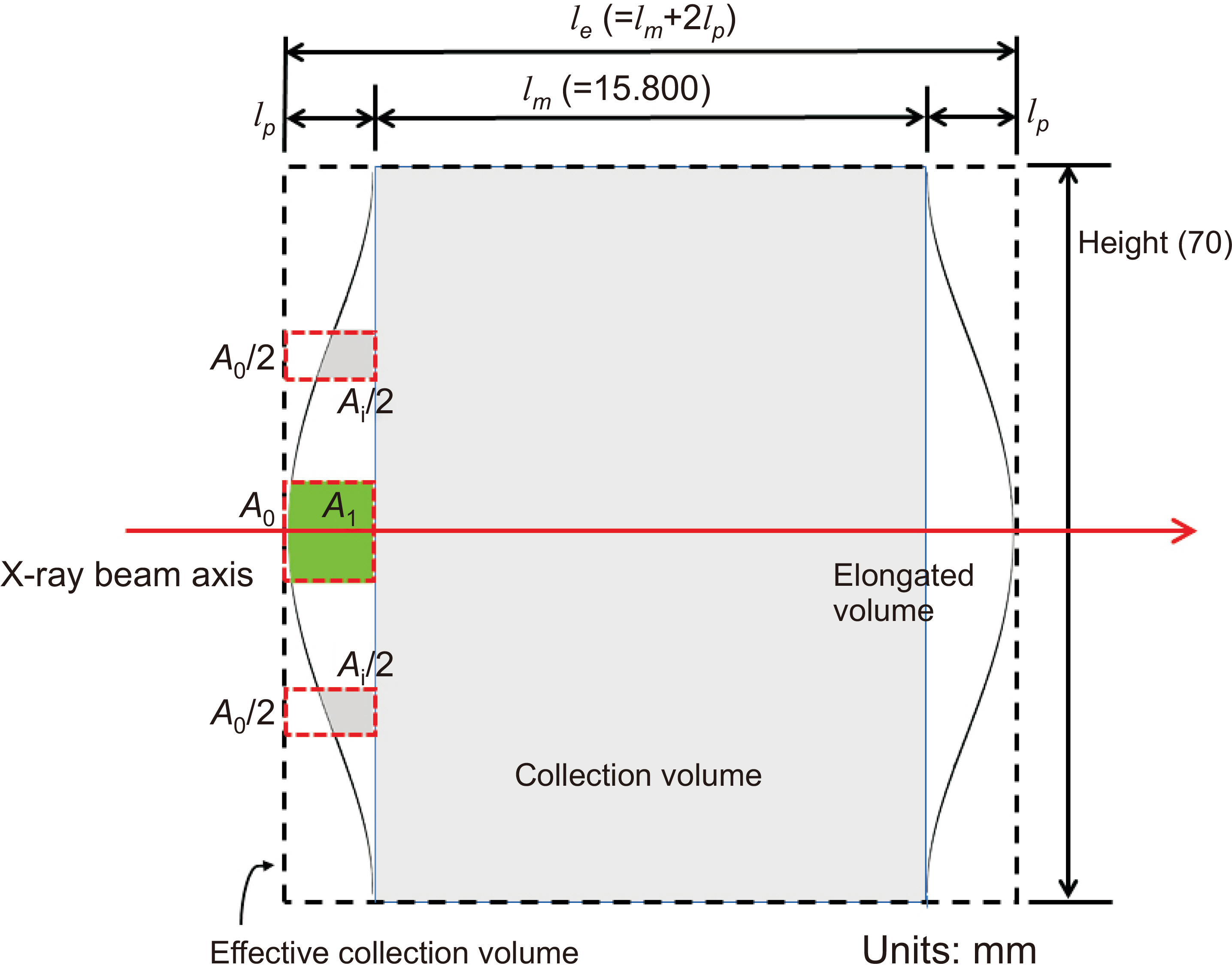
The mechanical dimensions were measured using a 3-dimensional coordinate mea suring machine (3-d CMM, Model UMM 500, Carl Zeiss). The diameter of the diaphragm was mea sured by a ring gauge calibrator (Model KRISS-DM1, KRISS). The elongation of the collector length due to electric field distortion was determined from the capacitance measurement of the KRISS FAC considering the result of the finite element method (FEM) analysis using the code QuickField v6.4.
Results
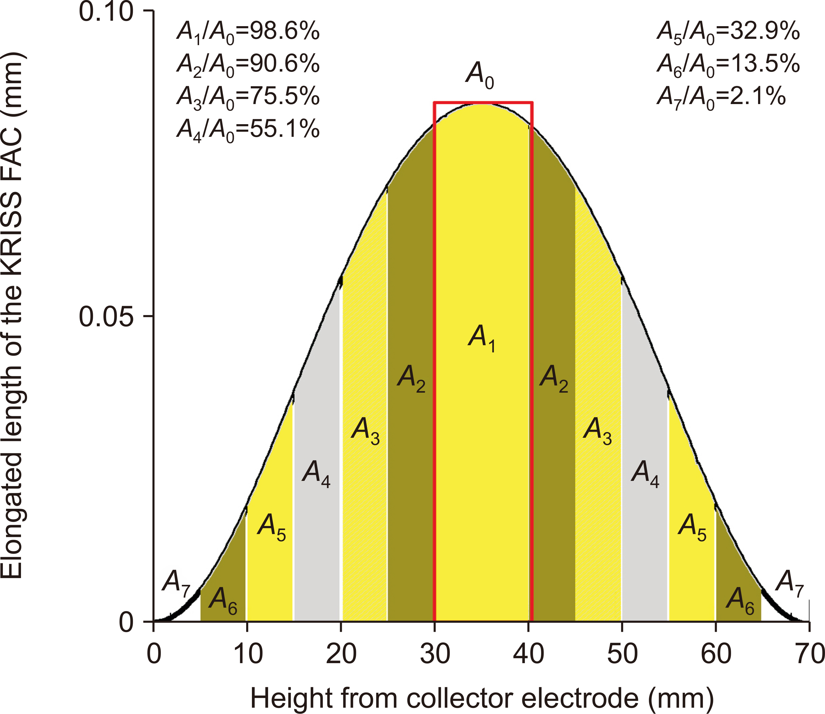
The measured length of the collector was 15.8003±0.0014 mm with a 68% confidence level (k=1). The aperture diameter of the diaphragm was 10.0021±0.0002 mm (k=1). The mechanical measurement volume of the KRISS FAC L1 was 1.2415±0.0006 cm 3 (k=1). The elongated length of the collector due to the electric field distortion was 0.170±0.021 mm. Considering the elongated length, the effective measurement volume of the KRISS FAC L1 was 1.2548±0.0019 cm3 (k=1).
Conclusions
The effective volume of the KRISS FAC L1 was determined from the mechanically measured value by adding the elongated volume due to the electric field distortion in the FAC. The effective volume will replace the existing mechanically determined volume in establishing and maintaining the primary standard of the low-energy X-ray.
Figure
Reference
-
References
1. International Standardization Organization (ISO). 2019. ISO Reference no. 4037-1. Radiological protection- X and gamma reference radiation for calibrating dosemeters and doserate meters and for determining their response as a function of photon energy. Part 1: Radiation characteristics and production methods. ISO;Geneva:2. International Electrotechnical Commission (IEC). 2005. IEC Reference no. 61267. Medical diagnostic X-ray equipment. Radiation conditions for use in the determination of characteristics. IEC;Geneva:3. International Commission on Radiation Units and Measurements (ICRU). 2014. ICRU Report 90. Key data for ionizing. Radiation dosimetry: measurement standards and applications. ICRU;Geneva:4. Kim JA, Kim JW, Kang CS, Eom TB. 2010; An interferometric Abbe-type comparator for the calibration of internal and external diameter standards. Meas Sci Technol. 21:075109. DOI: 10.1088/0957-0233/21/7/075109.
Article5. Korea Institute of Standards and Science (KRISS). 2012. KRISS calibration procedure no. C-01-1-0100-2000-1. Calibration procedure of cylindrical diameter standards using a laser interferometric comparator. KRISS;Daejeon:6. Korea Institute of Standards and Science (KRISS). 2005. KRISS calibration procedure no. C-26-1-1000-01. Calibration procedure of electrometers. KRISS;Daejeon:7. Lee RD, Kim HJ. Semenov YuP. 2001; New stable 10 pF gas-dielectric capacitors. IEEE Trans Instrum Meas. 50:294–297. DOI: 10.1109/19.918125.
Article8. Yi CY, Kim YH, Kim IJ. 2021; Temperataure dependence of effective area and depth of extrapolation chamber in dosimetry. Metrologia. 58:065001. DOI: 10.1088/1681-7575/ac2295.9. Kawrakow I, Mainegra-Hing E, Rogers DWO, Tessier F, Walters BRB. 2015. NRCC Report PIRS-701. The EGSnrc code system: Monte Carlo simulation of electron and photon transport. National Republican Congressional Committee;Washington, D.C.:10. Lye JE, Butler DJ, Webb DV. 2010; Monte Carlo correction factors for the ARPANSA kilovoltage free-air chambers and the effect of moving the limiting aperture. Metrologia. 47:11–20. DOI: 10.1088/0026-1394/47/1/002.
Article11. Kurosawa T, Takata N. 2005; Estimation of electron-loss and photon-scattering corrections for parallel-plate free-air chambers. J Nucl Sci Technol. 42:1077–1080. DOI: 10.1080/18811248.2005.9711060.
Article12. Burns DT, Kessler C, Allisy PJ. 2009; Re-evaluation of the BIPM international standards for air kerma in x-rays. Metrologia. 46:L21–L23. DOI: 10.1088/0026-1394/46/5/N01.
Article
- Full Text Links
- Actions
-
Cited
- CITED
-
- Close
- Share
- Similar articles
-
- Applying policy and health effects of air pollution in South Korea: focus on ambient air quality standards
- Chamber to Chamber Variations of a Cylindrical Ionization Chamber for the Calibration of an (192)Ir Brachytherapy Source Based on an Absorbed Dose to Water Standards
- Air Cavity Effects on the Absorbed Dose for 4-, 6- and 10-MV X-ray Beams: Larynx Model
- Authorship Policy of the Journal of Korean Medical Science
- Accuracy of sonographic diagnosis of pneumoperitoneum using the enhanced peritoneal stripe sign in beagle dogs

