Localization of infraorbital foramen and accessory infraorbital foramen with reference to facial bony landmarks: predictive method and its accuracy
- Affiliations
-
- 1Department of Anatomy, Faculty of Medicine, Chulalongkorn University, Bangkok, Thailand
- KMID: 2527669
- DOI: http://doi.org/10.5115/acb.21.208
Abstract
- The infraorbital nerve block is used for mid-facial anesthesia. We aim to determine the location of infraorbital foramen (IOF) and accessory infraorbital foramen (AIOF) with reference to anterior nasal spine (ANS) and the lowest point of zygomaticomaxillary junction (Z) and assess the accuracy of the predictive method. Two hundred and sixteen dry skulls were examined. A reference line was drawn from ANS to Z (line A) and its length was measured (distance A). The location of IOF was predicted by using the mean vertical distance from IOF to line A (line B) which was 15.14±1.99 mm and the mean ratio of the distance between ANS and the intersecting point of line B and line A (distance D) to distance A (D:A) which was 63.35%±3.90%. Eighty-six AIOFs were found. Most of them located superomedial to IOF, except for 3 AIOFs which located in the inferolateral position. For localization the AIOF, the mean vertical distance was 19.34±3.36 mm and the mean ratio was 51.8%±5.90%. No statistically significant difference of the predicted distances for both foramens was found between sex and sides. The accuracy of the predictive method was assessed in 15 embalmed cadavers. Predicted IOFs were 50% accurate and the mean distance error of the predicted IOF was 1.10±1.44 mm lateral and 0.59±1.39 mm inferior to the exact IOF. Therefore, this study provides an alternative method for localization of IOF and AIOF which could be useful in clinical settings.
Figure
-
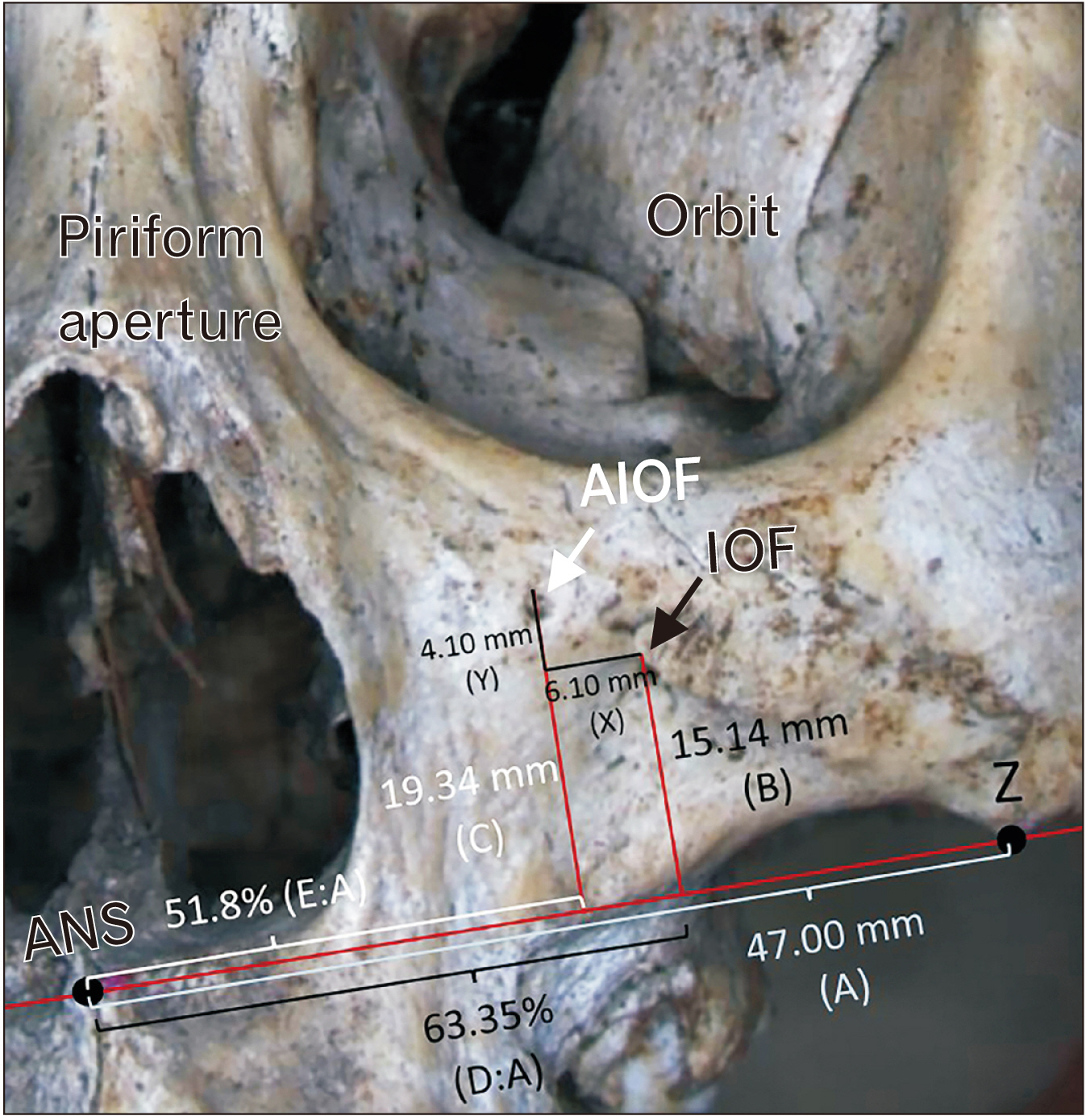
Fig. 1 Picture of skull illustrates the value of main parameters of IOF and AIOF with reference to the horizontal line from ANS to Z (unit: millimeters). A, distance from ANS to Z; AIOF, accessory infraorbital foramen; ANS, anterior nasal spine; B, vertical distance from the middle-upper edge of IOF to the line A; C, vertical distance from the middle-upper edge of AIOF to line A; D, horizontal distance from ANS to the intersecting point of the vertical line from IOF with line A; D:A, percentage of the ratio of distance D to distance A; E, horizontal distance from ANS to the intersecting point of the vertical line from AIOF with line A; E:A, percentage of the ratio of distance E to distance A; IOF, infraorbital foramen; X, horizontal distance between IOF and AIOF; Y, vertical distance between IOF and AIOF; Z, the lowest point of zygomaticomaxillary junction.
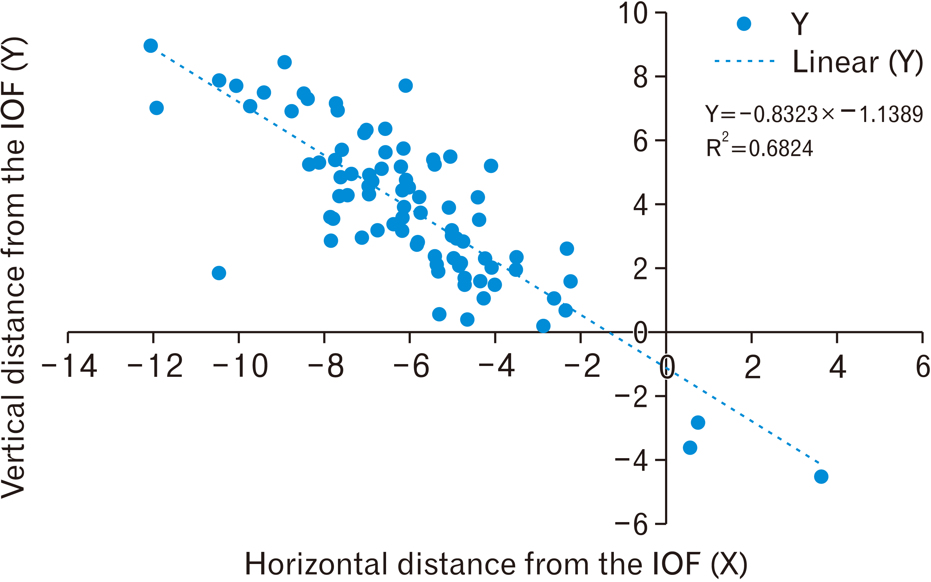
Fig. 2 Scatter plotted graph shows the location of the accessory infraorbital foramen (blue dot) with reference to the infraorbital foramen. AIOF, accessory infraorbital foramen; IOF, infraorbital foramen; X, horizontal distance between IOF and AIOF; Y, vertical distance between IOF and AIOF.
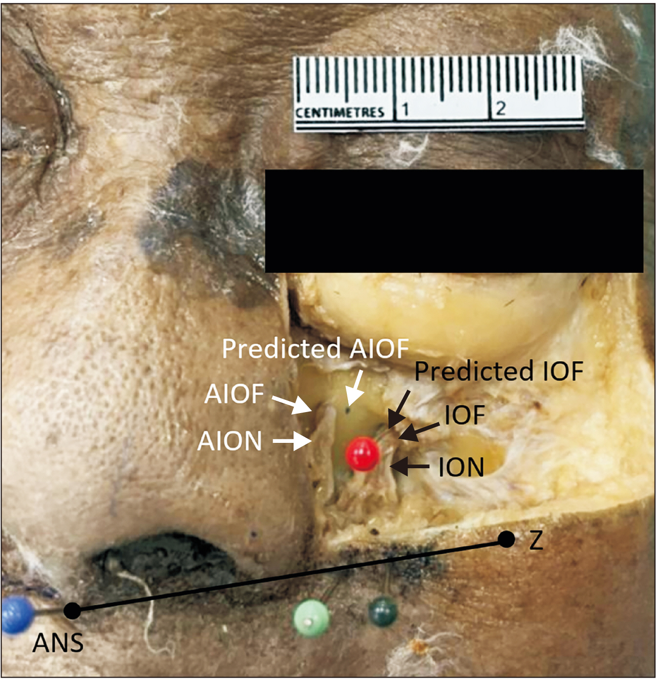
Fig. 3 Picture of left midface of a male cadaver showing the same location of both predicted IOF and the exact IOF (tip of red pin). The predicted AIOF was lateral to the exact AIOF. Dark green pin, location of the predicted distance D (distance A multiplied with the percentage of D:A); light green pin, location of the predicted distance E (distance A multiplied with the percentage of E:A); red pin, location of the predicted IOF; blue dot, the location of the predicted AIOF; blue pin, location of the anterior nasal spine. AIOF, accessory infraorbital foramen; AION, accessory infraorbital nerve; ANS, anterior nasal spine; IOF, infraorbital foramen; ION, infraorbital nerve; Z, the lowest point of zygomaticomaxillary junction; black line, the line between ANS and the lowest point of zygomaticomaxillary junction.

Fig. 4 Illustration describing the predictive method for localization of IOF in clinical practice. (A) Mark the location of ANS at the uppermost part of philtrum at the level of nostrils and Z at the lowest bony prominence of cheek. Draw line A between ANS and Z. (B) Mark at the medial two-thirds of line A (black dot line). (C) Draw perpendicularly with and above line A at approximately 15 mm. (D) Mark the location of predicted IOF. A, distance from ANS to Z; ANS, anterior nasal spine; IOF, infraorbital foramen; Z, the lowest point of the zygomaticomaxillary junction.
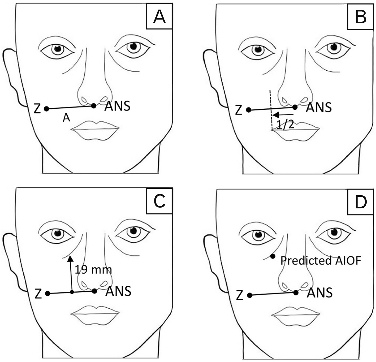
Fig. 5 Illustration describing the predictive method for localization of AIOF in clinical practice. (A) Mark the location of ANS at the uppermost part of philtrum at the level of nostrils and Z at the lowest bony prominence of cheek. Draw line A between ANS and Z. (B) Mark at the middle of line A (black dot line). (C) Draw perpendicularly with and above line A at approximately 19 mm. (D) Mark the location of predicted AIOF. A, distance from ANS to Z; AIOF, accessory infraorbital foramen; ANS, anterior nasal spine; Z, the lowest point of the zygomaticomaxillary junction.
Reference
-
References
1. Elhadi AM, Zaidi HA, Yagmurlu K, Ahmed S, Rhoton AL Jr, Nakaji P, Preul MC, Little AS. 2016; Infraorbital nerve: a surgically relevant landmark for the pterygopalatine fossa, cavernous sinus, and anterolateral skull base in endoscopic transmaxillary approaches. J Neurosurg. 125:1460–8. DOI: 10.3171/2015.9.JNS151099. PMID: 26943844.
Article2. Hu KS, Kwak HH, Song WC, Kang HJ, Kim HC, Fontaine C, Kim HJ. 2006; Branching patterns of the infraorbital nerve and topography within the infraorbital space. J Craniofac Surg. 17:1111–5. DOI: 10.1097/01.scs.0000236436.97720.5f. PMID: 17119413.
Article3. Hu KS, Kwak J, Koh KS, Abe S, Fontaine C, Kim HJ. 2007; Topographic distribution area of the infraorbital nerve. Surg Radiol Anat. 29:383–8. DOI: 10.1007/s00276-007-0227-z. PMID: 17585363.
Article4. Bösenberg AT, Kimble FW. 1995; Infraorbital nerve block in neonates for cleft lip repair: anatomical study and clinical application. Br J Anaesth. 74:506–8. DOI: 10.1093/bja/74.5.506. PMID: 7772421.
Article5. Choi H, Jung SH, Hong JM, Joo YH, Kim Y, Hong SH. 2019; Effects of bilateral infraorbital and infratrochlear nerve block on emergence agitation after septorhinoplasty: a randomized controlled trial. J Clin Med. 8:769. DOI: 10.3390/jcm8060769. PMID: 31151239. PMCID: PMC6616642.
Article6. Molliex S, Navez M, Baylot D, Prades JM, Elkhoury Z, Auboyer C. 1996; Regional anaesthesia for outpatient nasal surgery. Br J Anaesth. 76:151–3. DOI: 10.1093/bja/76.1.151. PMID: 8672358.
Article7. Spinelli G, Rocchetta D, Carnevali G, Valente D, Conti M, Agostini T. 2014; Infraorbital nerve block for isolated orbital floor fractures repair: review of 135 consecutive cases. Plast Reconstr Surg Glob Open. 2:e97. DOI: 10.1097/GOX.0000000000000039. PMID: 25289294. PMCID: PMC4174218.8. Cok OY, Deniz S, Eker HE, Oguzkurt L, Aribogan A. 2017; Management of isolated infraorbital neuralgia by ultrasound-guided infraorbital nerve block with combination of steroid and local anesthetic. J Clin Anesth. 37:146–8. DOI: 10.1016/j.jclinane.2016.12.007. PMID: 28235509.
Article9. Perloff MD, Chung JS. 2018; Urgent care peripheral nerve blocks for refractory trigeminal neuralgia. Am J Emerg Med. 36:2058–60. DOI: 10.1016/j.ajem.2018.08.019. PMID: 30119988.
Article10. Boselli E, Bouvet L, Augris-Mathieu C, Bégou G, Diot-Junique N, Rahali N, Vertu-Ciolino D, Gérard C, Pivot C, Disant F, Allaouchiche B. 2016; Infraorbital and infratrochlear nerve blocks combined with general anaesthesia for outpatient rhinoseptoplasty: a prospective randomised, double-blind, placebo-controlled study. Anaesth Crit Care Pain Med. 35:31–6. DOI: 10.1016/j.accpm.2015.09.002. PMID: 26549134.
Article11. McAdam D, Muro K, Suresh S. 2005; The use of infraorbital nerve block for postoperative pain control after transsphenoidal hypophysectomy. Reg Anesth Pain Med. 30:572–3. DOI: 10.1016/j.rapm.2005.07.192. PMID: 16326343.
Article12. Iwanaga J, Kikuta S, Kusukawa J, Tomaszewski KA, Walocha JA, Tubbs RS. 2020; Anatomic study of accessory infraorbital nerves and foramina: application for a better understanding of complications of le fort fractures and osteotomy. J Oral Maxillofac Surg. 78:717–23. DOI: 10.1016/j.joms.2020.01.004. PMID: 32035836.
Article13. Blanton PL, Jeske AH. 2003; The key to profound local anesthesia: neuroanatomy. J Am Dent Assoc. 134:753–60. DOI: 10.14219/jada.archive.2003.0262. PMID: 12839412.14. Gupta T. 2008; Localization of important facial foramina encountered in maxillo-facial surgery. Clin Anat. 21:633–40. DOI: 10.1002/ca.20688. PMID: 18773483.
Article15. Singh R. 2011; Morphometric analysis of infraorbital foramen in Indian dry skulls. Anat Cell Biol. 44:79–83. DOI: 10.5115/acb.2011.44.1.79. PMID: 21519552. PMCID: PMC3080011.
Article16. Pascal J, Charier D, Perret D, Navez M, Auboyer C, Molliex S. 2005; Peripheral blocks of trigeminal nerve for facial soft-tissue surgery: learning from failures. Eur J Anaesthesiol. 22:480–2. DOI: 10.1017/S0265021505260817. PMID: 15991518.17. Kazkayasi M, Ergin A, Ersoy M, Bengi O, Tekdemir I, Elhan A. 2001; Certain anatomical relations and the precise morphometry of the infraorbital foramen--canal and groove: an anatomical and cephalometric study. Laryngoscope. 111(4 Pt 1):609–14. DOI: 10.1097/00005537-200104000-00010. PMID: 11359128.
Article18. Chrcanovic BR, Abreu MH, Custódio AL. 2011; A morphometric analysis of supraorbital and infraorbital foramina relative to surgical landmarks. Surg Radiol Anat. 33:329–35. DOI: 10.1007/s00276-010-0698-1. PMID: 20625730.
Article19. Aggarwal A, Kaur H, Gupta T, Tubbs RS, Sahni D, Batra YK, Sondekoppam RV. 2015; Anatomical study of the infraorbital foramen: a basis for successful infraorbital nerve block. Clin Anat. 28:753–60. DOI: 10.1002/ca.22558. PMID: 26119635.20. Agthong S, Huanmanop T, Chentanez V. 2005; Anatomical variations of the supraorbital, infraorbital, and mental foramina related to gender and side. J Oral Maxillofac Surg. 63:800–4. DOI: 10.1016/j.joms.2005.02.016. PMID: 15944977.
Article21. Song WC, Kim SH, Paik DJ, Han SH, Hu KS, Kim HJ, Koh KS. 2007; Location of the infraorbital and mental foramen with reference to the soft-tissue landmarks. Plast Reconstr Surg. 120:1343–7. DOI: 10.1097/01.prs.0000279558.86727.5a. PMID: 17898610.
Article22. Zheng WX, Guo JL, Song BX, Liu XL, Lv DL, Tian Y, Li YQ, Cheng FB. 2012; Location of the supraorbital and infraorbital foramen with references to the soft tissue landmarks in a Chinese population. J Craniofac Surg. 23:1154–5. DOI: 10.1097/SCS.0b013e31824e2bd0. PMID: 22801112.
Article23. Ercikti N, Apaydin N, Kirici Y. 2017; Location of the infraorbital foramen with reference to soft tissue landmarks. Surg Radiol Anat. 39:11–5. DOI: 10.1007/s00276-016-1683-0. PMID: 27146295.
Article24. Pickering RB, Bachman D. 1997. The use of forensic anthropology. CRC Press;Boca Raton: p. 84–6. DOI: 10.1201/9781439834329.25. Williams BA, Rogers T. 2006; Evaluating the accuracy and precision of cranial morphological traits for sex determination. J Forensic Sci. 51:729–35. DOI: 10.1111/j.1556-4029.2006.00177.x. PMID: 16882212.
Article26. White TD, Folkens PA. 2005. The human bone manual. Elsevier Academic;Amsterdam: p. 388–9.27. Hwang K, Lee SJ, Kim SY, Hwang SW. 2015; Frequency of existence, numbers, and location of the accessory infraorbital foramen. J Craniofac Surg. 26:274–6. DOI: 10.1097/SCS.0000000000001375. PMID: 25490578.
Article28. Polo CL, Abdelkarim AZ, von Arx T, Lozanoff S. 2019; The morphology of the infraorbital nerve and foramen in the presence of an accessory infraorbital foramen. J Craniofac Surg. 30:244–53. DOI: 10.1097/SCS.0000000000004889. PMID: 30394975.
Article29. Nam Y, Bahk S, Eo S. 2017; Anatomical study of the infraorbital nerve and surrounding structures for the surgery of orbital floor fractures. J Craniofac Surg. 28:1099–104. DOI: 10.1097/SCS.0000000000003416. PMID: 28145925.
Article30. Dagistan S, Miloglu Ö, Altun O, Umar EK. 2017; Retrospective morphometric analysis of the infraorbital foramen with cone beam computed tomography. Niger J Clin Pract. 20:1053–64. DOI: 10.4103/1119-3077.217247. PMID: 29072226.
Article31. Gour KK, Nair S, Trivedi GN, Gupta SD. 2012; Anthropometric measurements of infra orbital foramen in dried human skulls. Int J Biol Med Res. 3:2003–6.32. Martins-Júnior PA, Rodrigues CP, De Maria ML, Nogueira LM, Silva JH, Silva MR. 2017; Analysis of anatomical characteristics and morphometric aspects of infraorbital and accessory infraorbital foramina. J Craniofac Surg. 28:528–33. DOI: 10.1097/SCS.0000000000003235. PMID: 27977492.
Article33. Tezer M, Öztürk A, Akgül M, Gayretli Ö, Kale A. 2011; Anatomic and morphometric features of the accessory infraorbital foramen. J Morphol Sci. 28:95–7.34. Ali IK, Sansare K, Karjodkar FR, Salve P. 2018; Cone beam computed tomography assessment of accessory infraorbital foramen and determination of infraorbital foramen position. J Craniofac Surg. 29:e124–6. DOI: 10.1097/SCS.0000000000004120. PMID: 29135734.
Article35. Shin KJ, Lee SH, Park MG, Shin HJ, Lee AG. 2020; Location of the accessory infraorbital foramen with reference to external landmarks and its clinical implications. Sci Rep. 10:8566. DOI: 10.1038/s41598-020-65330-4. PMID: 32444685. PMCID: PMC7244752.
Article36. Rai AR, Rai R, Vadgaonkar R, Madhyastha S, Rai RK, Alva D. 2013; Anatomical and morphometric analysis of accessory infraorbital foramen. J Craniofac Surg. 24:2124–6. DOI: 10.1097/SCS.0b013e31828f2fa6. PMID: 24220421.
Article
- Full Text Links
-
- Actions
-
Cited
- CITED
-
- Close
- Share
-
- Similar articles
-
- Non-Metrical Morphologic Variations of Korean Skull Foramina
- Morphometric analysis of infraorbital foramen in Indian dry skulls
- A clinical and anatomical study on the infraorbital foramen and infraorbital canal in Korean
- Accessory infraorbital foramen location using cone-beam computed tomography
- Localization of the Mental and Infraorbital Foramen with related to the Soft-tissue Landmarks
