Direct Removal of Fourth Ventricle Hematoma in Massive Intraventricular Hemorrhage
- Affiliations
-
- 1Department of Neurosurgery, Chonnam National University Hospital, Chonnam National University Medical School, Gwangju, Korea
- KMID: 2527189
- DOI: http://doi.org/10.3340/jkns.2021.0122
Abstract
- Various grading systems and surgical techniques have been developed for the treatment of intraventricular hemorrhage (IVH); however, little attention has been paid to the fourth ventricle hematoma. Nonetheless, hemorrhagic dilation of the fourth ventricle may lead to catastrophic consequences for patients with massive IVH. We present two cases of massive IVH accompanied by massive fourth ventricle hematoma which was successfully removed with combination of suboccipital craniotomy for fourth ventricle hematoma and intraventricular fibrinolysis for supratentorial hematoma.
Figure
-
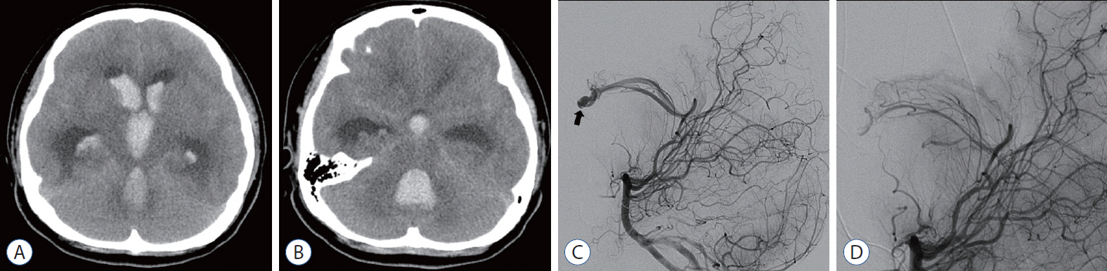
Fig. 1. Case 1. A and B : Initial computed tomography scan showed massive intraventricular hemorrhage with a fourth ventricle hematoma compressing the brainstem. C and D : The intranidal aneurysm was successfully obliterated in the forniceal arteriovenous malformation using Histoacryl® (B. Braun, Melsungen, Germany). Thick arrow indicates intranidal aneurysm.
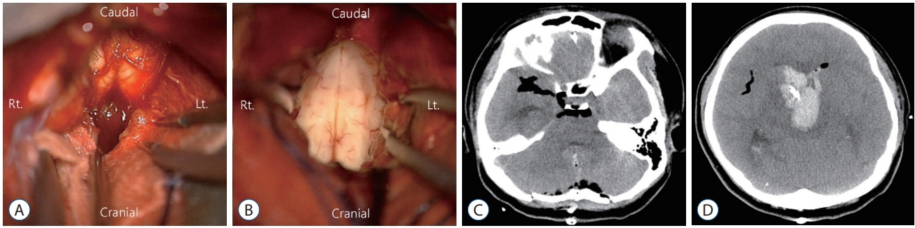
Fig. 2. Case 1. A and B : Removal of fourth ventricle hematoma was performed via midline suboccipital craniotomy. C and D : Postoperative computed tomography scan revealed complete removal of the fourth ventricle hematoma and slight improvement of the hydrocephalus.

Fig. 3. Case 1. A : The intraventricular hemorrhage was almost completely resolved following eight doses of intraventricularly administered tissue plasminogen activator given over 4 consecutive days. B : Normal ventricle size was maintained by a shunt during follow-up computed tomography scan at 18 months after surgery.
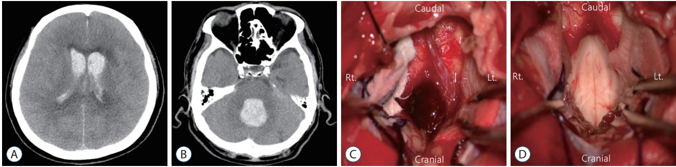
Fig. 4. Case 2. A and B : An initial computed tomography scan revealed massive intraventricular hemorrhage with a fourth ventricle hematoma compressing the brainstem. C and D : The fourth ventricle hematoma was removed via midline suboccipital craniotomy.

Fig. 5. Case 2. A and B : A postoperative computed tomography scan showed complete removal of the fourth ventricle hematoma and improvement of the hydrocephalus. C and D : The intraventricular hemorrhage was almost completely resolved following six doses of intraventricularly administered tissue plasminogen activator given over 3 consecutive days.
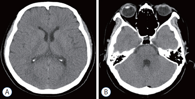
Fig. 6. Case 2. A and B : The ventricle’s normal size was maintained without a shunt during follow-up computed tomography scan at 12 months after surgery.
Reference
-
References
1. Di Rienzo A, Colasanti R, Esposito D, Della Costanza M, Carrassi E, Capece M, et al. Endoscope-assisted microsurgical evacuation versus external ventricular drainage for the treatment of cast intraventricular hemorrhage: results of a comparative series. Neurosurg Rev. 43:695–708. 2020.
Article2. Donauer E, Loew F, Faubert C, Alesch F, Schaan M. Prognostic factors in the treatment of cerebellar haemorrhage. Acta Neurochir (Wien). 131:59–66. 1994.
Article3. Feletti A, Basaldella L, Fiorindi A. How I do it: flexible endoscopic aspiration of intraventricular hemorrhage. Acta Neurochir (Wien). 162:3141–3146. 2020.
Article4. Findlay JM, Grace MG, Weir BK. Treatment of intraventricular hemorrhage with tissue plasminogen activator. Neurosurgery. 32:941–947. discussion 947. 1993.
Article5. Gaberel T, Magheru C, Emery E. Management of non-traumatic intraventricular hemorrhage. Neurosurg Rev. 35:485–494. discussion 494-495. 2012.
Article6. Graeb DA, Robertson WD, Lapointe JS, Nugent RA, Harrison PB. Computed tomographic diagnosis of intraventricular hemorrhage. Etiology and prognosis. Radiology. 143:91–96. 1982.
Article7. Hamada H, Hayashi N, Kurimoto M, Umemura K, Nagai S, Kurosaki K, et al. Neuroendoscopic removal of intraventricular hemorrhage combined with hydrocephalus. Minim Invasive Neurosurg. 51:345–349. 2008.
Article8. LeRoux PD, Haglund MM, Newell DW, Grady MS, Winn HR. Intraventricular hemorrhage in blunt head trauma: an analysis of 43 cases. Neurosurgery. 31:678–684. discussion 684-685. 1992.9. Li Y, Zhang H, Wang X, She L, Yan Z, Zhang N, et al. Neuroendoscopic surgery versus external ventricular drainage alone or with intraventricular fibrinolysis for intraventricular hemorrhage secondary to spontaneous supratentorial hemorrhage: a systematic review and meta-analysis. PLoS One. 8:e80599. 2013.
Article10. Martí-Fàbregas J, Piles S, Guardia E, Martí-Vilalta JL. Spontaneous primary intraventricular hemorrhage: clinical data, etiology and outcome. J Neurol. 246:287–291. 1999.
Article11. Mei L, Fengqun M, Qian H, Dongpo S, Zhenzhong G, Tong C. Exploration of efficacy and safety of interventions for intraventricular hemorrhage: a network meta-analysis. World Neurosurg. 136:382–389.e6. 2020.12. Morgan TC, Dawson J, Spengler D, Lees KR, Aldrich C, Mishra NK, et al. The modified Graeb score: an enhanced tool for intraventricular hemorrhage measurement and prediction of functional outcome. Stroke. 44:635–641. 2013.13. Shapiro SA, Campbell RL, Scully T. Hemorrhagic dilation of the fourth ventricle: an ominous predictor. J Neurosurg. 80:805–809. 1994.
Article14. Song P, Duan FL, Cai Q, Wu JL, Chen XB, Wang Y, et al. Endoscopic surgery versus external ventricular drainage surgery for severe intraventricular hemorrhage. Curr Med Sci. 38:880–887. 2018.
Article15. Tuhrim S, Horowitz DR, Sacher M, Godbold JH. Volume of ventricular blood is an important determinant of outcome in supratentorial intracerebral hemorrhage. Crit Care Med. 27:617–621. 1999.
Article16. Weisberg LA. Acute cerebellar hemorrhage and CT evidence of tight posterior fossa. Neurology. 36:858–860. 1986.
Article17. Yilmazlar S, Abas F, Korfali E. Comparison of ventricular drainage in poor grade patients after intracranial hemorrhage. Neurol Res. 27:653–656. 2005.
Article
- Full Text Links
-
- Actions
-
Cited
- CITED
-
- Close
- Share
-
- Similar articles
-
- Spontaneous Cerebellar Hemorrhage with the Fourth Ventricular Hemorrhage : Risk Factors Associated with Ventriculoperitoneal Shunt
- A Case of Intraventricular Meningioma Acompanied by Intraventricular Hematoma and Subarachnoid Hemorrhage: Case Report
- Clinical-Computed Tomographic Correlation of Spontaneous Intraventricular Hemorrhage Patients
- Prognostic Factors in Spontaneous Thalamic Hemorrhage
- An Experimental Study about the Effect of Urokinase Injected into the Ventricle on Intraventricular Hemorrhage
