J Korean Neurosurg Soc.
2022 Mar;65(2):307-314. 10.3340/jkns.2021.0135.
A Cadaveric Study of Thread Cubital Tunnel Release with Newly Developed Threads
- Affiliations
-
- 1Department of Rehabilitation Medicine, Seoul St. Mary’s Hospital, College of Medicine, The Catholic University of Korea, Seoul, Korea
- 2Department of Anatomy, Institute for Applied Anatomy, College of Medicine, The Catholic University of Korea, Seoul, Korea
- 3Department of Anatomy, College of Korean Medicine, Dongshin University, Naju, Korea
- 4Department of Rehabilitation Medicine, Howareyou Rehabilitation Clinic, Seoul, Korea
- 5Department of Research & Development, Ultra V Co. Ltd., Seoul, Korea
- 6Department of Rehabilitation Medicine, Incheon St. Mary`s Hospital, College of Medicine, The Catholic University of Korea, Seoul, Korea
- KMID: 2527187
- DOI: http://doi.org/10.3340/jkns.2021.0135
Abstract
Objective
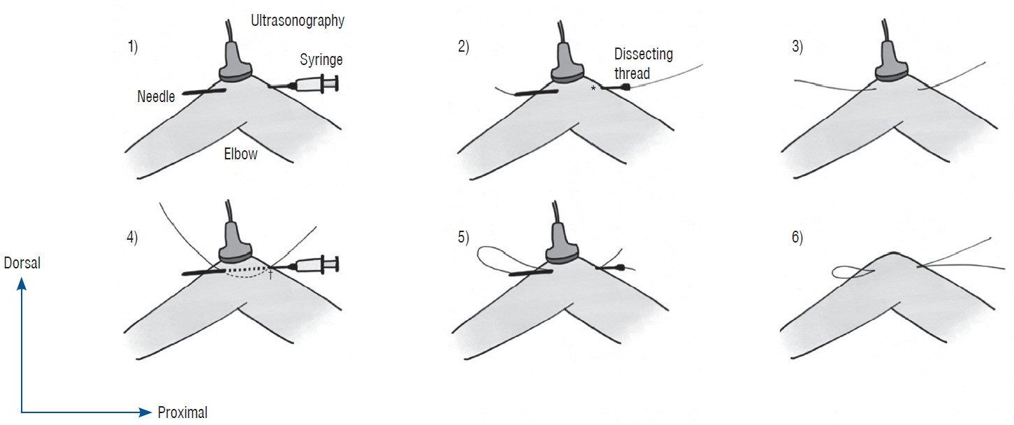
: The percutaneous thread transection technique is a surgical dissecting method using a dissecting thread inserted through a needle under ultrasound guidance without skin incision. As the new dissecting threads were developed domestically, this cadaver study was conducted to compare the effectiveness and safety between the new threads (ultra V sswire and smartwire-01) and a pre-existing commercial dissecting thread (loop & shear) by demonstrating a modified looped thread cubital tunnel release.
Methods
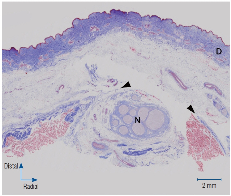
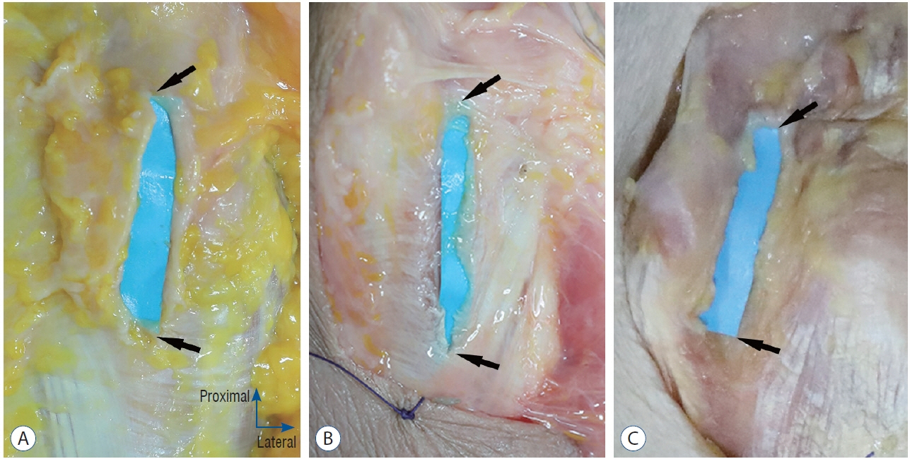
: The percutaneous cubital tunnel release procedure was performed on 29 fresh cadaveric upper extremities. The preexisting commercial thread was used in 5 upper extremities. The two newly developed threads were used in 24 upper extremities. Two practitioners performed the procedures separately. After the modified looped thread cubital release, anatomical and histological analyses were performed by a blinded anatomist. The presence of the dissected cubital tunnel and damaged adjacent soft tissue was assessed.
Results
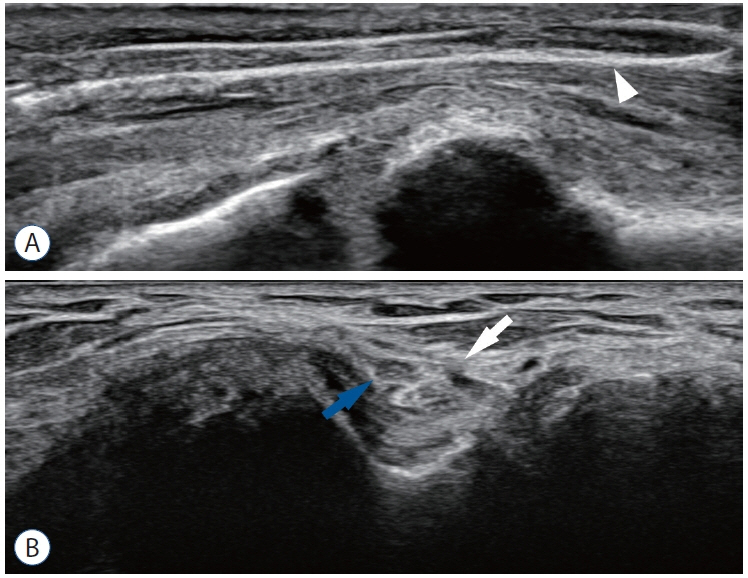
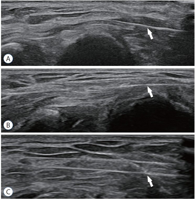
: Out of the 29 cadaveric upper extremities, 27 specimens showed complete dissection of the Osborne ligament and the proximal fascia of the flexor carpi ulnaris muscle. One specimen was incompletely dissected in each of the ultra V sswire and smartwire-01 groups. There were no injuries of adjacent structures including the ulnar nerve, ulnar artery, medial antebrachial cutaneous nerve, or flexor tendon with either the commercial thread or the newly developed threads. The anatomical analysis revealed clear and sharp incisional margins of the cubital tunnel in the Smartwire-01 and loop & shear groups. All three kinds of threads maintained proper linear elasticity for easy handling during the procedure. The smartwire-01 provided higher visibility in ultrasound than the other threads.
Conclusion
: The newly developed threads were effective and safe for use in the thread cubital tunnel release procedure.
Figure
Reference
-
References
1. Andrews K, Rowland A, Pranjal A, Ebraheim N. Cubital tunnel syndrome: anatomy, clinical presentation, and management. J Orthop. 15:832–836. 2018.
Article2. Boone S, Gelberman RH, Calfee RP. The management of cubital tunnel syndrome. J Hand Surg Am. 40:1897–1904. quiz 1904. 2015.
Article3. Buchanan PJ, Chieng LO, Hubbard ZS, Law TY, Chim H. Endoscopic versus open in situ cubital tunnel release: a systematic review of the literature and meta-analysis of 655 patients. Plast Reconstr Surg. 141:679–684. 2018.4. Guo D, Guo D, Guo J, Schmidt SC, Lytie RM. A clinical study of the modified thread carpal tunnel release. Hand (N Y). 12:453–460. 2017.
Article5. Guo D, Guo D, Harrison R, McCool L, Wang H, Tonkin B, et al. A cadaveric study using the ultra-minimally invasive thread transection technique to decompress the superficial peroneal nerve in the lower leg. Acta Neurochir (Wien). 161:2133–2139. 2019.
Article6. Guo D, Kliot M, McCool L, Senk A, Tonkin B, Guo D. Percutaneous cubital tunnel release with a dissection thread: a cadaveric study. J Hand Surg Eur Vol. 44:920–924. 2019.
Article7. Kim JH, Won SJ, Rhee WI, Park HJ, Hong HM. Diagnostic cutoff value for ultrasonography in the ulnar neuropathy at the elbow. Ann Rehabil Med. 39:170–175. 2015.
Article8. McCool L, Guo D, Guo D, Harrison R, Tonkin B, Senk A, et al. Thread common peroneal nerve release-a cadaveric validation study. Acta Neurochir (Wien). 161:1931–1936. 2019.
Article9. Smeraglia F, Del Buono A, Maffulli N. Endoscopic cubital tunnel release: a systematic review. Br Med Bull. 116:155–163. 2015.
Article10. Staples JR, Calfee R. Cubital tunnel syndrome: current concepts. J Am Acad Orthop Surg. 25:e215–e224. 2017.
- Full Text Links
- Actions
-
Cited
- CITED
-
- Close
- Share
- Similar articles
-
- Cadaveric Study of Thread Carpal Tunnel Release Using Newly Developed Thread, With a Histologic Perspective
- Ulnar neuropathy
- Cubital tunnel syndrome caused by an intraneural ganglion cyst treated with epineurectomy: a report of three cases
- Cubital Tunnel Syndrome Caused by Osteochondroma: A Case Report
- Real-Time Visualization of Ultrasonography Guided Cubital Tunnel Injection: A Cadaveric Study

