Korean J Gastroenterol.
2022 Jan;79(1):35-40. 10.4166/kjg.2021.139.
Primary Hepatic Neuroendocrine Tumor Arising at a Young Age: Rare Case Report and Literature Review
- Affiliations
-
- 1Department of Surgery, Chonnam National University Hwasun Hospital, Chonnam National University Medical School, Hwasun, Korea
- KMID: 2525521
- DOI: http://doi.org/10.4166/kjg.2021.139
Abstract
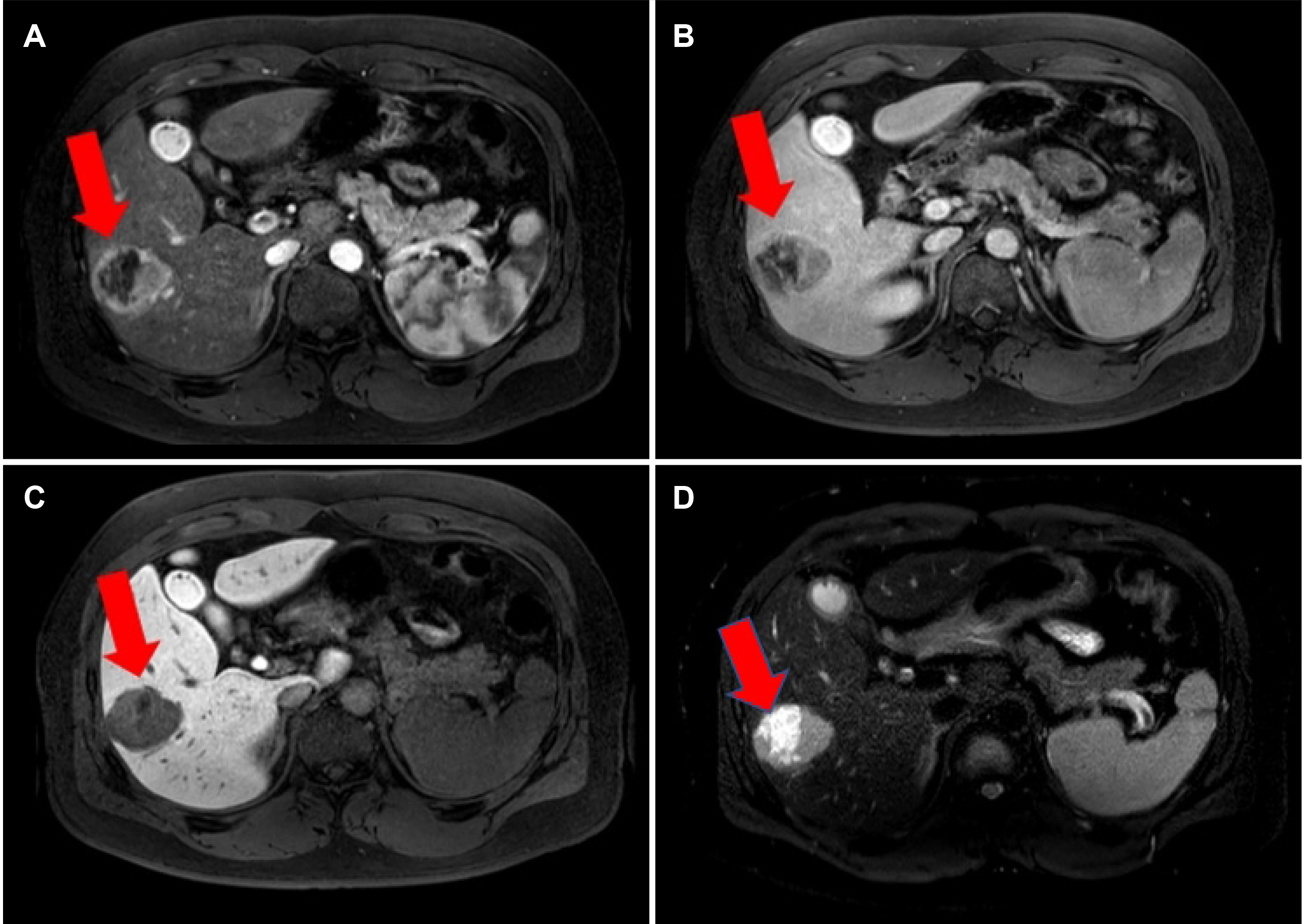
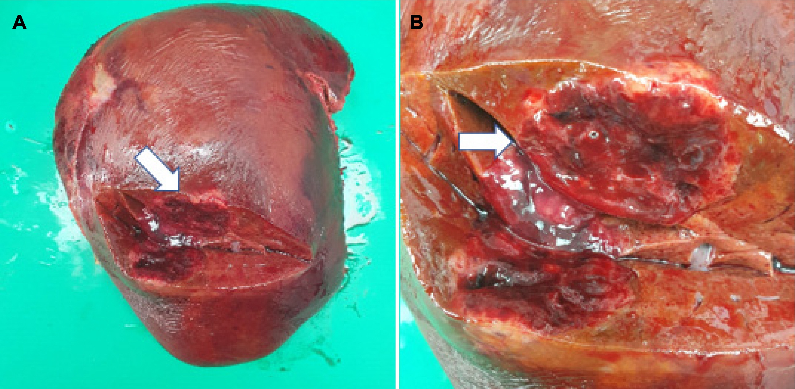
- Neuroendocrine tumors (NETs) are low-grade malignancies arising from neuroendocrine cells. Primary hepatic neuroendocrine tumors (PHNETs) are extremely rare and difficult to differentiate from other liver tumors, such as hepatocellular carcinoma (HCC) or cholangiocarcinoma. A 22-year-old male presented with intermittent abdominal pain. A preoperative imaging study revealed a 5.1cm-sized heterogeneously enhancing mass in S6 of the liver, suggesting HCC. Laparoscopic right hepatectomy was performed, and a well-demarcated brown solid mass was found. The pathology report revealed a neuroendocrine tumor of the liver. 18F-fluorodeoxyglucose–positron emission tomography/computed tomography was performed postoperatively to exclude extrahepatic lesions, and no lesions were found. This is a rare case of PHNET that developed at a young age and was misdiagnosed as HCC preoperatively. This suggests that PHNET should be considered one of the differential diagnoses when a non-specific enhanced hepatic tumor is found, even when the patient is young.
Keyword
Figure
Reference
-
1. Maggard MA, O'Connell JB, Ko CY. 2004; Updated population-based review of carcinoid tumors. Ann Surg. 240:117–122. DOI: 10.1097/01.sla.0000129342.67174.67. PMID: 15213627. PMCID: PMC1356383.
Article2. Park CH, Chung JW, Jang SJ, et al. 2012; Clinical features and outcomes of primary hepatic neuroendocrine carcinomas. J Gastroenterol Hepatol. 27:1306–1311. DOI: 10.1111/j.1440-1746.2012.07117.x. PMID: 22414232.
Article3. Quartey B. 2011; Primary hepatic neuroendocrine tumor: what do we know now? World J Oncol. 2:209–216. DOI: 10.4021/wjon341w. PMID: 29147250. PMCID: PMC5649681.
Article4. Mousavi SR, Ahadi M. 2015; Primary neuroendocrine tumor of liver (rare tumor of liver). Iran J Cancer Prev. 8:e3144. DOI: 10.17795/ijcp-3144. PMID: 26855717. PMCID: PMC4736067.
Article5. Li R, Tang CL, Yang D, et al. 2016; Primary hepatic neuroendocrine tumors: clinical characteristics and imaging features on contrast-enhanced ultrasound and computed tomography. Abdom Radiol (NY). 41:1767–1775. DOI: 10.1007/s00261-016-0770-3. PMID: 27156080.
Article6. Ichiki M, Nishida N, Furukawa A, Kanasaki S, Ohta S, Miki Y. 2014; Imaging findings of primary hepatic carcinoid tumor with an emphasis on MR imaging: case study. Springerplus. 3:607. DOI: 10.1186/2193-1801-3-607. PMID: 25392779. PMCID: PMC4210452.
Article7. Baek SH, Yoon JH, Kim KW. 2013; Primary hepatic neuroendocrine tumor: gadoxetic acid (Gd-EOB-DTPA)-enhanced magnetic resonance imaging. Acta Radiol Short Rep. 2:2047981613482897. DOI: 10.1177/2047981613482897. PMID: 23986857. PMCID: PMC3736966.
Article8. Yalav O, Ülkü A, Akçam TA, Demiryürek H, Doran F. 2012; Primary hepatic neuroendocrine tumor: five cases with different preoperative diagnoses. Turk J Gastroenterol. 23:272–278. DOI: 10.4318/tjg.2012.0465. PMID: 22798119.
Article9. Shi C, Zhao Q, Dai B, Xie F, Yang J. 2018; Primary hepatic neuroendocrine neoplasm: long-time surgical outcome and prognosis. Medicine (Baltimore). 97:e11764. DOI: 10.1097/MD.0000000000011764. PMID: 30075602. PMCID: PMC6081183.10. Song JE, Kim BS, Lee CH. 2016; Primary hepatic neuroendocrine tumor: a case report and literature review. World J Clin Cases. 4:243–247. DOI: 10.12998/wjcc.v4.i8.243. PMID: 27574614. PMCID: PMC4983697.
Article11. Knox CD, Anderson CD, Lamps LW, Adkins RB, Pinson CW. 2003; Long-term survival after resection for primary hepatic carcinoid tumor. Ann Surg Oncol. 10:1171–1175. DOI: 10.1245/ASO.2003.04.533. PMID: 14654473.
Article12. Yao KA, Talamonti MS, Nemcek A, et al. 2001; Indications and results of liver resection and hepatic chemoembolization for metastatic gastrointestinal neuroendocrine tumors. Surgery. 130:677–685. DOI: 10.1067/msy.2001.117377. PMID: 11602899.
Article13. Gamblin TC, Christians K, Pappas SG. 2011; Radiofrequency ablation of neuroendocrine hepatic metastasis. Surg Oncol Clin N Am. 20:273–viii. DOI: 10.1016/j.soc.2010.11.002. PMID: 21377583.
Article14. Li YF, Zhang QQ, Wang WL. 2020; Clinicopathological characteristics and survival outcomes of primary hepatic neuroendocrine tumor: a surveillance, epidemiology, and end results (SEER) population-based study. Med Sci Monit. 26:e923375. DOI: 10.12659/MSM.923375. PMID: 32651994. PMCID: PMC7370587.
Article15. Jung J, Hwang S, Hong SM, et al. 2019; Long-term postresection prognosis of primary neuroendocrine tumors of the liver. Ann Surg Treat Res. 97:176–183. DOI: 10.4174/astr.2019.97.4.176. PMID: 31620391. PMCID: PMC6779954.
Article16. Oh YH, Kang GH, Kim OJ. 1998; Primary hepatic carcinoid tumor with a paranuclear clear zone: a case report. J Korean Med Sci. 13:317–320. DOI: 10.3346/jkms.1998.13.3.317. PMID: 9681813. PMCID: PMC3054491.
Article17. Kim JM, Kim SY, Kwon CH, et al. 2013; Primary hepatic neuroendocrine carcinoma. Korean J Hepatobiliary Pancreat Surg. 17:34–37. DOI: 10.14701/kjhbps.2013.17.1.34. PMID: 26155210. PMCID: PMC4304509.
Article18. Kim JM, Lee WA, Shin HD, Song IH, Kim SB. 2021; Cystic primary hepatic neuroendocrine tumor. Korean J Gastroenterol. 78:300–304. DOI: 10.4166/kjg.2021.125. PMID: 34824189.
Article19. Lee SH, Kim KA, Lee JS, et al. 2006; A case of primary neuroendocrine carcinoma of liver presenting with liver abscess. Korean J Gastroenterol. 48:277–280. PMID: 17060722.20. Lee GJ, Jo KW, Kim J, Park IY. 2015; Metastatic brain neuroendocrine tumor originating from the liver. J Korean Neurosurg Soc. 58:550–553. DOI: 10.3340/jkns.2015.58.6.550. PMID: 26819691. PMCID: PMC4728094.
Article
- Full Text Links
- Actions
-
Cited
- CITED
-
- Close
- Share
- Similar articles
-
- Primary Small Cell Neuroendocrine Carcinoma of the Breast: A Case Report With Literature Review
- Ovarian Large Cell Neuroendocrine Carcinoma Associated with Endocervical-like Mucinous Borderline Tumor: A Case Report and Literature Review
- Primary Neuroendocrine Carcinoma of the Breast: A Case Report and Literature Review
- A Carcinoid Tumor Arising from a Normal Kidney in a Young Man
- Primary multifocal cystic signet ring neuroendocrine tumor of liver: a case report


