Vps34 Inhibits Hepatocellular Carcinoma Invasion by Regulating Endosome-Lysosome Trafficking via Rab7-RILP and Rab11
- Affiliations
-
- 1Department of Pathology, School of Basic Medical Sciences, Shanghai Medical College, Fudan University, Shanghai, China
- 2Department of Pathology, Huashan Hospital, Fudan University, Shanghai, China
- 3Department of Pathology, Yantai Yuhuangding Hospital of Qingdao University, Yantai, China
- KMID: 2524599
- DOI: http://doi.org/10.4143/crt.2020.578
Abstract
- Purpose
The role of vacuolar protein sorting 34 (Vps34), an indispensable protein required for cell vesicular trafficking, in the biological behavior of hepatocellular carcinoma (HCC) has yet to be studied.
Materials and Methods
In the present study, the expression of Vps34 in HCC and the effect of Vps34 on HCC cell invasion was detected both in vivo and in vitro. Furthermore, by modulating the RILP and Rab11, which regulate juxtanuclear lysosome aggregation and recycling endosome respectively, the underlying mechanism was investigated.
Results
Vps34 was significantly decreased in HCC and negatively correlated with the HCC invasiveness both in vivo and in vitro. Moreover, Vps34 could promote lysosomal juxtanuclear accumulation, reduce the invasive ability of HCC cells via the Rab7-RILP pathway. In addition, the deficiency of Vps34 in HCC cells affected the endosome-lysosome system, resulting in enhanced Rab11 mediated endocytic recycling of cell surface receptor and increased invasion of HCC cells.
Conclusion
Our study reveals that Vps34 acts as an invasion suppressor in HCC cells, and more importantly, the endosome-lysosome trafficking regulated by Vps34 has the potential to become a target pathway in HCC treatment.
Keyword
Figure
-

Fig. 1 Vacuolar protein sorting 34 (Vps34) was decreased in hepatocellular carcinoma (HCC) and inversely correlated with the invasion ability. (A) Immunochemistry staining of Vps34 in peri-carcinoma tissue or carcinoma tissue of HCC specimens. Scoring mentioned in materials and methods. The expression levels of Vps34 were divided into four grades (−, +, ++, and +++) and defined as low or high expression group. (B) Quantification of high or low Vps34 cases in peri-carcinoma tissue or carcinoma tissue of HCC specimens (n=73), respectively. (C) Representative images of H&E and immunohistochemistry staining of Vps34 in HCC specimens that have both carcinoma tissue and peri-carcinoma tissue. (D) Vps34 scores of carcinoma tissue in patients with or without microvascular invasion (MVI). (E) Transwell assay of the hepatocyte cell line (L-02) and HCC cell lines (HepG2, Hep3B, SMMC-7721, Huh-7, MHCC97-H, Bel-7402, and HCC-LM3). (F) Western blot demonstrated the MMP-2 protein level in above cell lines. (G, H) Protein (G) and mRNA (H) levels of Vps34 in hepatocyte cell line (L-02), HCC cell lines with lower invasive potentials (HepG2 and Hep3B) or HCC cell lines with higher invasive abilities (SMMC-7721, Huh-7, MHCC97-H, Bel-7402, and HCC-LM3). MMP-2, matrix metalloproteinase 2. Data were showed as means±standard error of mean. **p < 0.01, ****p < 0.0001.

Fig. 2 Vacuolar protein sorting 34 (Vps34) regulated the invasive ability of hepatocellular carcinoma (HCC) cells. (A) H&E staining of HepG2 cells with transfection of shRNA against Vps34 (shVps34) or the non-target shRNA control (shNT). (B) Western blot demonstrated the efficiency of Vps34 knockdown and the protein expression of epithelial marker (E-cadherin), mesenchymal marker (Vimentin) and MMP-2 in HepG2 cells with shVps34 or shNT transfection. (C) Transwell assay of HepG2 cells after shVps34 transfection. (D) Wound healing assay of HepG2 cells expressing shNT or shVps34. (E) Western blot examined the protein level of Vps34, E-cadherin, vimentin, and matrix metalloproteinase 2 (MMP-2) in SMMC-7721 HCC cells expressing vector control (Vector) or exogenous upregulation (Vps34). (F, G) Transwell assay (F) and wound healing assay of SMMC-7721 cells (G) with Vps34 or vector transfection. Data were showed as means±standard error of mean. *p < 0.05, **p < 0.01, ***p < 0.001, ****p < 0.0001.

Fig. 3 Vacuolar protein sorting 34 (Vps34) regulated the lysosome distribution in hepatocellular carcinoma (HCC) cells. (A) Immunohistochemistry (IHC) staining of Lamp1 (lysosomal marker) in carcinoma and peri-carcinoma tissue showed that juxtanuclear lysosome aggregation (JLA) existed in peri-carcinoma tissue, but not in carcinoma tissue. (B) Immunofluorescence (IF) staining of Lamp1 (green) and DAPI (blue) in normal hepatocyte cell line (L-02) and HCC cell line with high invasive ability (SMMC-7721). Red arrows indicated the JLA. (C) Immunofluorescence staining of Lamp1 showed the lysosome distribution in L-02 cells expressing shNT or shVps34, and in SMMC7721 expressing blank vector or exogenous Vps34. Arrows indicated the juxtanuclear lysosome aggregation. (D) Immunohistochemistry staining of Rab7-interacting lysosomal protein (RILP) in HCC patient specimens containing both carcinoma and peri-carcinoma regions (n=10). Data were showed as means±standard error of mean. ****p < 0.0001.

Fig. 4 Vacuolar protein sorting 34 (Vps34) inhibited the hepatocellular carcinoma invasion through the Rab7–Rab7-interacting lysosomal protein (RILP) pathway mediated lysosomal trafficking. (A) Western blot examined the protein level of Vps34, Rab7, RILP, and Lamp1 in SMMC-7721 cells with exogenous Vps34 or vector control. (B) Immunoprecipitation of Rab7 and RILP and quantification of RILP pulled down by Rab7 from SMMC-7721 cells with Vps34 overexpression and vector control. Western blot examined the input and immunoprecipitated level of RILP. (C) Double immunofluorescence staining of Lamp1 (red) and β-tubulin (green) in SMMC-7721 cells with or without exogenous Vps34 and/or shRILP transfection. (D, E) Transwell invasion assay (D) and western blots of the TSG101 and CD63 (exosome markers) from the exosome extract (E) of SMMC-7721 cells with or without exogenous Vps34 and/or shRILP transfection. IF, immunofluorescence. Data were showed as means±standard error of mean. *p < 0.05.

Fig. 5 Vacuolar protein sorting 34 (Vps34) deficiency affected the endosome-lysosome system and epidermal growth factor (EGF) induced epidermal growth factor receptor (EGFR) signaling in hepatocellular carcinoma cells. (A) Western blot examined the protein level of Vps34 in HepG2 cells with shVps34 or shNT transfection, and representative images of Vps34 in HepG2 cells with shVps34 or shNT transfection by an inverted microscope. (B) Immunofluorescence (IF) staining of the late endosomal marker Rab7 (red) and Lamp1 (green) in HepG2 cells with shVps34 or shNT transfection, and in HepG2 cells with Vps34-IN1(4 μM, 24 hours) treatment, a Vps34 inhibitor, and DMSO control treatment as well. Arrow indicated the abnormal accumulation of macrovesicles. (C) Western blot examined the protein level of Vps34, EGFR, and p-EGFR (Y1068) in HepG2 cells with shVps34 or shNT transfection after stimulating with EGF (100 ng/mL) for 0, 15, 30, 60, and 120 minutes. (D) Immunofluorescence staining of EGFR (red) in SMMC-7721 cells with Vps34-IN1 or DMSO control treatment, after stimulating with EGF (100 ng/mL) for 15 or 60 minutes. GAPDH, glyceraldehyde 3-phosphate dehydrogenase. Data were showed as mean±standard error of mean. *p < 0.05.
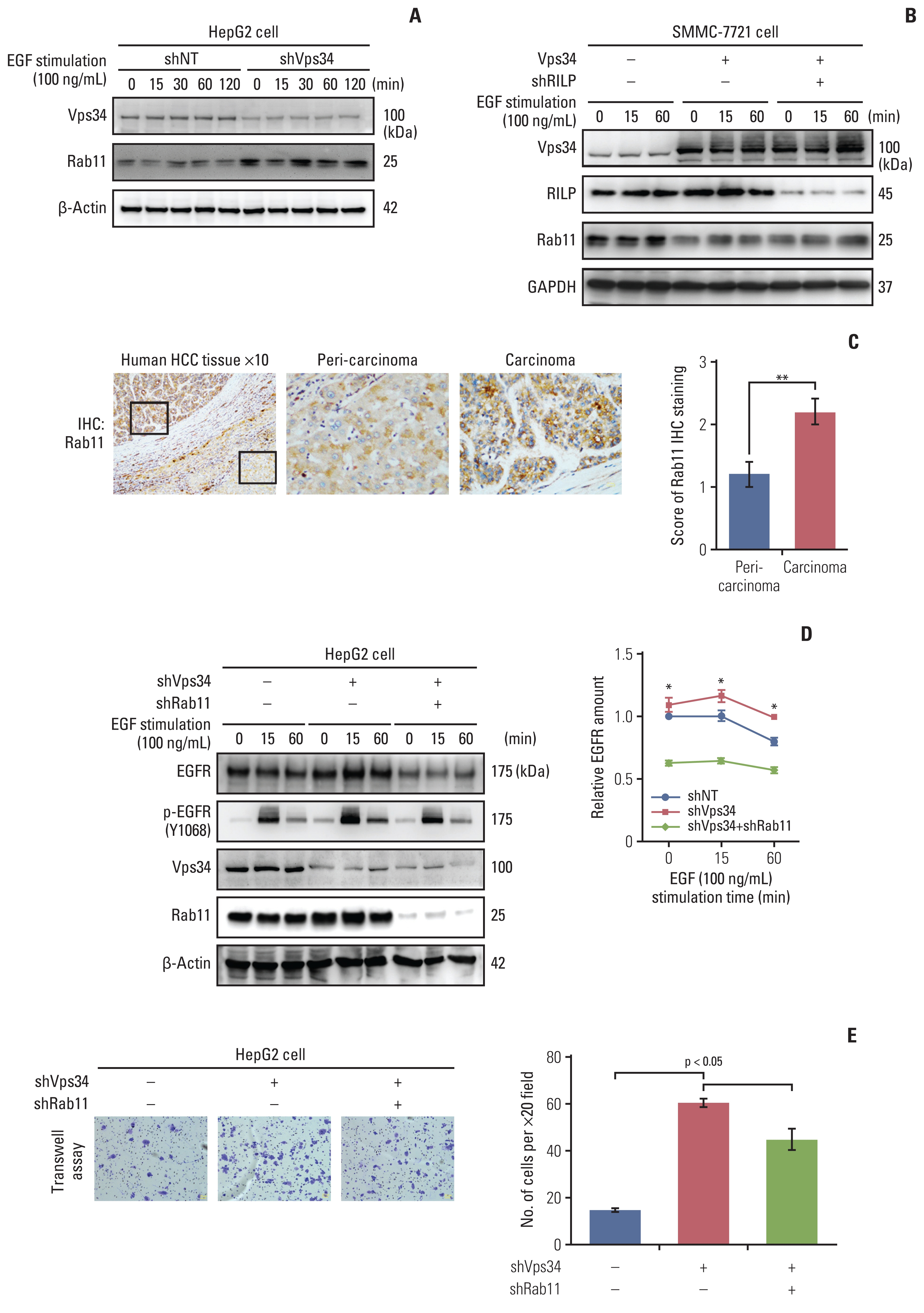
Fig. 6 Vacuolar protein sorting 34 (Vps34) affected the invasion ability of hepatocellular carcinoma (HCC) cells by regulating Rab11 mediated endocytic recycling. (A) Western blot examined the protein level of Vps34 and Rab11 in HepG2 cells with shVps34 or shNT transfection after stimulating with epidermal growth factor (EGF) (100 ng/mL) for 0, 15, 30, 60, and 120 minutes. (B) Western blot examined the protein level of Vps34, Rab7-interacting lysosomal protein (RILP) and Rab11 in SMMC-7721 cells with or without exogenous Vps34 and/or shRILP transfection after stimulating with EGF (100 ng/mL) for 0, 15, and 60 minutes. (C) Immunohistochemistry staining of Rab11 in HCC patient specimens containing both carcinoma and peri-carcinoma regions (n=5). (D) Western blot examined the protein level of Vps34, Rab11, EGFR, and p-EGFR (Y1068) in HepG2 cells with or without shVps34 and/or shRab11 transfection after stimulating with EGF (100 ng/mL) for 0, 15, and 60 minutes. (E) Representative images and quantification of Transwell assay in HepG2 cells with or without shVps34 and/or shRab11 transfection. Data were presented as mean±standard error of mean. *p < 0.05, **p < 0.01.
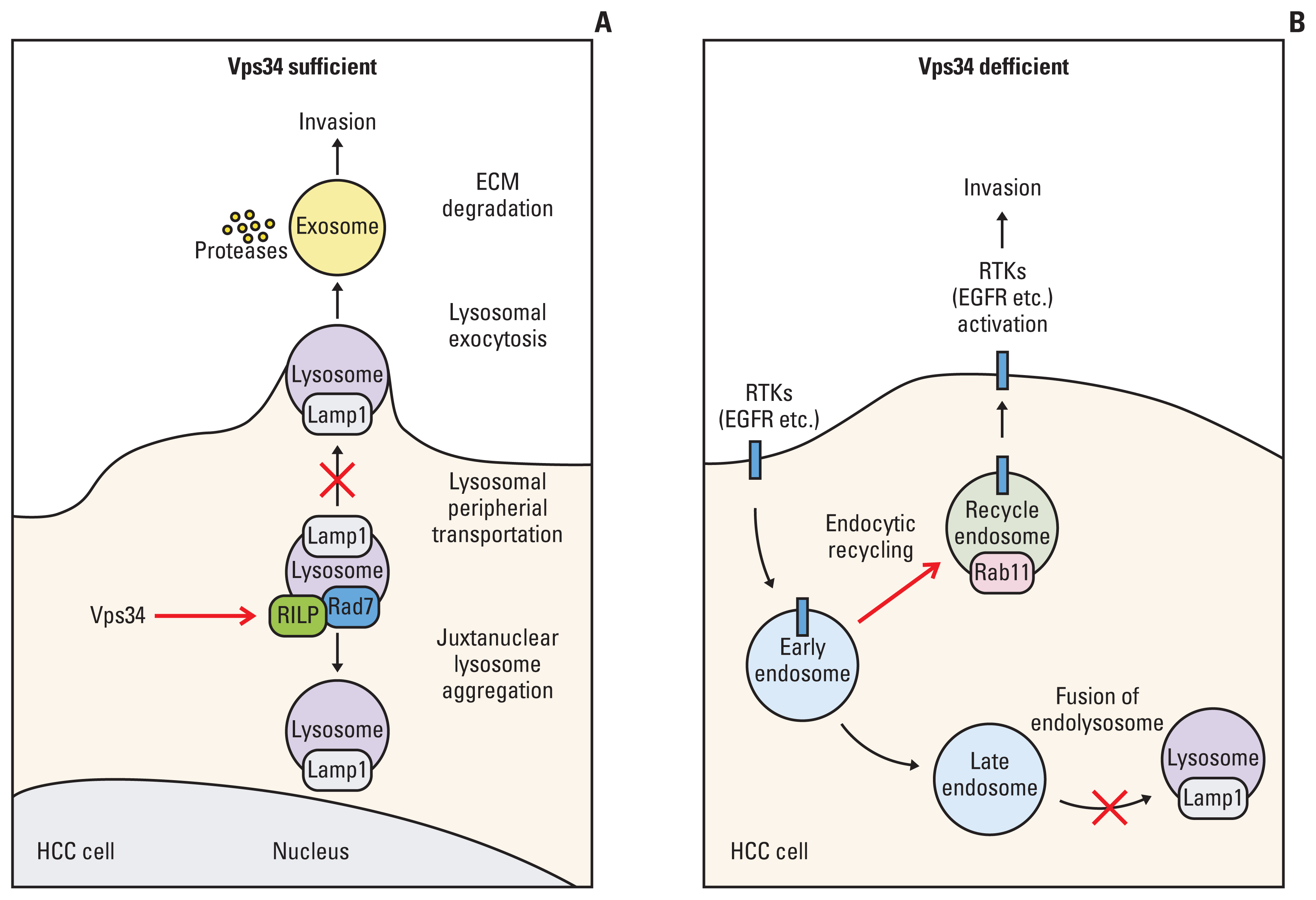
Fig. 7 Signaling pathway underlying the regulation of vacuolar protein sorting 34 (Vps34) on hepatocellular carcinoma (HCC) invasion. A schematic drawing of the two mechanisms of Vps34 in inhibiting the HCC invasion by regulating the endosome-lysosome system. (A) When Vps34 is sufficient. Vps34 could promote lysosomal juxtanuclear accumulation through Rab7–Rab7-interacting lysosomal protein (RILP) pathway (red arrow), thus less lysosomal peripheral transportation reduced the secretion of exosomes, degradation of extracellular matrix (ECM) and invasion (red cross). (B) When Vps34 is deficient. Vps34 deficiency reduced endocytic degradation (red cross) and promoted the Rab11 mediated endocytic recycling of cell surface receptors, such as epidermal growth factor receptor (EGFR) (red arrow), inducing the activation of downstream signaling and invasion. RTK, receptor tyrosine kinase.
Reference
-
References
1. Lindmo K, Stenmark H. Regulation of membrane traffic by phosphoinositide 3-kinases. J Cell Sci. 2006; 119:605–14.
Article2. Nobukuni T, Joaquin M, Roccio M, Dann SG, Kim SY, Gulati P, et al. Amino acids mediate mTOR/raptor signaling through activation of class 3 phosphatidylinositol 3OH-kinase. Proc Natl Acad Sci U S A. 2005; 102:14238–43.
Article3. Lamb CA, Dooley HC, Tooze SA. Endocytosis and autophagy: shared machinery for degradation. Bioessays. 2013; 35:34–45.
Article4. Perera RM, Zoncu R. The lysosome as a regulatory hub. Annu Rev Cell Dev Biol. 2016; 32:223–53.
Article5. Mellman I, Yarden Y. Endocytosis and cancer. Cold Spring Harb Perspect Biol. 2013; 5:a016949.
Article6. Yu L, McPhee CK, Zheng L, Mardones GA, Rong Y, Peng J, et al. Termination of autophagy and reformation of lysosomes regulated by mTOR. Nature. 2010; 465:942–6.
Article7. Wirth M, Joachim J, Tooze SA. Autophagosome formation: the role of ULK1 and Beclin1-PI3KC3 complexes in setting the stage. Semin Cancer Biol. 2013; 23:301–9.8. Liang XH, Jackson S, Seaman M, Brown K, Kempkes B, Hibshoosh H, et al. Induction of autophagy and inhibition of tumorigenesis by beclin 1. Nature. 1999; 402:672–6.
Article9. LI M, Xiong J. Interpretation of guidelines for diagnosis and treatment of primary liver cancer (2017 edition). Chin J Gen Surg. 2019; 28:785–9.10. van Zijl F, Krupitza G, Mikulits W. Initial steps of metastasis: cell invasion and endothelial transmigration. Mutat Res. 2011; 728:23–34.
Article11. Ma Y, Ma L, Guo Q, Zhang S. Expression of bone morphogenetic protein-2 and its receptors in epithelial ovarian cancer and their influence on the prognosis of ovarian cancer patients. J Exp Clin Cancer Res. 2010; 29:85.
Article12. Ye QH, Zhu WW, Zhang JB, Qin Y, Lu M, Lin GL, et al. GOLM1 modulates EGFR/RTK cell-surface recycling to drive hepatocellular carcinoma metastasis. Cancer Cell. 2016; 30:444–58.
Article13. Yan Y, Backer JM. Regulation of class III (Vps34) PI3Ks. Biochem Soc Trans. 2007; 35:239–41.
Article14. Hutagalung AH, Novick PJ. Role of Rab GTPases in membrane traffic and cell physiology. Physiol Rev. 2011; 91:119–49.
Article15. Al-Akhrass H, Naves T, Vincent F, Magnaudeix A, Durand K, Bertin F, et al. Sortilin limits EGFR signaling by promoting its internalization in lung cancer. Nat Commun. 2017; 8:1182.
Article16. Bechtel W, Helmstadter M, Balica J, Hartleben B, Kiefer B, Hrnjic F, et al. Vps34 deficiency reveals the importance of endocytosis for podocyte homeostasis. J Am Soc Nephrol. 2013; 24:727–43.
Article17. Jaber N, Dou Z, Chen JS, Catanzaro J, Jiang YP, Ballou LM, et al. Class III PI3K Vps34 plays an essential role in autophagy and in heart and liver function. Proc Natl Acad Sci U S A. 2012; 109:2003–8.
Article18. Zhou X, Wang L, Hasegawa H, Amin P, Han BX, Kaneko S, et al. Deletion of PIK3C3/Vps34 in sensory neurons causes rapid neurodegeneration by disrupting the endosomal but not the autophagic pathway. Proc Natl Acad Sci U S A. 2010; 107:9424–9.
Article19. Korolchuk VI, Saiki S, Lichtenberg M, Siddiqi FH, Roberts EA, Imarisio S, et al. Lysosomal positioning coordinates cellular nutrient responses. Nat Cell Biol. 2011; 13:453–60.
Article20. Sabeh F, Shimizu-Hirota R, Weiss SJ. Protease-dependent versus -independent cancer cell invasion programs: three-dimensional amoeboid movement revisited. J Cell Biol. 2009; 185:11–9.
Article21. Jordens I, Fernandez-Borja M, Marsman M, Dusseljee S, Janssen L, Calafat J, et al. The Rab7 effector protein RILP controls lysosomal transport by inducing the recruitment of dynein-dynactin motors. Curr Biol. 2001; 11:1680–5.
Article22. Funderburk SF, Wang QJ, Yue Z. The Beclin 1-VPS34 complex: at the crossroads of autophagy and beyond. Trends Cell Biol. 2010; 20:355–62.23. Itakura E, Mizushima N. Atg14 and UVRAG: mutually exclusive subunits of mammalian Beclin 1-PI3K complexes. Autophagy. 2009; 5:534–6.
Article24. Muller PA, Trinidad AG, Timpson P, Morton JP, Zanivan S, van den Berghe PV, et al. Mutant p53 enhances MET trafficking and signalling to drive cell scattering and invasion. Oncogene. 2013; 32:1252–65.
Article25. Muller PA, Caswell PT, Doyle B, Iwanicki MP, Tan EH, Karim S, et al. Mutant p53 drives invasion by promoting integrin recycling. Cell. 2009; 139:1327–41.
Article26. Ullrich O, Reinsch S, Urbe S, Zerial M, Parton RG. Rab11 regulates recycling through the pericentriolar recycling endosome. J Cell Biol. 1996; 135:913–24.
Article27. Dong Q, Fu L, Zhao Y, Du Y, Li Q, Qiu X, et al. Rab11a promotes proliferation and invasion through regulation of YAP in non-small cell lung cancer. Oncotarget. 2017; 8:27800–11.
Article28. Gong X, Liu J, Zhang X, Dong F, Liu Y, Wang P. Rab11 Functions as an oncoprotein via nuclear factor kappa B (NF-kappaB) signaling pathway in human bladder carcinoma. Med Sci Monit. 2018; 24:5093–101.29. Zhu W, Li MC, Wang FR, Mackenzie GG, Oteiza PI. The inhibitory effect of ECG and EGCG dimeric procyanidins on colorectal cancer cells growth is associated with their actions at lipid rafts and the inhibition of the epidermal growth factor receptor signaling. Biochem Pharmacol. 2020; 175:113923.
Article30. Sheng W, Shi X, Lin Y, Tang J, Jia C, Cao R, et al. Musashi2 promotes EGF-induced EMT in pancreatic cancer via ZEB1-ERK/MAPK signaling. J Exp Clin Cancer Res. 2020; 39:16.
Article
- Full Text Links
-
- Actions
-
Cited
- CITED
-
- Close
- Share
-
- Similar articles
-
- Regulatory Mechanisms Governing the Autophagy-Initiating VPS34 Complex and Its inhibitors
- Hepatitis B virus X Protein Promotes Liver Cancer Progression through Autophagy Induction in Response to TLR4 Stimulation
- Hepatitis B virus X Protein Promotes Liver Cancer Progression through Autophagy Induction in Response to TLR4 Stimulation
- Bile Duct Invasion of Hepatocellular Carcinoma
- Inferior Vena Cava Invasion of Hepatocellular Carcinoma
