Anesth Pain Med.
2021 Oct;16(4):398-402. 10.17085/apm.21015.
Repeated endotracheal tube cuff tears during nasotracheal intubation due to nasal cavity orthodontic micro-implant - A case report -
- Affiliations
-
- 1Department of Anesthesiology and Pain Medicine, Chung-Ang University College of Medicine, Seoul, Korea
- KMID: 2524454
- DOI: http://doi.org/10.17085/apm.21015
Abstract
- Background
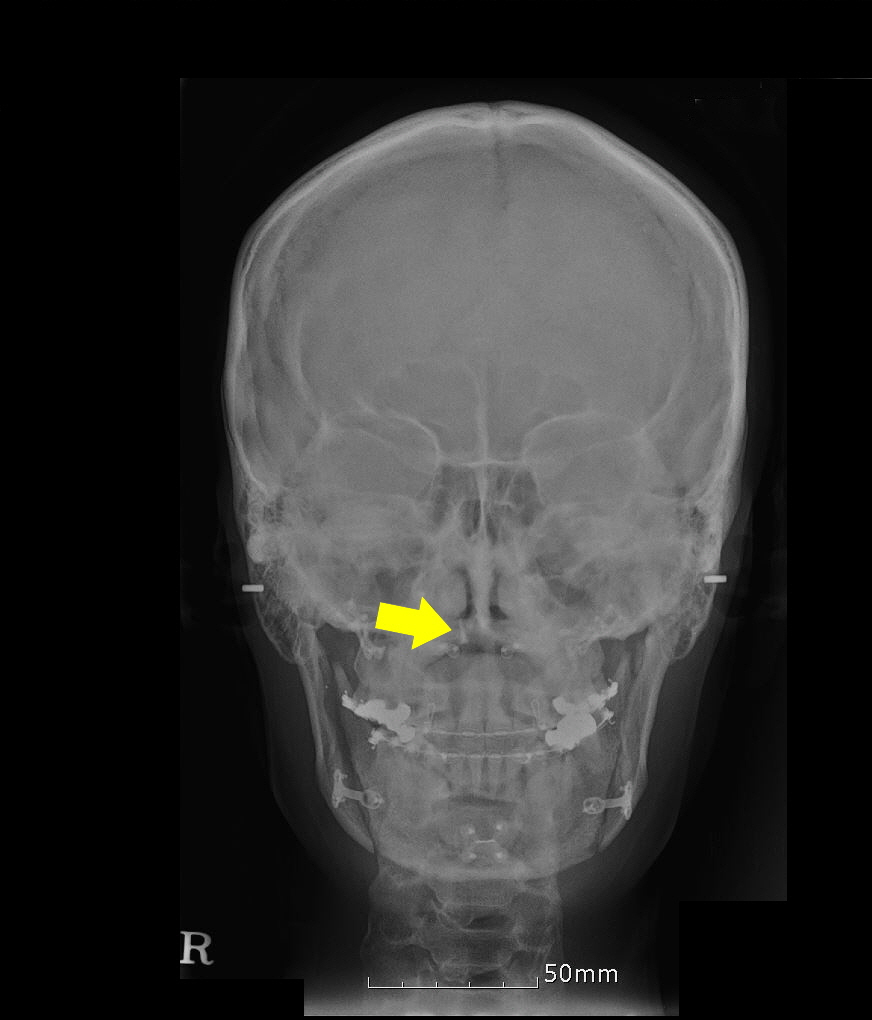
Nasotracheal intubation is generally performed for intraoral surgery. Case: A 34-year-old female patient who underwent orthognathic surgery exhibited repeated endotracheal tube cuff tears during nasotracheal intubation. After intubation, leaks developed, and torn endotracheal cuff was observed in the removed endotracheal tube. Subsequently, re-intubation through the same nasal cavity was performed immediately, but leakage from the torn endotracheal tube cuff was re-observed. A leakage test of the extubated tube revealed air bubbles and leaks near the tube cuff due to the tear. Nasotracheal intubation was performed through the other nasal cavity, and there were no leakage findings or abnormalities. During the course of the surgery, the surgeon noticed that the orthodontic micro-implant deposited in the mid-tube cavity was exposed to the nasal cavity.
Conclusions
We aimed to emphasize caution and discuss the possibility that orthodontic micro-implants that are not confirmed during preoperative evaluation may cause repeated endotracheal tube cuff tears.
Keyword
Figure
Reference
-
1. Stauffer JL, Olson DE, Petty TL. Complications and consequences of endotracheal intubation and tracheotomy. A prospective study of 150 critically ill adult patients. Am J Med. 1981; 70:65–76.2. Zwillich CW, Pierson DJ, Creagh CE, Sutton FD, Schatz E, Petty TL. Complications of assisted ventilation. A prospective study of 354 consecutive episodes. Am J Med. 1974; 57:161–70.3. El-Orbany M, Salem MR. Endotracheal tube cuff leaks: causes, consequences, and management. Anesth Analg. 2013; 117:428–34.4. Chen CH, Chang CS, Hsieh CH, Tseng YC, Shen YS, Huang IY, et al. The use of microimplants in orthodontic anchorage. J Oral Maxillofac Surg. 2006; 64:1209–13.5. Hall CE, Shutt LE. Nasotracheal intubation for head and neck surgery. Anaesthesia. 2003; 58:249–56.6. Prasanna D, Bhat S. Nasotracheal intubation: an overview. J Maxillofac Oral Surg. 2014; 13:366–72.7. Kim DR, Jung YH, Kang H, Oh JI, Park Y. Accidental middle turbinectomy by nasotracheal intubation −a case report−. Anesth Pain Med. 2016; 11:217–9.8. Nakamura S, Watanabe T, Hiroi E, Sasaki T, Matsumoto N, Hori T. [Cuff damage during naso-tracheal intubation for general anesthesia in oral surgery]. Masui. 1997; 46:1508–14. Japanese.9. Scamman FL, Babin RW. An unusual complication of nasotracheal intubation. Anesthesiology. 1983; 59:352–3.10. Ahmed-Nusrath A, Tong JL, Smith JE. Pathways through the nose for nasal intubation: a comparison of three endotracheal tubes. Br J Anaesth. 2008; 100:269–74.11. Sahin-Yilmaz A, Naclerio RM. Anatomy and physiology of the upper airway. Proc Am Thorac Soc. 2011; 8:31–9.12. Williamson R. Nasotracheal intubation for head and neck surgery. Anaesthesia. 2003; 58:1129–30; author reply 1130-1. .13. Abraham ST, Paul MMC. Micro implants for orthodontic anchorage: a review of complications and management. J Dent Implant. 2013; 3:165–7.14. Nojima K, Komatsu K, Isshiki Y, Ikumoto H, Hanai J, Saito C. The use of an osseointegrated implant for orthodontic anchorage to a Class II Div 1 malocclusion. Bull Tokyo Dent Coll. 2001; 42:177–83.
- Full Text Links
- Actions
-
Cited
- CITED
-
- Close
- Share
- Similar articles
-
- Cuff tear of endotracheal tube induced by a palatal orthodontic device during nasotracheal intubation: a case report
- Clinical Applications of the Balloon Dilation Technique during the Insertion of the Nasotracheal Tube
- Air leakage due to the cuff hanging on the vocal cords during nasotracheal intubation: a case report
- Protecting the tracheal tube cuff: a novel solution
- Effective removal of epistaxis during nasotracheal intubation utilizing a fiberoptic scope in a difficult airway: A case report


