Osseous Erosion by Spinoglenoid Ganglion Cyst in Adolescent Baseball Player: A Case Report
- Affiliations
-
- 1Department of Orthopaedic Surgery, Good Samsun Hospital, Busan, Korea
- KMID: 2523234
- DOI: http://doi.org/10.5763/kjsm.2021.39.4.188
Abstract
- Spinoglenoid notch cysts, a certain expansion form of paralabral ganglion cyst, are often associated with superior labrum anterior to posterior (SLAP) lesions in overhead athletes. We report a unique case of spinoglenoid notch cyst that extended to posterosuperior bony glenoid in a 16-year-old high school male baseball fielder. Magnetic resonance imaging showed multilobulated spinoglenoid notch ganglion cyst associated with posterosuperior SLAP lesion, and computed tomography (CT) revealed distinct osseous erosion of posterosuperior glenoid. The cyst was enlarged on serial follow-up imaging, and his symptoms were continued, arthroscopic decompression was performed via posterosuperior capsulotomy. The concomitant SLAP lesion was not repaired, but only marginal debridement was performed. At 6 months after surgery, he returned to game without symptoms, and the bony glenoid lesion was almost remodeled on follow-up CT. In adolescent athletes, significant osseous erosion by spinoglenoid notch cyst may be accentuated due to the skeletal immaturity of posterosuperior glenoid.
Keyword
Figure
-
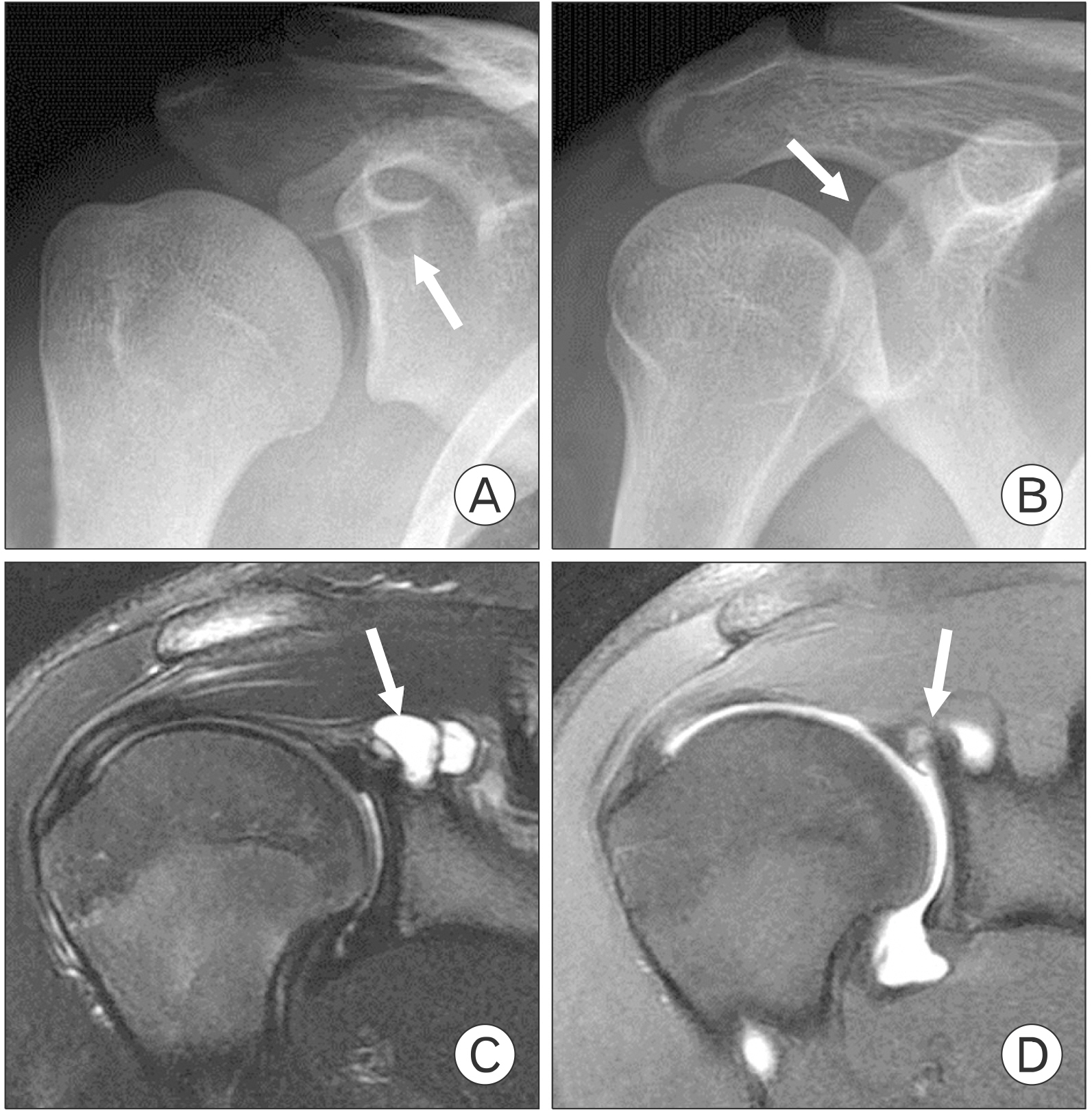
Fig. 1 Anteroposterior radiographs in (A) neutral and (B) internal rotation show oval radiolucent cystic lesion (arrows) in posterosuperior glenoid. (C) T2-weighted coronal magnetic resonance (MR) image demonstrates multi-lobulated homogenous high signal intensity paralabral cyst (arrow). (D) Fat-suppressed coronal MR arthrography shows superior labrum anterior to posterior lesion (arrow) communicated with paralabral cyst.

Fig. 2 Preoperative magnetic resonance images 8 months after initial presentation. (A) T2-weighted coronal image demonstrates enlargement of previous cyst (arrow) and (B) axial and (C) oblique sagittal images show anteroinferior osseous erosion (arrows) by adjacent extension of spinoglenoid ganglion cyst. (D) T1-weighted image shows significant scalloping of posterosuperior glenoid maintaining low-signal cortical continuity (arrow).
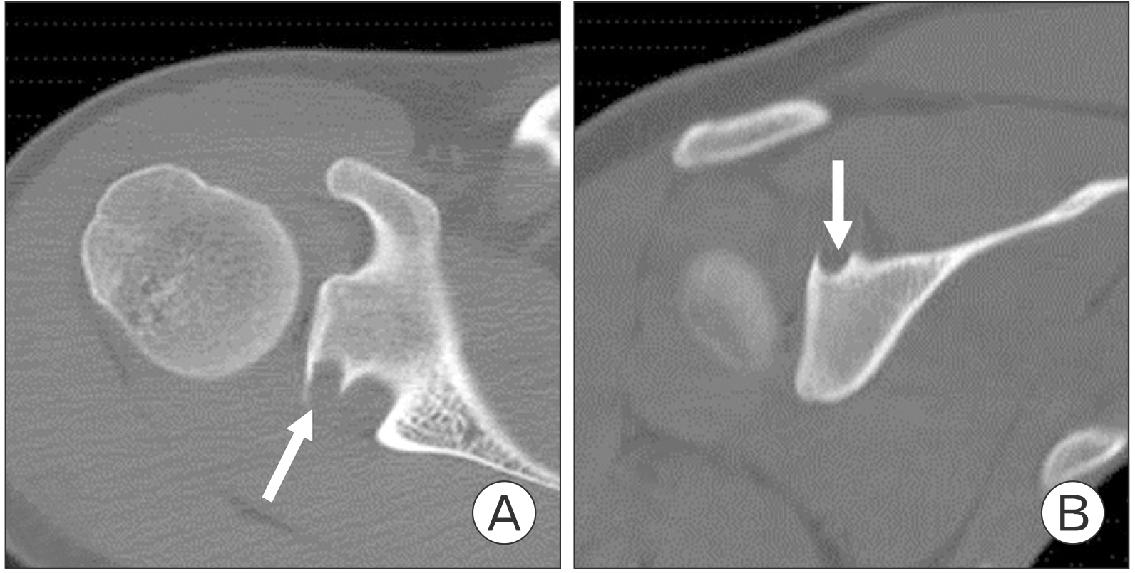
Fig. 3 Preoperative (A) axial and (B) coronal computed tomography images show punched-out osseous erosion (arrows) of posterosuperior glenoid in lateral portion of the spinoglenoid notch.

Fig. 4 Intraoperative arthroscopic images during spinoglenoid cyst decompression show (A) posterosuperior labral tear from anterior viewing portal and (B) characteristic amber-colored, gelatinous content within the cyst after posterosuperior capsulotomy. During cyst decompression, (C) posterosuperior glenoid cortex was maintained intact (arrows) and (D) stable posterosuperior labrum was established after debridement.

Fig. 5 Six months after cyst decompression, anteroposterior radiographs in (A) neutral and (B) internal rotation show significant resolution of preoperative radiolucent lesion (arrows) and (C) axial and (D) coronal computed tomography images demonstrate remodeling of eroded posterosuperior glenoid (arrows).
Reference
-
1. Ferrick MR, Marzo JM. 1997; Ganglion cyst of the shoulder associated with a glenoid labral tear and symptomatic glenohumeral instability: a case report. Am J Sports Med. 25:717–9. DOI: 10.1177/036354659702500523. PMID: 9302483.2. Lichtenberg S, Magosch P, Habermeyer P. 2004; Compression of the suprascapular nerve by a ganglion cyst of the spinoglenoid notch: the arthroscopic solution. Knee Surg Sports Traumatol Arthrosc. 12:72–9. DOI: 10.1007/s00167-003-0443-y. PMID: 14595536.
Article3. Jeong AK, Kim SM, Kim KS, Shin MJ, Ahn JM, Chun JM. 1999; Ganglionic cysts related to the scapula: MR findings. J Korean Radiol Soc. 41:171–5. DOI: 10.3348/jkrs.1999.41.1.171.
Article4. Schroder CP, Lundgreen K, Kvakestad R. 2018; Paralabral cysts of the shoulder treated with isolated labral repair: effect on pain and radiologic findings. J Shoulder Elbow Surg. 27:1283–9. DOI: 10.1016/j.jse.2017.12.022. PMID: 29449084.5. Park JY, Jeon SH, Oh KS, Chung SW, Lim JJ, Bang JY. 2015; Compressive partial neuropathy of axillary nerve resulting from antero-inferior paralabral cyst in an adolescent overhead athlete. Korean J Sports Med. 33:34–9. DOI: 10.5763/kjsm.2015.33.1.34.
Article6. Tirman PF, Feller JF, Janzen DL, Peterfy CG, Bergman AG. 1994; Association of glenoid labral cysts with labral tears and glenohumeral instability: radiologic findings and clinical significance. Radiology. 190:653–8. DOI: 10.1148/radiology.190.3.8115605. PMID: 8115605.
Article7. Perdikakis E, Skiadas V. 2013; MRI characteristics of cysts and "cyst-like" lesions in and around the knee: what the radiologist needs to know. Insights Imaging. 4:257–72. DOI: 10.1007/s13244-013-0240-1. PMID: 23479129. PMCID: PMC3675245.
Article8. Kambolis C, Bullough PG, Jaffe HI. 1973; Ganglionic cystic defects of bone. J Bone Joint Surg Am. 55:496–505. DOI: 10.2106/00004623-197355030-00005. PMID: 4703202.
Article9. Zember JS, Rosenberg ZS, Kwong S, Kothary SP, Bedoya MA. 2015; Normal skeletal maturation and imaging pitfalls in the pediatric shoulder. Radiographics. 35:1108–22. DOI: 10.1148/rg.2015140254. PMID: 26172355.
Article10. Caldwell PE 3rd, Dyer DC, Pearson SE. 2016; Arthroscopic debridement of the thrower's shoulder: less is more. Arthrosc Tech. 5:e1381–6. DOI: 10.1016/j.eats.2016.08.006. PMID: 28149736. PMCID: PMC5263853.
Article
- Full Text Links
-
- Actions
-
Cited
- CITED
-
- Close
- Share
-
- Similar articles
-
- The Follow Up Results of Residual Spinoglenoid Ganglion Cyst after Arthroscopic Decompression and Superior Labral Repair: Cases Report
- Nonunion of a Stress Fracture Through the Olecranon Epiphyseal Plate in an Adolescent Judo Player: A Case Report
- Synovial Osteochondromatosis of the Subtalar Joint in an Adolescent Baseball Player
- Suprascapular Nerve Injury Restricted to the Infraspinatus Muscle by a Ganglion Cyst: A case report
- Cubital Tunnel Syndrome by a Ganglion Cyst in an Amateur Tennis Player
