Usefulness of Color Doppler Ultrasonography for the Preoperative Evaluation of Thin Anterolateral Thigh Flap Perforators
- Affiliations
-
- 1Department of Plastic and Reconstructive Surgery, Gwangmyeong Sungae General Hospital, Gwangmyeong, Korea
- 2Department of Radiology, Gwangmyeong Sungae General Hospital, Gwangmyeong, Korea
- KMID: 2522625
- DOI: http://doi.org/10.12790/ahm.21.0105
Abstract
- Purpose
The anterolateral thigh flap is commonly applied to various body sites for reconstruction. However, surgeons often struggle against unexpected locations and the nature of perforator vessels during surgery. Thus, this study aimed to assess the accuracy and usefulness of color Doppler ultrasonography as a preoperative tool for the perforator position and course of anterolateral thigh flaps.
Methods
A prospective study involving 77 anterolateral thigh flaps was conducted between March 2016 and February 2021. Among them, 37 perforators (group A) were detected using the preoperative color Doppler ultrasound, and the other 40 perforators (group B) were tested using a hand-held Doppler only. All patients in group A underwent color Doppler ultrasonography performed by a radiologist at our institution. The nature and course of the perforator vessels were recorded, and their precise locations were plotted in an orthonormal coordinate system.
Results
A total of 37 anterolateral thigh perforator flaps (group A) were successfully dissected. The median distance between the preoperative color Doppler ultrasonography and the real location during surgery of the perforators was 7.50 mm, which was statistically smaller than 10 mm (p<0.001). This preoperative ultrasound test also had a success rate of 94.6% to determine the nature of the perforators (musculocutaneous type vs. septocutaneous type).
Conclusion
Preoperative color Doppler ultrasonography provides a harmless, reliable, and accurate technique for visualizing the vascular anatomy of anterolateral thigh flaps. It has a high correlation with surgical findings, allowing surgeons to cope with variable vascular anatomy.
Figure
-
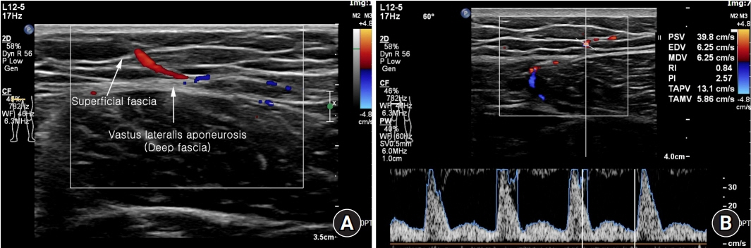
Fig. 1. Color Doppler ultrasound preoperative assessment (perforator 33). (A) The location and course of the perforator surges through the superficial fascia (left arrow) from the vastus lateralis aponeurosis (deep fascia, right arrow). (B) The flow of the perforator is measured using the color Doppler flowmeter.
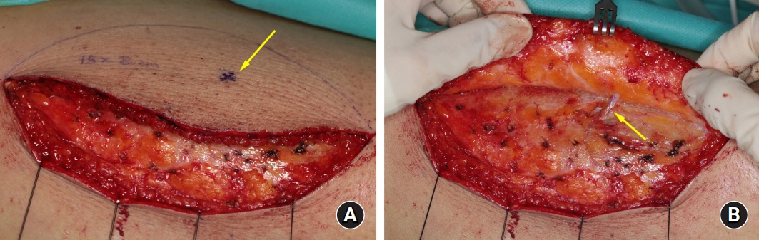
Fig. 2. Intraoperative photographs of harvesting a thin anterolateral thigh (ALT) flap (perforator 33). (A) The ALT flap is harvesting on the plane of the superficial fascia, being careful for the predicted perforator (arrow). (B) The perforator (arrow) is saved, and recorded in the orthonormal coordinate system. The patient provided written informed consent for the publication and the use of his images.
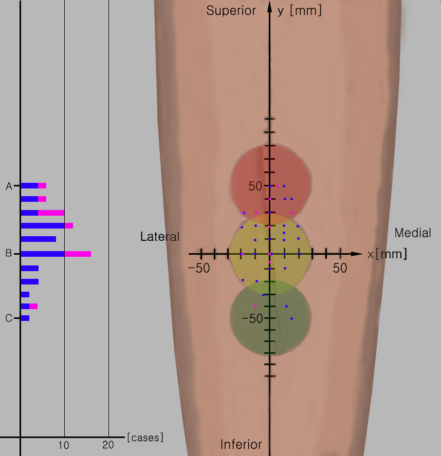
Fig. 3. Overall distribution of perforators during surgery. A total of 37 perforators during surgery were plotted in the orthonormal coordinate system. Area B has the most perforators (22), followed by areas A and C. Blue, musculocutaneous type; pink, septocutaneous type.
Reference
-
1. Song YG, Chen GZ, Song YL. The free thigh flap: a new free flap concept based on the septocutaneous artery. Br J Plast Surg. 1984; 37:149–59.
Article2. Yu P, Sanger JR, Matloub HS, Gosain A, Larson D. Anterolateral thigh fasciocutaneous island flaps in perineoscrotal reconstruction. Plast Reconstr Surg. 2002; 109:610–8.
Article3. Wei FC, Suominen S, Cheng MH, Celik N, Lai YL. Anterolateral thigh flap for postmastectomy breast reconstruction. Plast Reconstr Surg. 2002; 110:82–8.
Article4. Wei FC, Celik N, Chen HC, Cheng MH, Huang WC. Combined anterolateral thigh flap and vascularized fibula osteoseptocutaneous flap in reconstruction of extensive composite mandibular defects. Plast Reconstr Surg. 2002; 109:45–52.
Article5. Adani R, Tarallo L, Marcoccio I, Cipriani R, Gelati C, Innocenti M. Hand reconstruction using the thin anterolateral thigh flap. Plast Reconstr Surg. 2005; 116:467–77.
Article6. Yu P. Reinnervated anterolateral thigh flap for tongue reconstruction. Head Neck. 2004; 26:1038–44.
Article7. Yu P, Robb GL. Pharyngoesophageal reconstruction with the anterolateral thigh flap: a clinical and functional outcomes study. Plast Reconstr Surg. 2005; 116:1845–55.
Article8. Lin SJ, Rabie A, Yu P. Designing the anterolateral thigh flap without preoperative Doppler or imaging. J Reconstr Microsurg. 2010; 26:67–72.
Article9. Blondeel PN, Beyens G, Verhaeghe R, et al. Doppler flowmetry in the planning of perforator flaps. Br J Plast Surg. 1998; 51:202–9.
Article10. Debelmas A, Camuzard O, Aguilar P, Qassemyar Q. Reliability of color Doppler ultrasound imaging for the assessment of anterolateral thigh flap perforators: a prospective study of 30 perforators. Plast Reconstr Surg. 2018; 141:762–6.11. Ibrahim RM, Gunnarsson GL, Akram J, Sørensen JA, Thomsen JB. Color Doppler ultrasonography targeted reconstruction using pedicled perforator flaps: a systematic review and meta-analysis. Eur J Plast Surg. 2018; 41:495–504.12. Hong JP, Chung IW. The superficial fascia as a new plane of elevation for anterolateral thigh flaps. Ann Plast Surg. 2013; 70:192–5.
Article13. Shen Y, Huang J, Dong MJ, Li J, Ye WM, Sun J. Application of computed tomography angiography mapping and located template for accurate location of perforator in head and neck reconstruction with anterolateral thigh perforator flap. Plast Reconstr Surg. 2016; 137:1875–85.
Article14. González Martínez J, Torres Pérez A, Gijón Vega M, Nuñez-Villaveiran T. Preoperative vascular planning of free flaps: comparative study of computed tomographic angiography, color Doppler ultrasonography, and hand-held Doppler. Plast Reconstr Surg. 2020; 146:227–37.
Article15. Ensat F, Babl M, Conz C, et al. The efficacy of color duplex sonography in preoperative assessment of anterolateral thigh flap. Microsurgery. 2012; 32:605–10.
Article16. Feng S, Min P, Grassetti L, et al. A prospective head-to-head comparison of color Doppler ultrasound and computed tomographic angiography in the preoperative planning of lower extremity perforator flaps. Plast Reconstr Surg. 2016; 137:335–47.
Article17. Malhotra K, Lian TS, Chakradeo V. Vascular anatomy of anterolateral thigh flap. Laryngoscope. 2008; 118:589–92.
Article
- Full Text Links
-
- Actions
-
Cited
- CITED
-
- Close
- Share
-
- Similar articles
-
- Reconstruction of the Tissue Defects of Extremities with Anterolateral Thigh Free Flap
- Total Tongue Reconstruction with Folded Anterolateral Thigh Free Flap
- The Regional Anatomy of Perforating artery and Pedicle for the Anterolateral Thigh Free Flap in the Korean
- Posteromedial Thigh Perforator Flap: An Anatomical Study and Clinical Applications
- Augmented reality and dynamic infrared thermography for perforator mapping in the anterolateral thigh
