J Korean Med Sci.
2021 Nov;36(43):e266. 10.3346/jkms.2021.36.e266.
Natural Progression of Ground-glass Nodules after Curative Resection for Non-small Cell Lung Cancer
- Affiliations
-
- 1Department of Thoracic and Cardiovascular Surgery, International St. Mary's Hospital, Catholic Kwandong University, College of Medicine, Incheon, Korea
- 2Department of Thoracic and Cardiovascular Surgery, Asan Medical Center, University of Ulsan College of Medicine, Seoul, Korea
- KMID: 2522285
- DOI: http://doi.org/10.3346/jkms.2021.36.e266
Abstract
- Background
This retrospective study investigated the natural course of synchronous groundglass nodules (GGNs) that remained after curative resection for non-small-cell lung cancer (NSCLC).
Methods
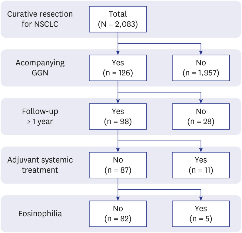
Prospectively collected retrospective data were reviewed concerning 2,276 patients who underwent curative resection for NSCLC between 2008 and 2017. High-resolution computed tomography or thin-section computed tomography data of 82 patients were included in the study. Growth in size was considered the most valuable outcome, and patients were grouped according to GGN size change. Patient demographic data (e.g., age, sex, and smoking history), perioperative data (e.g., GGN characteristics, histopathology and pathological stage of the resected tumours), and other medical history were evaluated in a risk factor analysis concerning GGN size change.
Results
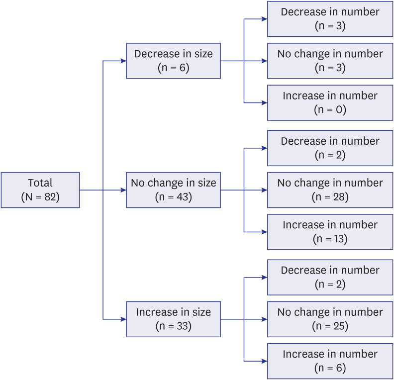
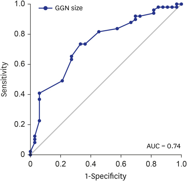
The median duration of follow-up was 36.0 months (interquartile range, 23.0–59.3 months). GGN size decreased in 6 patients (7.3%), was stationary in 43 patients (52.4%), and increased in 33 patients (40.2%). In univariate analysis, male sex, the GGN size on initial CT, part-solid GGN and smoking history (≥ 10 pack-years) were significant risk factors. Among them, multivariate analysis revealed that lager GGN size, part-solid GGN and smoking history were independent risk factors.
Conclusion
During follow-up, 40.2% of GGNs increased in size, emphasising that patients with larger GGNs, part-solid GGN or with a smoking history should be observed.
Figure
Reference
-
1. National Lung Screening Trial Research Team. Aberle DR, Adams AM, Berg CD, Black WC, Clapp JD, et al. Reduced lung-cancer mortality with low-dose computed tomographic screening. N Engl J Med. 2011; 365(5):395–409. PMID: 21714641.
Article2. Pastorino U, Silva M, Sestini S, Sabia F, Boeri M, Cantarutti A, et al. Prolonged lung cancer screening reduced 10-year mortality in the MILD trial: new confirmation of lung cancer screening efficacy. Ann Oncol. 2019; 30(10):1672. PMID: 31168572.
Article3. De Koning H, Van Der Aalst C, Ten Haaf K, Oudker M. PL02.05 Effects of volume CT lung cancer screening: mortality results of the NELSON randomised-controlled population based trial. J Thorac Oncol. 2018; 13(10):S185.
Article4. Henschke CI, Lee IJ, Wu N, Farooqi A, Khan A, Yankelevitz D, et al. CT screening for lung cancer: prevalence and incidence of mediastinal masses. Radiology. 2006; 239(2):586–590. PMID: 16641357.5. Takx RA, Išgum I, Willemink MJ, van der Graaf Y, de Koning HJ, Vliegenthart R, et al. Quantification of coronary artery calcium in nongated CT to predict cardiovascular events in male lung cancer screening participants: results of the NELSON study. J Cardiovasc Comput Tomogr. 2015; 9(1):50–57. PMID: 25533223.
Article6. Park CM, Goo JM, Lee HJ, Lee CH, Chun EJ, Im JG. Nodular ground-glass opacity at thin-section CT: histologic correlation and evaluation of change at follow-up. Radiographics. 2007; 27(2):391–408. PMID: 17374860.
Article7. Nakata M, Saeki H, Takata I, Segawa Y, Mogami H, Mandai K, et al. Focal ground-glass opacity detected by low-dose helical CT. Chest. 2002; 121(5):1464–1467. PMID: 12006429.
Article8. Sone S, Takashima S, Li F, Yang Z, Honda T, Maruyama Y, et al. Mass screening for lung cancer with mobile spiral computed tomography scanner. Lancet. 1998; 351(9111):1242–1245. PMID: 9643744.
Article9. Cho J, Kim ES, Kim SJ, Lee YJ, Park JS, Cho YJ, et al. Long-term follow-up of small pulmonary ground-glass nodules stable for 3 years: implications of the proper follow-up period and risk factors for subsequent growth. J Thorac Oncol. 2016; 11(9):1453–1459. PMID: 27287413.
Article10. Godoy MC, Naidich DP. Subsolid pulmonary nodules and the spectrum of peripheral adenocarcinomas of the lung: recommended interim guidelines for assessment and management. Radiology. 2009; 253(3):606–622. PMID: 19952025.
Article11. Henschke CI, Yankelevitz DF, Mirtcheva R, McGuinness G, McCauley D, Miettinen OS, et al. CT screening for lung cancer: frequency and significance of part-solid and nonsolid nodules. AJR Am J Roentgenol. 2002; 178(5):1053–1057. PMID: 11959700.12. Kobayashi Y, Mitsudomi T. Management of ground-glass opacities: should all pulmonary lesions with ground-glass opacity be surgically resected? Transl Lung Cancer Res. 2013; 2(5):354–363. PMID: 25806254.13. MacMahon H, Naidich DP, Goo JM, Lee KS, Leung AN, Mayo JR, et al. Guidelines for management of incidental pulmonary nodules detected on CT images: from the Fleischner Society 2017. Radiology. 2017; 284(1):228–243. PMID: 28240562.14. Hiramatsu M, Inagaki T, Inagaki T, Matsui Y, Satoh Y, Okumura S, et al. Pulmonary ground-glass opacity (GGO) lesions-large size and a history of lung cancer are risk factors for growth. J Thorac Oncol. 2008; 3(11):1245–1250. PMID: 18978558.
Article15. Goldstraw P, Chansky K, Crowley J, Rami-Porta R, Asamura H, Eberhardt WE, et al. The IASLC Lung Cancer Staging Project: proposals for revision of the TNM stage groupings in the forthcoming (Eighth) edition of the TNM classification for lung cancer. J Thorac Oncol. 2016; 11(1):39–51. PMID: 26762738.16. Travis WD, Brambilla E, Nicholson AG, Yatabe Y, Austin JHM, Beasley MB, et al. The 2015 World Health Organization classification of lung tumors: impact of genetic, clinical and radiologic advances since the 2004 classification. J Thorac Oncol. 2015; 10(9):1243–1260. PMID: 26291008.17. Beasley MB, Brambilla E, Travis WD. The 2004 World Health Organization classification of lung tumors. Semin Roentgenol. 2005; 40(2):90–97. PMID: 15898407.
Article18. Gu B, Burt BM, Merritt RE, Stephanie S, Nair V, Hoang CD, et al. A dominant adenocarcinoma with multifocal ground glass lesions does not behave as advanced disease. Ann Thorac Surg. 2013; 96(2):411–418. PMID: 23806231.
Article19. Shimada Y, Saji H, Otani K, Maehara S, Maeda J, Yoshida K, et al. Survival of a surgical series of lung cancer patients with synchronous multiple ground-glass opacities, and the management of their residual lesions. Lung Cancer. 2015; 88(2):174–180. PMID: 25758554.
Article20. Chang B, Hwang JH, Choi YH, Chung MP, Kim H, Kwon OJ, et al. Natural history of pure ground-glass opacity lung nodules detected by low-dose CT scan. Chest. 2013; 143(1):172–178. PMID: 22797081.
Article21. Lee JH, Park CM, Kim H, Hwang EJ, Park J, Goo JM. Persistent part-solid nodules with solid part of 5 mm or smaller: can the ‘follow-up and surgical resection after interval growth’ policy have a negative effect on patient prognosis? Eur Radiol. 2017; 27(1):195–202. PMID: 27126519.22. Kim HK, Choi YS, Kim J, Shim YM, Lee KS, Kim K. Management of multiple pure ground-glass opacity lesions in patients with bronchioloalveolar carcinoma. J Thorac Oncol. 2010; 5(2):206–210. PMID: 19901852.
Article23. Kim HK, Choi YS, Kim K, Shim YM, Jeong SY, Lee KS, et al. Management of ground-glass opacity lesions detected in patients with otherwise operable non-small cell lung cancer. J Thorac Oncol. 2009; 4(10):1242–1246. PMID: 19687762.
Article
- Full Text Links
- Actions
-
Cited
- CITED
-
- Close
- Share
- Similar articles
-
- Guidelines for the Investigation and Management of Ground Glass Nodules
- Pulmonary Subsolid Nodules: An Overview & Management Guidelines
- Lobectomy versus Sublobar Resection in Non-Lepidic Small-Sized Non-Small Cell Lung Cancer
- The Clinical Approach to Nodular Ground Glass Opacity in the Lung
- What Should Thoracic Surgeons Consider during Surgery for Ground-Glass Nodules?: Lymph Node Dissection


