Developing an Interactive Computer Program for Integrated Dental Education
- Affiliations
-
- 1School of Dentistry, Faculty of Medicine and Dentistry, College of Health Sciences, University of Alberta, Edmonton, Canada
- KMID: 2522215
- DOI: http://doi.org/10.4258/hir.2021.27.4.335
Abstract
Objectives
The knowledge of anatomy is an integral part of dental and medical education that builds the foundations of pathology, physiology, and other related disciplines. Traditional three-dimensional (3D) models used to teach anatomy cannot represent dynamic physiological processes and lack structural detail in the oral regions relevant for dental education. We developed an interactive computer program to teach oral anatomy, pathology, and microbiology in an integrated manner to improve students’ learning experiences.
Methods
The computer program, Jawnatomy, was developed as a 3D human head. Cognitive load theory guided the design of the tool, with the goal of reducing the heavy cognitive load of learning anatomy and integrating this knowledge with pathology and microbiology. Keller’s attention, relevance, confidence, and satisfaction (ARCS) model of motivational design was used while creating the tool to improve learners’ motivation and engagement. Blender was used to create the graphics, and Unity 3D was used to incorporate interactivity in the program. The 3D renderings of oral anatomy and progression of tooth decay were created with the input of content experts.
Results
Jawnatomy will be launched in our institution’s dentistry and dental hygiene program to support project- and team-based learning. This program will also be introduced to students as a self-directed learning tool to help them practice and strengthen their anatomical knowledge at their own pace.
Conclusions
Surveys and focus groups will be conducted to evaluate and further improve the computer program. We believe that Jawnatomy will become an invaluable teaching tool for dental education.
Figure
-
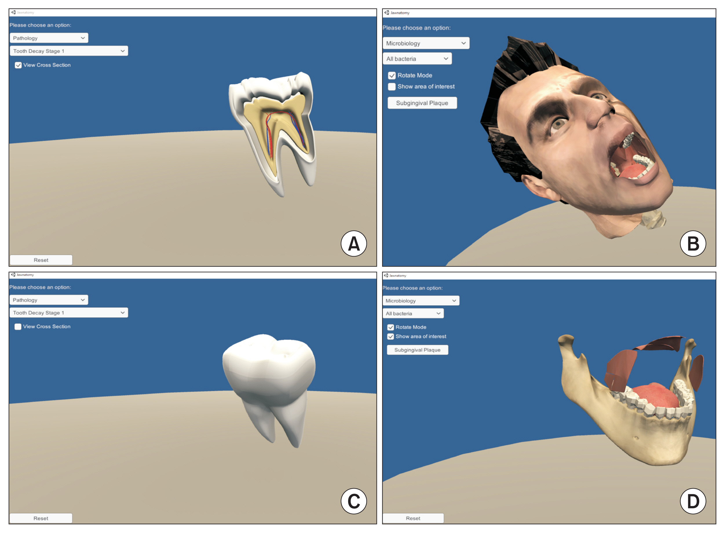
Figure 1 Learning modules of Jawnatomy. Along with a model of the physiological condition, Jawnatomy offers two other learning modules: (A) pathology and (B) microbiology. Jawnatomy allows learners to view the anatomical structure of a healthy tooth in both its surface view (C) and cross-sectional view (A). (D) Students can also remove external facial structures to visualize internal oral structures to learn oral anatomy.
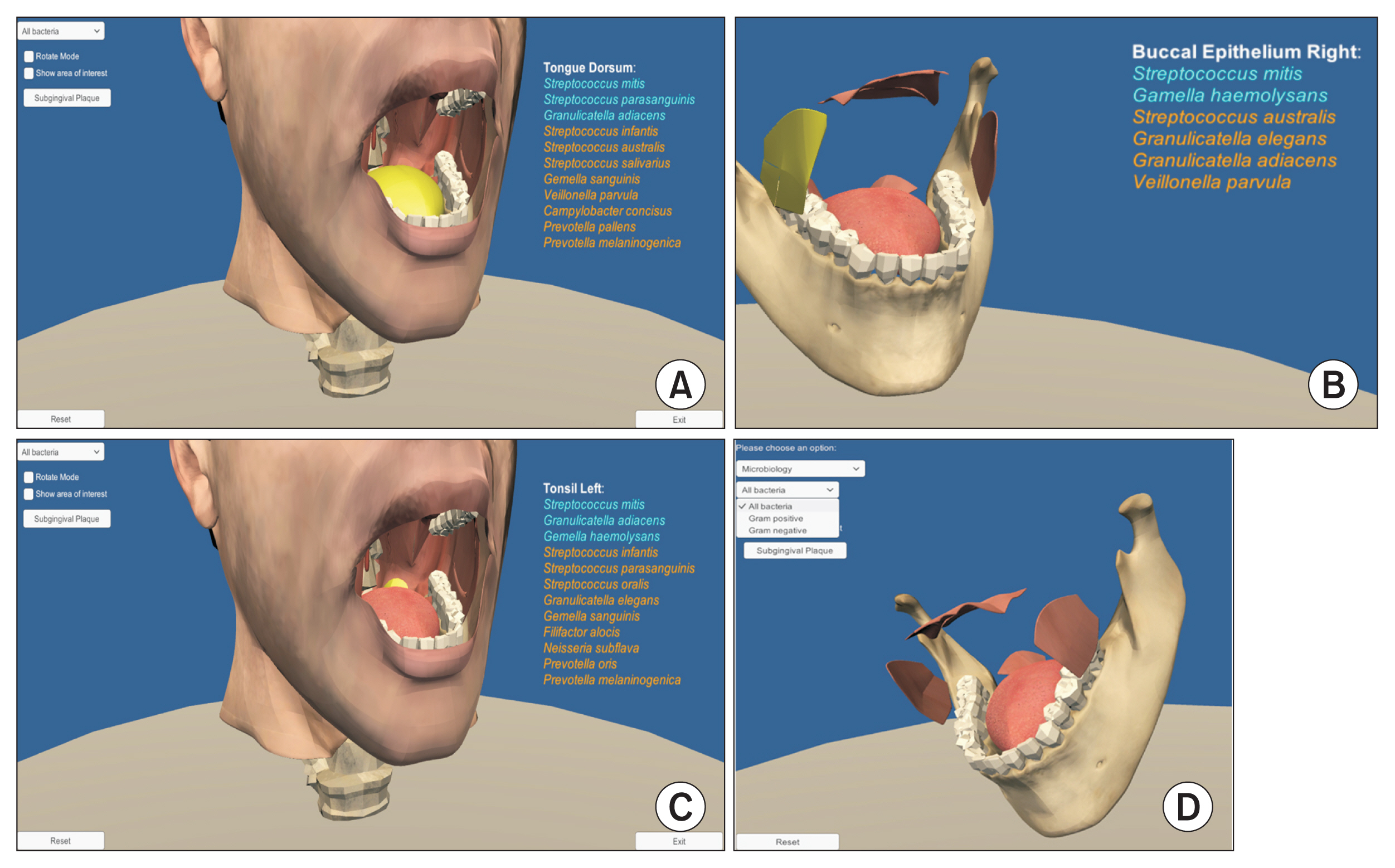
Figure 2 Microbiology module of Jawnatomy. Jawnatomy allows the learner the explore the normal microbial flora of the oral cavity. When selected, each oral cavity region shows the list of bacterial species found in that area. The lists of bacteria colonizing the tongue dorsum, buccal epithelium, and tonsil are shown in (A), (B), and (C), respectively. The list of bacteria is color-coded according to their prevalence, with those in blue being more abundant in that region. (D) Students can also select and group bacteria according to their cell wall properties (Gram-positive and Gram-negative).
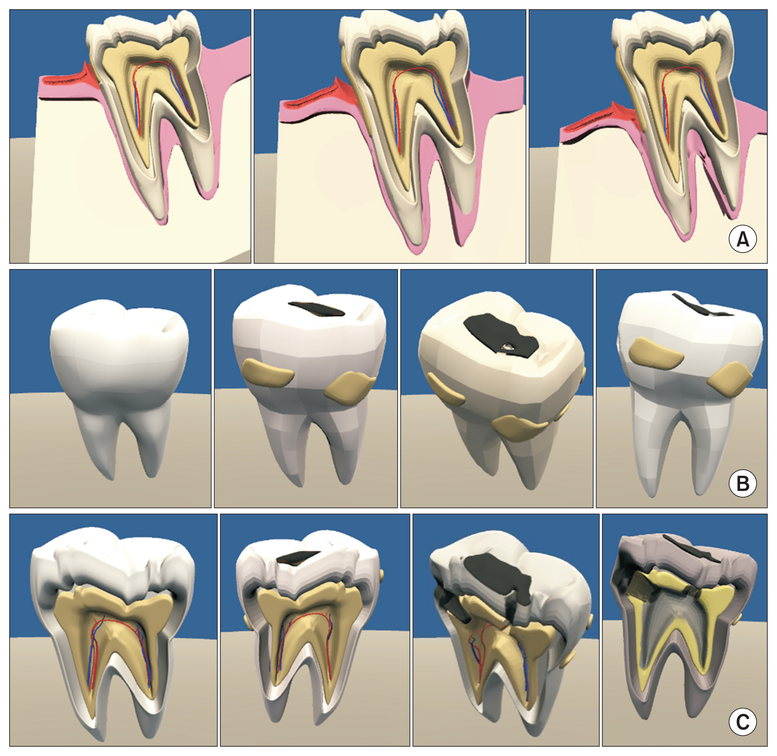
Figure 3 Pathology module of Jawnatomy. The pathology module of Jawnatomy allows learners to visualize the pathological processes observed in (A) periodontal disease and (B, C) dental caries. Periodontal disease is a chronic inflammatory disease that starts with bacterial plaque accumulation in the subgingival region. (A) Students can observe the progression of gingival inflammation and gradual loss of underlying alveolar bone to understand the development of periodontal disease. (B) Learners can also visualize plaque accumulation and dental caries formation on a molar tooth. They can rotate the tooth model to different angles and magnify areas to observe structural details. (C) Students can switch to the cross-sectional mode to analyze the effect of dental caries on the tooth’s internal structure. This module allows students to understand the gradual progression of the disease and compare it with healthy teeth and oral structures.
Reference
-
References
1. Alraddadi A. Literature review of anatomical variations: clinical significance, identification approach, and teaching strategies. Cureus. 2021; 13(4):e14451.
Article2. Han ER, Yeo S, Kim MJ, Lee YH, Park KH, Roh H. Medical education trends for future physicians in the era of advanced technology and artificial intelligence: an integrative review. BMC Med Educ. 2019; 19(1):460.
Article3. Harden RM. Ten key features of the future medical school-not an impossible dream. Med Teach. 2018; 40(10):1010–5.
Article4. Frenk J, Chen L, Bhutta ZA, Cohen J, Crisp N, Evans T, et al. Health professionals for a new century: transforming education to strengthen health systems in an inter-dependent world. Lancet. 2010; 376(9756):1923–58.
Article5. Grainger R, Liu Q, Geertshuis S. Learning technologies: a medium for the transformation of medical education? Med Educ. 2021; 55(1):23–9.
Article6. Dahle LO, Brynhildsen J, Behrbohm Fallsberg M, Rundquist I, Hammar M. Pros and cons of vertical integration between clinical medicine and basic science within a problem-based undergraduate medical curriculum: examples and experiences from Linköping, Sweden. Med Teach. 2002; 24(3):280–5.
Article7. Brauer DG, Ferguson KJ. The integrated curriculum in medical education: AMEE Guide No. 96. Med Teach. 2015; 37(4):312–22.
Article8. Aas JA, Paster BJ, Stokes LN, Olsen I, Dewhirst FE. Defining the normal bacterial flora of the oral cavity. J Clin Microbiol. 2005; 43(11):5721–32.
Article9. Wilbert SA, Mark Welch JL, Borisy GG. Spatial ecology of the human tongue dorsum microbiome. Cell Rep. 2020; 30(12):4003–4015e3.
Article10. Blender: a 3D modelling and rendering package [Internet]. Amsterdam, Netherlands: Stichting Blender Foundation;2018. [cited at 2021 Sep 30]. Available from: http://www.blender.org .11. Unity: using the UI tools [Internet]. San Francisco (CA): Unity Technologies;c2021. [cited at 2021 Sep 30]. Available from: https://unity.com/ .12. Khalil MK, Paas F, Johnson TE, Payer AF. Design of interactive and dynamic anatomical visualizations: the implication of cognitive load theory. Anat Rec B New Anat. 2005; 286(1):15–20.
Article13. Sweller J. Cognitive load theory and educational technology. Educ Technol Res Dev. 2020; 68(1):1–16.
Article14. Martens R, Gulikers J, Bastiaens T. The impact of in trinsic motivation on e-learning in authentic computer tasks. J Comput Assist Learn. 2004; 20(5):368–76.15. Keller JM. Motivational design for learning and performance: the ARCS model approach. New York (NY): Springer;2010.16. Li K, Keller JM. Use of the ARCS model in education: a literature review. Computers & Education. 2018; 122:54–62.
Article17. Milman NB, Wessmiller J. Motivating the online learner using Keller’s ARCS model. Distance Learn. 2016; 13(2):67–71.18. Bandura A. Social learning theory. Englewood Cliffs (NJ): Prentice-Hall;1977.19. Knowles MS. Self-directed learning: a guide for learners and teachers. New York (NY): Association Press;1975.20. Azer SA. A multimedia CD-ROM tool to improve student understanding of bile salts and bilirubin metabolism: evaluation of its use in a medical hybrid PBL course. Adv Physiol Educ. 2005; 29(1):40–50.
Article21. Rosenberg H, Grad HA, Matear DW. The effectiveness of computer-aided, self-instructional programs in dental education: a systematic review of the literature. J Dent Educ. 2003; 67(5):524–32.
Article22. Revell SM, McCurry MK. Engaging millennial learners: effectiveness of personal response system technology with nursing students in small and large classrooms. J Nurs Educ. 2010; 49(5):272–5.
Article23. Janssen A, Shaw T, Goodyear P, Kerfoot BP, Bryce D. A little healthy competition: using mixed methods to pilot a team-based digital game for boosting medical student engagement with anatomy and histology content. BMC Med Educ. 2015; 15:173.
Article24. Thompson ME, Ford R, Webster A. Effectiveness of interactive, online games in learning neuroscience and students’ perception of the games as learning tools: a pre-experimental study. J Allied Health. 2011; 40(3):150–5.
- Full Text Links
-
- Actions
-
Cited
- CITED
-
- Close
- Share
-
- Similar articles
-
- Development of Interactive Multimedia Learning in Aging Education
- Development of Online Sex Education Programs Using Interactive Human-Computer Dialogue Technology
- Development and Evaluation of a Computerized Multimedia Approach to Educate Older Adults about Safe Medication
- A study on the Korean dental education system II: the need for integrated education system
- Perceptions of dental undergraduates in India of a clinical induction program
