Posterior Thoracic Cage Interbody Fusion Offers Solid Bone Fusion with Sagittal Alignment Preservation for Decompression and Fusion Surgery in Lower Thoracic and Thoracolumbar Spine
- Affiliations
-
- 1Department of Neurological Surgery, Asan Medical Center, University of Ulsan College of Medicine, Seoul, Korea
- 2Department of Neurological Surgery, National Police Hospital, Seoul, Korea
- 3Department of Neurological Surgery, Dongtan Sacred Heart Hospital, College of Medicine, Hallym University, Hwaseong, Korea
- KMID: 2521982
- DOI: http://doi.org/10.3340/jkns.2020.0311
Abstract
Objective
: It is challenging to make solid fusion by posterior screw fixation and laminectomy with posterolateral fusion (PLF) in thoracic and thoracolumbar (TL) diseases. In this study, we report our experience and follow-up results with a new surgical technique entitled posterior thoracic cage interbody fusion (PTCIF) for thoracic and TL spine in comparison with conventional PLF.
Methods
: After institutional review board approval, a total of 57 patients who underwent PTCIF (n=30) and conventional PLF (n=27) for decompression and fusion in thoracic and TL spine between 2004 and 2019 were analyzed. Clinical outcomes and radiological parameters, including bone fusion, regional Cobb angle, and proximal junctional Cobb angle, were evaluated.
Results
: In PTCIF and conventional PLF, the mean age was 61.2 and 58.2 years (p=0.46), and the numbers of levels fused were 2.8 and 3.1 (p=0.46), respectively. Every patient showed functional improvement except one case of PTCIF. Postoperative hematoma as a perioperative complication occurred in one and three cases, respectively. The mean difference in the regional Cobb angle immediately after surgery compared with that of the last follow-up was 1.4° in PTCIF and 7.6° in conventional PLF (p=0.003), respectively. The mean durations of postoperative follow-up were 35.6 months in PTCIF and 37.3 months in conventional PLF (p=0.86).
Conclusion
: PTCIF is an effective fusion method in decompression and fixation surgery with good clinical outcomes for various spinal diseases in the thoracic and TL spine. It provides more stable bone fusion than conventional PLF by anterior column support.
Keyword
Figure
-

Fig. 1. A flowchart of the study. TL : thoracolumbar, PTCIF : posterior thoracic cage interbody fusion, PLF : posterolateral fusion.
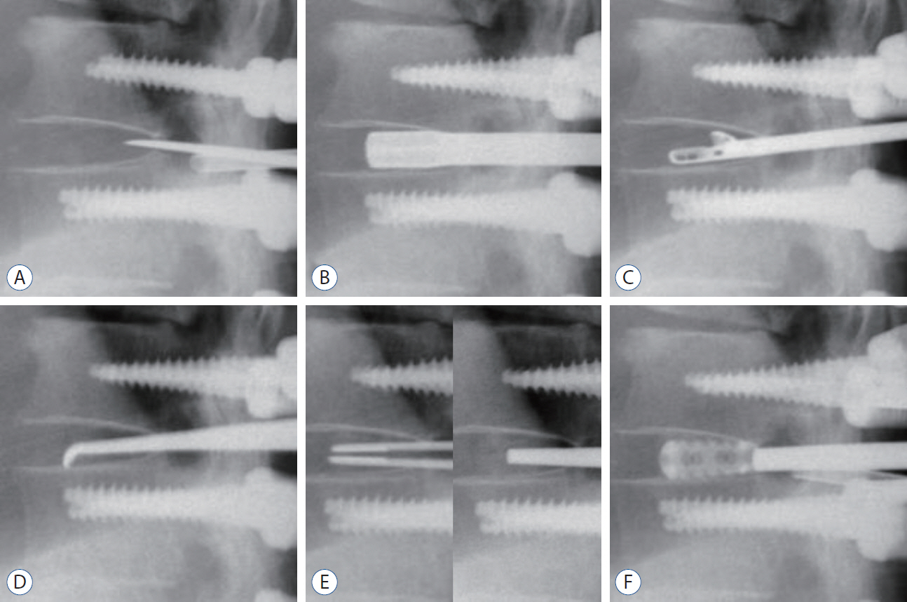
Fig. 2. Intraoperative C-arm images in posterior thoracic cage interbody fusion procedures. A : Discectomy was performed after the incision of the annulus using knife and sharp osteotome. B : A shaver was inserted and rotated to scrub the endplate. C : The disc material was removed using a pituitary forcep. D : The remnant disc material of the endplate was curetted with an angled curette. E : Autologous bone chips were inserted into the disc space and packed by a sharp impactor to enhance bone fusion before cage insertion. F : Thereafter, cages packed with autologous bone chips were inserted.
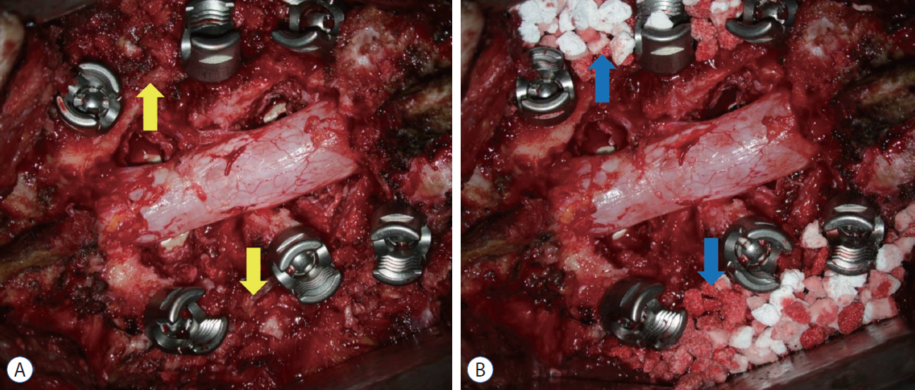
Fig. 3. Images of conventional posterolateral fusion after posterior screw fixation and laminectomy images at surgical field are shown. Bone chips were applied onto the fusion bed obtained through extensive lateral muscle dissection. A : The yellow arrows indicate the local autologous bone chips. B : Allograft or hydroxyapatite bone chips can be used along with autologous bone chips. The blue arrows indicate the hydroxyapatite bone chips.
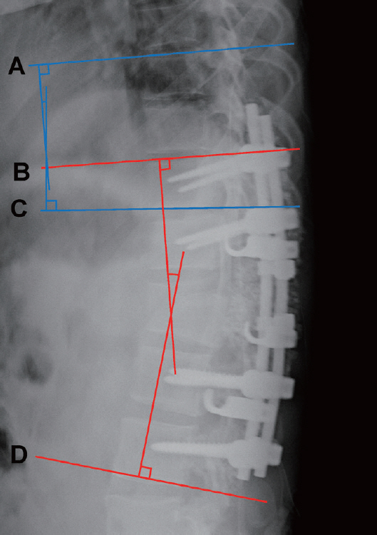
Fig. 4. Radiological measurement of the sagittal X-ray. The regional Cobb angle is the angle between the red tangential line to the cephalad endplate line of the upper instrumented vertebrae (UIV) (B) and the red tangential line to the caudal endplate line of the lower instrumented vertebrae (D). The proximal junctional Cobb angle is the angle between the blue tangential line to the cephalad endplate line of the two supraadjacent vertebrae above the UIV (A) and the blue tangential line to the caudal endplate line of the UIV (C).
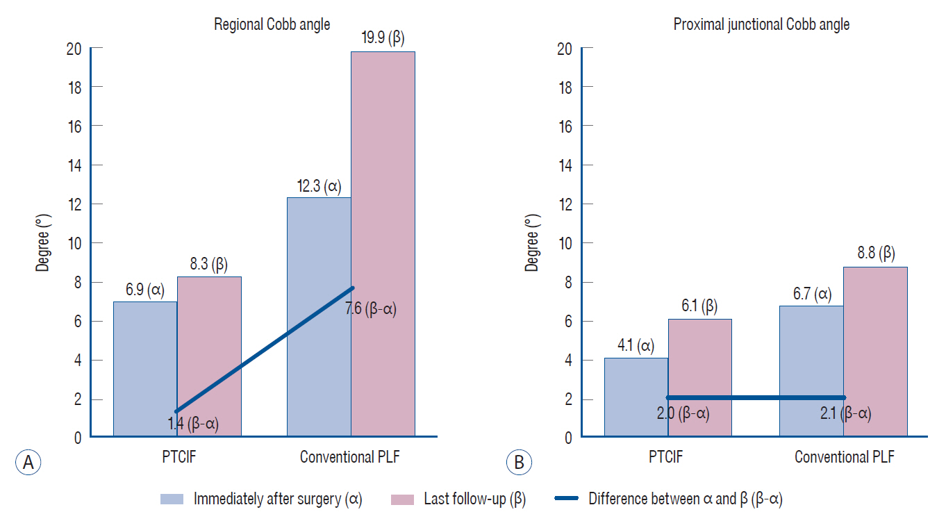
Fig. 5. The changes of radiological parameters of the two groups during follow-up. A : The mean difference in the regional Cobb angle immediately after surgery compared with that of the last follow-up was 1.4° in posterior thoracic cage interbody fusion (PTCIF) and 7.6° in conventional posterolateral fusion (PLF), which showed significant difference between the two groups (p=0.003). B : The mean difference in the proximal junctional Cobb angle immediately after surgery compared with that of the last follow-up was 2.0° in PTCIF and 2.1° in conventional PLF, which showed no significant difference (p=0.97).
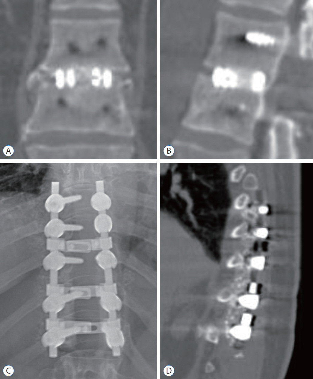
Fig. 6. Bony continuity within and circumjacent the cages between the upper and lower endplates are well identified in coronal (A) and sagittal (B) CT images 3 months after PTCIF at T12–L1, whereas it is complex to verify the bony continuity in simple spine X-ray (C) and sagittal CT image (D) 3 months after conventional PLF. PTCIF : posterior thoracic cage interbody fusion, PLF : posterolateral fusion, CT : computed tomography.
Reference
-
References
1. Agazzi S, Reverdin A, May D. Posterior lumbar interbody fusion with cages: an independent review of 71 cases. J Neurosurg. 91:186–192. 1999.
Article2. Alsaleh KA, Tougas CA, Roffey DM, Wai EK. Osteoconductive bone graft extenders in posterolateral thoracolumbar spinal fusion: a systematic review. Spine (Phila Pa 1976). 37:E993–E1000. 2012.3. Bono CM, Lee CK. Critical analysis of trends in fusion for degenerative disc disease over the past 20 years: influence of technique on fusion rate and clinical outcome. Spine (Phila Pa 1976). 29:455–63. discussion Z5. 2004.4. Bransford R, Zhang F, Bellabarba C, Konodi M, Chapman JR. Early experience treating thoracic disc herniations using a modified transfacet pedicle-sparing decompression and fusion. J Neurosurg Spine. 12:221–231. 2010.
Article5. Cheng L, Nie L, Zhang L. Posterior lumbar interbody fusion versus posterolateral fusion in spondylolisthesis: a prospective controlled study in the Han nationality. Int Orthop. 33:1043–1047. 2009.
Article6. Cloward RB. The treatment of ruptured lumbar intervertebral discs by vertebral body fusion. I. Indications, operative technique, after care. J Neurosurg. 10:154–168. 1953.
Article7. Feng Y, Chen L, Gu Y, Zhang ZM, Yang HL, Tang TS. Restoration of the spinopelvic sagittal balance in isthmic spondylolisthesis: posterior lumbar interbody fusion may be better than posterolateral fusion. Spine J. 15:1527–1535. 2015.
Article8. Glattes RC, Bridwell KH, Lenke LG, Kim YJ, Rinella A, Edwards C 2nd. Proximal junctional kyphosis in adult spinal deformity following long instrumented posterior spinal fusion: incidence, outcomes, and risk factor analysis. Spine (Phila Pa 1976). 30:1643–1649. 2005.
Article9. Kim KT, Lee SH, Lee YH, Bae SC, Suk KS. Clinical outcomes of 3 fusion methods through the posterior approach in the lumbar spine. Spine (Phila Pa 1976). 31:1351–1357. discussion 1358. 2006.
Article10. Kim M, Oh SK, Choi I, Seo DK, Roh SW, Jeon SR. Clinical outcomes of posterior thoracic cage interbody fusion (PTCIF) to treat trauma and degenerative disease of the thoracic and thoracolumbar junctional spine. J Clin Neurosci. 60:117–123. 2019.
Article11. Kirshblum SC, Burns SP, Biering-Sorensen F, Donovan W, Graves DE, Jha A, et al. International standards for neurological classification of spinal cord injury (revised 2011). J Spinal Cord Med. 34:535–546. 2011.
Article12. Lakshmanan P, Jones A, Mehta J, Ahuja S, Davies PR, Howes JP. Recurrence of kyphosis and its functional implications after surgical stabilization of dorsolumbar unstable burst fractures. Spine J. 9:1003–1009. 2009.
Article13. Liu XY, Qiu GX, Weng XS, Yu B, Wang YP. What is the optimum fusion technique for adult spondylolisthesis-PLIF or PLF or PLIF plus PLF? A meta-analysis from 17 comparative studies. Spine (Phila Pa 1976). 39:1887–1898. 2014.
Article14. McDonnell MF, Glassman SD, Dimar JR 2nd, Puno RM, Johnson JR. Perioperative complications of anterior procedures on the spine. J Bone Joint Surg Am. 78:839–847. 1996.
Article15. Ohnishi K, Miyamoto K, Kanamori Y, Kodama H, Hosoe H, Shimizu K. Anterior decompression and fusion for multiple thoracic disc herniation. J Bone Joint Surg Br. 87:356–360. 2005.
Article16. Okuyama K, Abe E, Suzuki T, Tamura Y, Chiba M, Sato K. Posterior lumbar interbody fusion: a retrospective study of complications after facet joint excision and pedicle screw fixation in 148 cases. Acta Orthop Scand. 70:329–334. 1999.
Article17. Pearcy MJ, Tibrewal SB. Axial rotation and lateral bending in the normal lumbar spine measured by three-dimensional radiography. Spine (Phila Pa 1976). 9:582–587. 1984.
Article18. Pourtaheri S, Issa K, Lord E, Ajiboye R, Drysch A, Hwang K, et al. Paraspinal muscle atrophy after lumbar spine surgery. Orthopedics. 39:e209–e214. 2016.
Article19. Ray CD. Threaded titanium cages for lumbar interbody fusions. Spine (Phila Pa 1976). 22:667–679. discussion 679-680. 1997.
Article20. Sengupta DK, Truumees E, Patel CK, Kazmierczak C, Hughes B, Elders G, et al. Outcome of local bone versus autogenous iliac crest bone graft in the instrumented posterolateral fusion of the lumbar spine. Spine (Phila Pa 1976). 31:985–991. 2006.
Article21. Seo DK, Kim MJ, Roh SW, Jeon SR. Morphological analysis of interbody fusion following posterior lumbar interbody fusion with cages using computed tomography. Medicine (Baltimore). 96:e7816. 2017.
Article22. Shah RR, Mohammed S, Saifuddin A, Taylor BA. Comparison of plain radiographs with CT scan to evaluate interbody fusion following the use of titanium interbody cages and transpedicular instrumentation. Eur Spine J. 12:378–385. 2003.
Article23. Sudo H, Oda I, Abumi K, Ito M, Kotani Y, Minami A. Biomechanical study on the effect of five different lumbar reconstruction techniques on adjacent-level intradiscal pressure and lamina strain. J Neurosurg Spine. 5:150–155. 2006.
Article24. van Dijk M, Smit TH, Sugihara S, Burger EH, Wuisman PI. The effect of cage stiffness on the rate of lumbar interbody fusion: an in vivo model using poly(l-lactic Acid) and titanium cages. Spine (Phila Pa 1976). 27:682–688. 2002.25. Xu R, Burgar A, Ebraheim NA, Yeasting RA. The quantitative anatomy of the laminas of the spine. Spine (Phila Pa 1976). 24:107–113. 1999.
Article26. Yamasaki R, Okuda S, Maeno T, Haku T, Iwasaki M, Oda T. Surgical outcomes of posterior thoracic interbody fusion for thoracic disc herniations. Eur Spine J. 22:2496–2503. 2013.
Article
- Full Text Links
-
- Actions
-
Cited
- CITED
-
- Close
- Share
-
- Similar articles
-
- Uniportal Endoscopic Lumbar Interbody Fusion
- Future Development of Interbody Fusion Cages
- Repeated Migration of a Fusion Cage after Posterior Lumbar Interbody Fusion
- Anterior Interbody Fusion using a Horizontal Cylinder Cage in a Degenerative Lumbar Spine
- Posterior Interbody Fusion using Cage for T4 Bursting Fracture
