Three-dimensional intraoperative computed tomography imaging for zygomatic fracture repair
- Affiliations
-
- 1Department of Otolaryngology Head and Neck Surgery and Maxillofacial Surgery, Tel Aviv Sourasky Medical Center, Sackler School of Medicine, Tel Aviv University, Tel Aviv, Israel,
- 2Department of Oral and Maxillofacial Surgery, Goldschleger School of Dental Medicine, Tel Aviv University, Tel Aviv, Israel
- KMID: 2521795
- DOI: http://doi.org/10.5125/jkaoms.2021.47.5.382
Abstract
Objectives
Zygomatic complex (ZMC) fractures comprise up to 40% of all facial fractures. Misaligned bone fragments and misplaced fixation hardware traditionally detected postoperatively on plain radiographs of the skull might require re-operation. The intraoperative O-Arm (Medtronic, USA) is a three-dimensional (3D) computed tomographic imaging system.
Materials and Methods
This retrospective single-center study evaluated the utility of O-Arm scanning during corrective surgeries for ZMC and zygomatic arch (ZA) fractures from 2018 to 2020. Three females and 16 males (mean age, 31.52 years; range, 22-48 years) were included. Fracture instability (n=6) and facial deformity (n=15) were the most frequent indications for intraoperative 3D O-Arm scan.
Results
The images demonstrated that all fracture lines were properly reduced and fixed. Another scan performed at the end of the fixation or reduction stage, however, revealed suboptimal results in five of the 19 cases, and further reduction and fixation of the fracture lines were required.
Conclusion
Implementation of an intraoperative O-Arm system in ZMC and ZA fracture surgeries assists in obtaining predictable and accurate results and obviates the need for revision surgeries. The device should be considered for precise operations such as ZMC fracture repairs.
Keyword
Figure
-
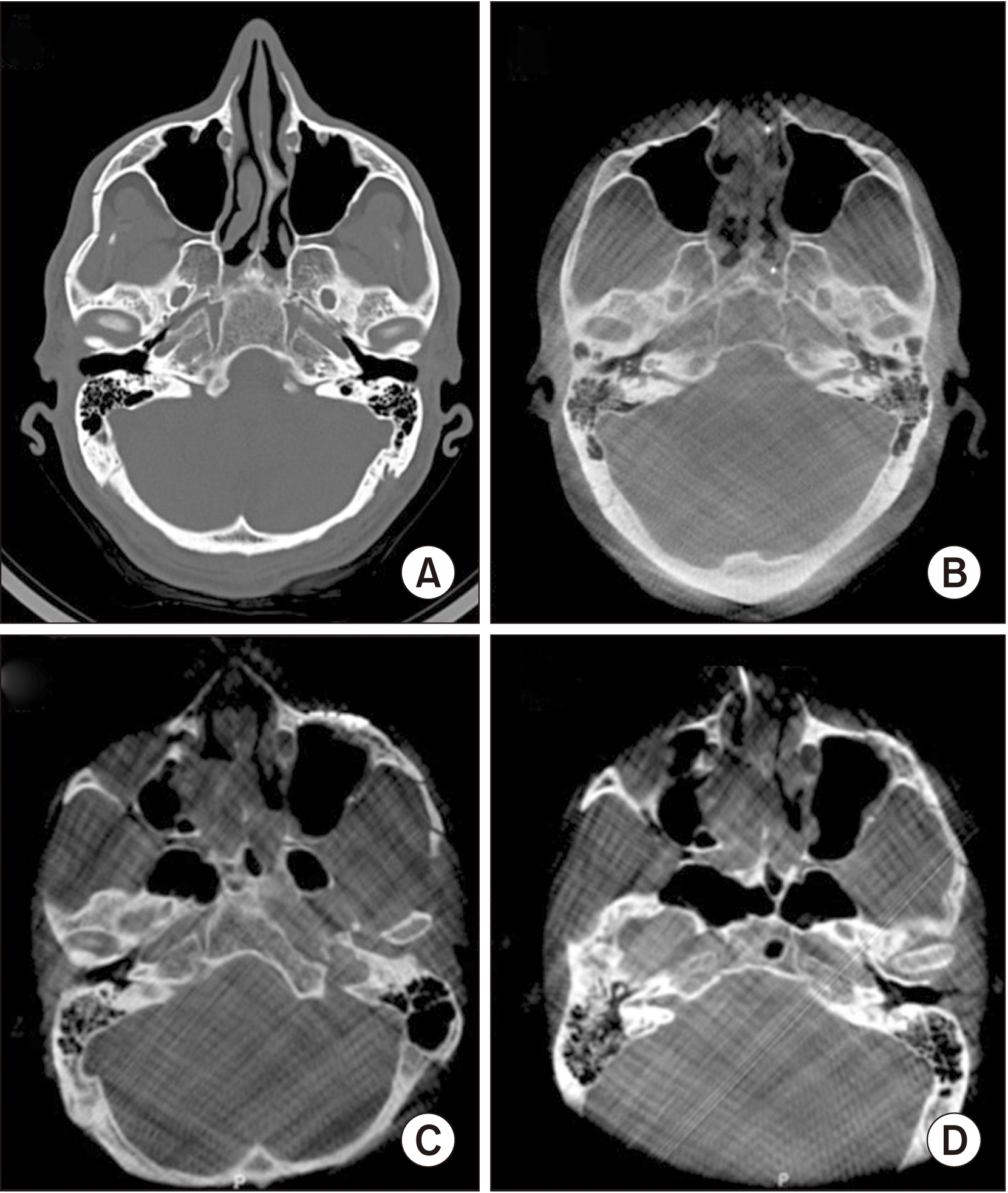
Fig. 1 A composite figure presenting two clinical cases. Case No. 1: a preoperative computed tomographic scan demonstrating a right zygomatic arch fracture (A) followed by an intraoperative O-Arm scan that demonstrates an adequate reduction of the zygomatic arch fracture (B). Case No. 2: an insufficient reduction of a left zygomatic complex (ZMC) fracture as demonstrated in an intraoperative O-Arm scan (C), and a second intraoperative O-Arm scan following the correction of the reduction and fixation of the left ZMC fracture lines (D).

Fig. 2 A composite figure presenting another case: a preoperative three-dimensional (3D) reconstruction computed tomographic scan demonstrates a left zygomatic complex fracture (A, B) followed by an intraoperative O-Arm scan that demonstrates a proper reduction and fixation of the zygomatic complex fracture (C), and a 3D reconstruction of the same intraoperative O-Arm scan presenting a good clinical result (D).
Reference
-
References
1. Kelley P, Hopper R, Gruss J. 2007; Evaluation and treatment of zygomatic fractures. Plast Reconstr Surg. 120(7 Suppl 2):5S–15S. https://doi.org/10.1097/01.prs.0000260720.73370.d7 . DOI: 10.1097/01.prs.0000260720.73370.d7. PMID: 18090725.
Article2. Bogusiak K, Arkuszewski P. 2010; Characteristics and epidemiology of zygomaticomaxillary complex fractures. J Craniofac Surg. 21:1018–23. https://doi.org/10.1097/scs.0b013e3181e62e47 . DOI: 10.1097/scs.0b013e3181e62e47. PMID: 20677370.
Article3. Marinho RO, Freire-Maia B. 2013; Management of fractures of the zygomaticomaxillary complex. Oral Maxillofac Surg Clin North Am. 25:617–36. https://doi.org/10.1016/j.coms.2013.07.011 . DOI: 10.1016/j.coms.2013.07.011. PMID: 24055372.
Article4. Gomes PP, Passeri LA, Barbosa JR. 2006; A 5-year retrospective study of zygomatico-orbital complex and zygomatic arch fractures in Sao Paulo State, Brazil. J Oral Maxillofac Surg. 64:63–7. https://doi.org/10.1016/j.joms.2005.09.012 . DOI: 10.1016/j.joms.2005.09.012. PMID: 16360858.
Article5. Adam AA, Zhi L, Bing LZ, Zhong Xing WU. 2012; Evaluation of treatment of zygomatic bone and zygomatic arch fractures: a retrospective study of 10 years. J Maxillofac Oral Surg. 11:171–6. https://doi.org/10.1007/s12663-011-0294-x . DOI: 10.1007/s12663-011-0294-x. PMID: 23730064. PMCID: PMC3386399.
Article6. Gülicher D, Krimmel M, Reinert S. 2006; The role of intraoperative ultrasonography in zygomatic complex fracture repair. Int J Oral Maxillofac Surg. 35:224–30. https://doi.org/10.1016/j.ijom.2005.10.005 . DOI: 10.1016/j.ijom.2005.10.005. PMID: 16364594.
Article7. Kim JS, Park YJ, Lee YJ, Kim NG, Lee KS. 2016; Reduction of zygomatic arch isolated fracture using ultra sound and needle marking. Arch Craniofac Surg. 17:198–201. https://doi.org/10.7181/acfs.2016.17.4.198 . DOI: 10.7181/acfs.2016.17.4.198. PMID: 28913283. PMCID: PMC5556836.
Article8. Ellis E 3rd, Perez D. 2014; An algorithm for the treatment of isolated zygomatico-orbital fractures. J Oral Maxillofac Surg. 72:1975–83. https://doi.org/10.1016/j.joms.2014.04.015 . DOI: 10.1016/j.joms.2014.04.015. PMID: 25053570.
Article9. Ostrofsky MK, Lownie JF. 1977; Zygomatico-coronoid ankylosis. J Oral Surg. 35:752–4. PMID: 267760.10. Khaqani MS, Tavosi F, Gholami M, Eftekharian HR, Khojastepour L. 2018; Analysis of facial symmetry after zygomatic bone fracture management. J Oral Maxillofac Surg. 76:595–604. https://doi.org/10.1016/j.joms.2017.10.005 . DOI: 10.1016/j.joms.2017.10.005. PMID: 29121488.
Article11. Manson PN, Clifford CM, Su CT, Iliff NT, Morgan R. 1986; Mechanisms of global support and posttraumatic enophthalmos: I. The anatomy of the ligament sling and its relation to intramuscular cone orbital fat. Plast Reconstr Surg. 77:193–202. PMID: 3945682.12. Barry C, Coyle M, Idrees Z, Dwyer MH, Kearns G. 2008; Ocular findings in patients with orbitozygomatic complex fractures: a retrospective study. J Oral Maxillofac Surg. 66:888–92. https://doi.org/10.1016/j.joms.2008.01.005 . DOI: 10.1016/j.joms.2008.01.005. PMID: 18423276.
Article13. Maheedhar AV, Ravindran C, Azariah ED. 2017; Use of C-arm to assess reduction of zygomatic complex fractures: a comparative study. Craniomaxillofac Trauma Reconstr. 10:35–43. https://doi.org/10.1055/s-0036-1592085 . DOI: 10.1055/s-0036-1592085. PMID: 28210406. PMCID: PMC5305307.
Article14. van Hout WM, Van Cann EM, Muradin MS, Frank MH, Koole R. 2014; Intraoperative imaging for the repair of zygomaticomaxillary complex fractures: a comprehensive review of the literature. J Craniomaxillofac Surg. 42:1918–23. https://doi.org/10.1016/j.jcms.2014.07.012 . DOI: 10.1016/j.jcms.2014.07.012. PMID: 25213198.
Article15. Jank S, Deibl M, Strobl H, Oberrauch A, Nicasi A, Missmann M, et al. 2005; Interrater reliability of sonographic examinations of orbital fractures. Eur J Radiol. 54:344–51. https://doi.org/10.1016/j.ejrad.2004.07.009 . DOI: 10.1016/j.ejrad.2004.07.009. PMID: 15899334.
Article16. Jenkins CN, Thuau H. 1997; Ultrasound imaging in assessment of fractures of the orbital floor. Clin Radiol. 52:708–11. https://doi.org/10.1016/s0009-9260(97)80037-2 . DOI: 10.1016/s0009-9260(97)80037-2. PMID: 9313738.
Article17. Hoelzle F, Klein M, Schwerdtner O, Lueth T, Albrecht J, Hosten N, et al. 2001; Intraoperative computed tomography with the mobile CT Tomoscan M during surgical treatment of orbital fractures. Int J Oral Maxillofac Surg. 30:26–31. https://doi.org/10.1054/ijom.2000.0014 . DOI: 10.1054/ijom.2000.0014. PMID: 11289617.
Article18. Hoffmann J, Krimmel M, Dammann F, Reinert S. 2002; [Feasibility of intraoperative diagnosis with a mobile computed tomography scanner]. Mund Kiefer Gesichtschir. 6:346–50. German. https://doi.org/10.1007/s10006-002-0406-3 . DOI: 10.1007/s10006-002-0406-3. PMID: 12448239.
Article19. Stanley RB Jr. 1999; Use of intraoperative computed tomography during repair of orbitozygomatic fractures. Arch Facial Plast Surg. 1:19–24. https://doi.org/10.1001/archfaci.1.1.19 . DOI: 10.1001/archfaci.1.1.19. PMID: 10937070.
Article20. Uneri A, Zhang X, Yi T, Stayman JW, Helm PA, Theodore N, et al. 2018; Image quality and dose characteristics for an O-arm intraoperative imaging system with model-based image reconstruction. Med Phys. 45:4857–68. https://doi.org/10.1002/mp.13167 . DOI: 10.1002/mp.13167. PMID: 30180274. PMCID: PMC6711149.
Article21. Sarwahi V, Payares M, Wendolowski S, Maguire K, Thornhill B, Lo Y, et al. 2017; Low-dose radiation 3D intraoperative imaging: how low can we go? An O-arm, CT scan, cadaveric study. Spine (Phila Pa 1976). 42:E1311–7. https://doi.org/10.1097/BRS.0000000000002154 . DOI: 10.1097/BRS.0000000000002154. PMID: 28296816.
Article22. Verhofste BP, Glotzbecker MP, Hresko MT, MacDougall RD, Birch CM, O'Neill NP, et al. 2020; Intraoperative use of O-arm in pediatric cervical spine surgery. J Pediatr Orthop. 40:e266–71. https://doi.org/10.1097/BPO.0000000000001416 . DOI: 10.1097/BPO.0000000000001416. PMID: 31192887.
Article23. Mettler FA Jr, Huda W, Yoshizumi TT, Mahesh M. 2008; Effective doses in radiology and diagnostic nuclear medicine: a catalog. Radiology. 248:254–63. https://doi.org/10.1148/radiol.2481071451 . DOI: 10.1148/radiol.2481071451. PMID: 18566177.
Article24. Wilde F, Lorenz K, Ebner AK, Krauss O, Mascha F, Schramm A. 2013; Intraoperative imaging with a 3D C-arm system after zygomatico-orbital complex fracture reduction. J Oral Maxillofac Surg. 71:894–910. https://doi.org/10.1016/j.joms.2012.10.031 . DOI: 10.1016/j.joms.2012.10.031. PMID: 23352428.
Article25. Kurita M, Okazaki M, Ozaki M, Tanaka Y, Tsuji N, Takushima A, et al. 2010; Patient satisfaction after open reduction and internal fixation of zygomatic bone fractures. J Craniofac Surg. 21:45–9. https://doi.org/10.1097/SCS.0b013e3181c36304 . DOI: 10.1097/SCS.0b013e3181c36304. PMID: 20061977.
Article
- Full Text Links
-
- Actions
-
Cited
- CITED
-
- Close
- Share
-
- Similar articles
-
- Closed Reduction of Zygomatic Arch Fracture with Intraoperative Mobile Computed Tomography Scan
- The Signficance of Intraoperative Radiograph in Treatment of Zygoma Fracture
- Endoscopic Reconstruction of Zygomatic Bone Fracture without Periorbital Incision
- Radiologic and Histological Study of Healing Process on Malunion Rat Model after Zygomatic Arch Fracture
- Three-dimensional imaging modalities in endodontics
