Anat Cell Biol.
2021 Sep;54(3):395-398. 10.5115/acb.21.042.
Bilateral venous sinuses of Kelch
- Affiliations
-
- 1Department of Neurosurgery, Tulane Center for Clinical Neurosciences, Tulane University School of Medicine, New Orleans, LA, USA.
- 2Department of Neurology, Tulane Center for Clinical Neurosciences, Tulane University School of Medicine, New Orleans, LA, USA.
- 3Department of Anatomical Sciences, St. George's University, St. George's, Grenada.
- 4Department of Structural & Cellular Biology, Tulane University School of Medicine, New Orleans, LA, USA.
- 5Department of Neurosurgery and Ochsner Neuroscience Institute, Ochsner Health System, New Orleans, LA, USA.
- 6Department of Surgery, Tulane University School of Medicine, New Orleans, LA, USA.
- 7University of Queensland, Brisbane, Australia.
- KMID: 2521052
- DOI: http://doi.org/10.5115/acb.21.042
Abstract
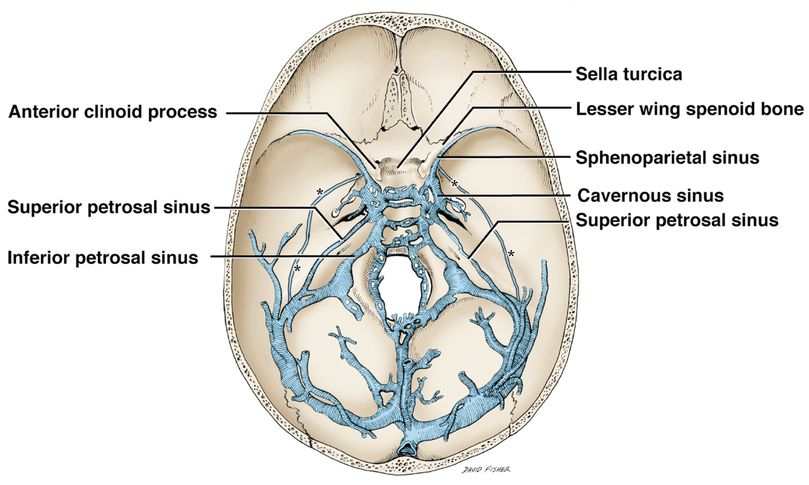
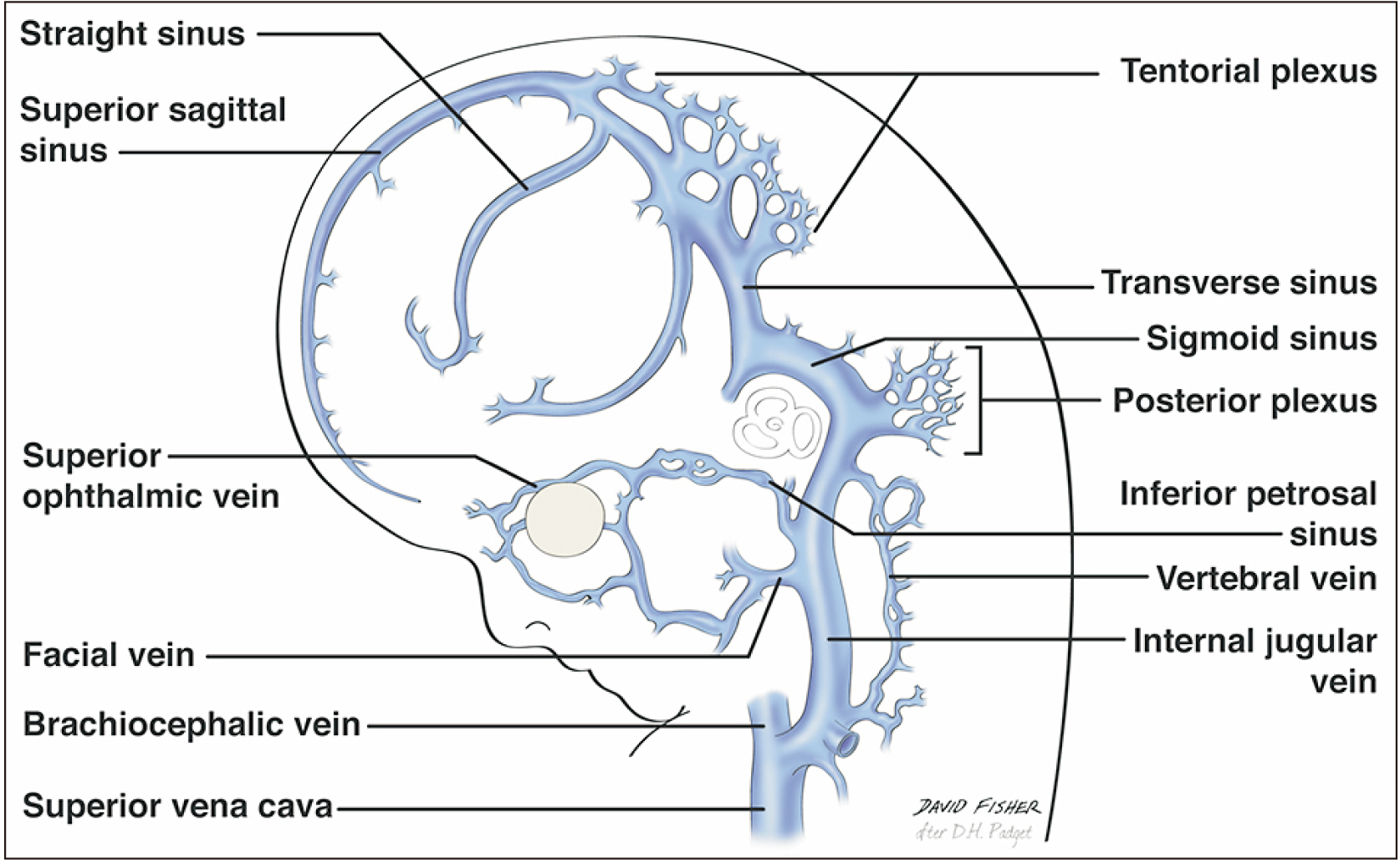
- Knowledge of the variant anatomy of the intradural venous sinuses is important to anatomists and clinicians alike. Herein, we report a cadaveric case of the rare venous sinus of Kelch, which some have believed is a remnant of the cranioorbital sinuses. To our knowledge, only one other cadaveric case has been reported in the extant medical literature. Clinically, knowledge of such a variant venous sinus can minimize misdiagnoses such as when anatomical variations are noted on imaging. Surgically, such an understanding can avoid intraoperative complications such as iatrogenic hemorrhage.
Figure
Reference
-
References
1. Klein NE. 1985; Catalog of human variations. Plast Reconstr Surg. 76:478. DOI: 10.1097/00006534-198509000-00033.
Article2. An YH, Wee JH, Han KH, Kim YH. 2011; Two cases of petrosquamosal sinus in the temporal bone presented as perioperative finding. Laryngoscope. 121:381–4. DOI: 10.1002/lary.21369. PMID: 21271593.3. Padget DH. 1956; The cranial venous system in man in reference to development, adult configuration, and relation to the arteries. Am J Anat. 98:307–55. DOI: 10.1002/aja.1000980302. PMID: 13362118.
Article4. Diamond MK. 1992; Homology and evolution of the orbitotemporal venous sinuses of humans. Am J Phys Anthropol. 88:211–44. DOI: 10.1002/ajpa.1330880209. PMID: 1605319.
Article5. Streeter GL. 1915; The development of the venous sinuses of the dura mater in the human embryo. Am J Anat. 18:145–78. DOI: 10.1002/aja.1000180202.
Article6. Knott JF. 1881; On the cerebral sinuses and their variations. J Anat Physiol. 16(Pt 1):27–42. PMID: 17231415. PMCID: PMC1310067.7. Hollinshead WH. 1982. Anatomy for surgeons. 3rd ed. Lippincott;Philadelphia:8. Tubbs RS. 2019. Anatomy, imaging and surgery of the intracranial dural venous sinuses. Elsevier;Philadelphia:9. Tubbs RS, Loukas M, Shoja MM, Oakes WJ. 2007; Accessory venous sinus of Hyrtl. Folia Morphol (Warsz). 66:198–9. PMID: 17985319.10. Cvetko E, Bosnjak R. 2014; Unilateral absence of foramen spinosum with bilateral ophthalmic origin of the middle meningeal artery: case report and review of the literature. Folia Morphol (Warsz). 73:87–91. DOI: 10.5603/FM.2014.0013. PMID: 24590529.
Article11. Umeh R, Loukas M, Oskouian RJ, Tubbs RS. 2017; Ectopic arachnoid granulation involving a rare intracranial venous sinus variant. Folia Morphol (Warsz). 76:319–21. DOI: 10.5603/FM.a2016.0062. PMID: 27813633.
Article12. Iwanaga J, Singh V, Ohtsuka A, Hwang Y, Kim HJ, Moryś J, Ravi KS, Ribatti D, Trainor PA, Sañudo JR, Apaydin N, Şengül G, Albertine KH, Walocha JA, Loukas M, Duparc F, Paulsen F, Del Sol M, Adds P, Hegazy A, Tubbs RS. 2021; Acknowledging the use of human cadaveric tissues in research papers: recommendations from anatomical journal editors. Clin Anat. 34:2–4. DOI: 10.1002/ca.23671. PMID: 32808702.
Article
- Full Text Links
- Actions
-
Cited
- CITED
-
- Close
- Share
- Similar articles
-
- Two Cases of Fungus Ball in Bilateral Paranasal Sinuses
- Endovascular Treatment of Idiopathic Intracranial Hypertension: A Case Report
- An Anatomical Study on the Variations of the Venous Sinuses at the Torcular Herophili
- A Case of Multiple Discrete Fungus Balls of the Bilateral Paranasal Sinuses
- A Unique Type of Dural Arteriovenous Fistula at Confluence of Sinuses Treated with Endovascular Embolization: A Case Report


