Sex estimation using radius in a Thai population
- Affiliations
-
- 1Department of Forensic Medicine, Faculty of Medicine, Chiang Mai University, Chiang Mai, Thailand
- 2Department of Anatomy, Faculty of Medicine, Chiang Mai University, Chiang Mai, Thailand
- 3Department of Statistics, Faculty of Science, Chiang Mai University, Chiang Mai, Thailand
- 4Excellence Center in Osteology Research and Training Center (ORTC), Chiang Mai University, Chiang Mai, Thailand
- KMID: 2521045
- DOI: http://doi.org/10.5115/acb.20.319
Abstract
- The estimation of sex is an essential component of forensic osteological analyses, and the potential of an incomplete radius for sex determination of human remains is investigated. The present study was conducted on 200 left-right pairs of radial bone from a northern Thai population (100 males and 100 females). The most dimorphic single parameter was maximum head diameter (MDH) with accuracies 92.0% for the right side and 90.5% for the left side. At the distal part of radius, the distal end width of the radius (RDEW) was the best sex indicator, in which the sex classification accuracies were 91.5% and 89.0%, for the right and left sides, respectively. Stepwise discriminant function analysis was performed for all measurements and specified separately to the proximal and distal radius. The circumference of the radial neck, headtuberosity length, MDH, and RDEW were selected for the stepwise procedure as these parameters produced the best correct classification results for both sides. The use of proximal radius for sex estimation was examined, with accuracies of 95.0% and 93.0% for the right and left sides, respectively. The sex classification functions for distal radius provided the accuracies of 92.5% and 89.5%, for the right and left sides, respectively. In summary, the fragments of radius indicated a high ability to estimate sex in the Northern Thai population.
Keyword
Figure
-
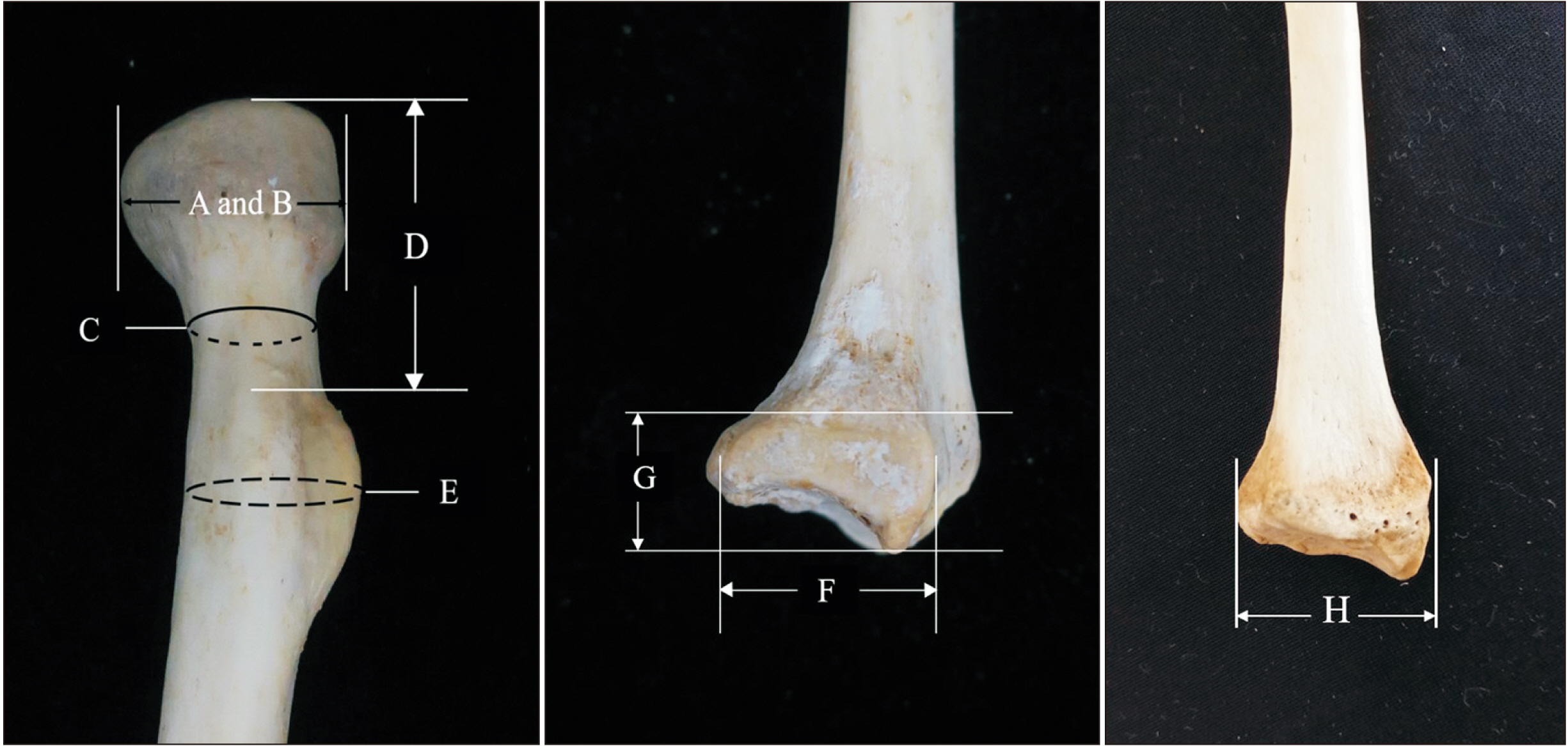
Fig. 1 Eight measurements of radius. A: maximum diameter of head, B: minimum diameter of head, C: circumference of neck, D: head-tuberosity length, E: circumference of tuberosity, F: ulnar notch length, G: ulnar notch width, H: distal end width of the radius.
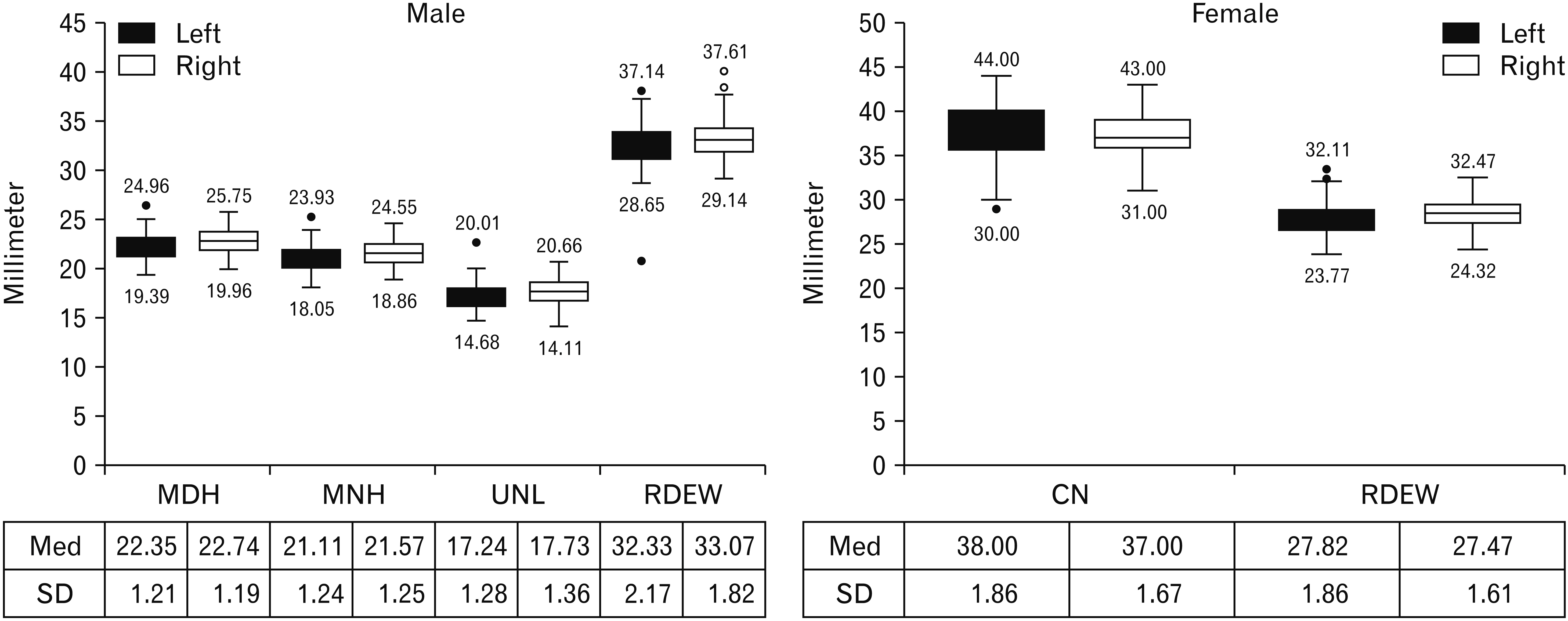
Fig. 2 The boxplot diagram shows median values (Med), outliers, and standard deviation (SD) of maximum diameter of head (MDH), minimum diameter of head (MNH), ulnar notch length (UNL), distal end width of the radius (RDEW) in male samples and circumference of neck (CN) and RDEW in female samples.
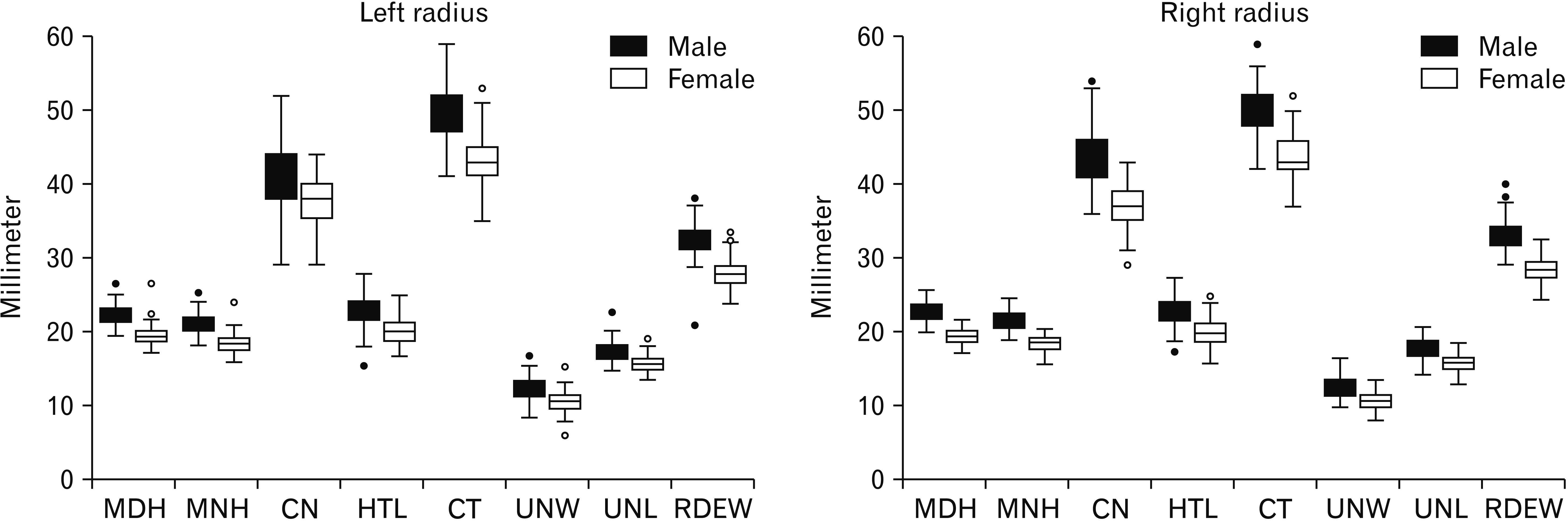
Fig. 3 The distribution of eight measurements of male and female samples for each side. MDH, maximum diameter of head; MNH, minimum diameter of head; CN, circumference of neck; HTL, head-tuberosity length; CT, circumference of tuberosity; UNW, ulnar notch width; UNL, ulnar notch length; RDEW, distal end width of the radius.
Reference
-
References
1. DiGangi EA, Moore MK. 2012. Research methods in human skeletal biology. Academic Press;London: DOI: 10.1016/B978-0-12-385189-5.00018-2.2. Phenice TW. 1969; A newly developed visual method of sexing the os pubis. Am J Phys Anthropol. 30:297–301. DOI: 10.1002/ajpa.1330300214. PMID: 5772048.
Article3. Christensen AM, Passalacqua NV, Bartelink EJ. 2014. Forensic anthropology: current methods and practice. Academic Press;Oxford: DOI: 10.1016/B978-0-12-418671-2.00005-7.4. Bruzek J. 2002; A method for visual determination of sex, using the human hip bone. Am J Phys Anthropol. 117:157–68. DOI: 10.1002/ajpa.10012. PMID: 11815949.
Article5. Spradley MK, Jantz RL. 2011; Sex estimation in forensic anthropology: skull versus postcranial elements. J Forensic Sci. 56:289–96. DOI: 10.1111/j.1556-4029.2010.01635.x. PMID: 21210801.
Article6. Safont S, Malgosa A, Subirà ME. 2000; Sex assessment on the basis of long bone circumference. Am J Phys Anthropol. 113:317–28. DOI: 10.1002/1096-8644(200011)113:3<317::AID-AJPA4>3.0.CO;2-J. PMID: 11042535.
Article7. Mall G, Hubig M, Büttner A, Kuznik J, Penning R, Graw M. 2001; Sex determination and estimation of stature from the long bones of the arm. Forensic Sci Int. 117:23–30. DOI: 10.1016/S0379-0738(00)00445-X. PMID: 11230943.
Article8. Suwanlikhid N, Mahakkanukrauh P. 2004; Northern Thai radius and sexing. Bull Chiang Mai Assoc Med Sci. 37:97–105.9. Barrier IL, L'Abbé EN. 2008; Sex determination from the radius and ulna in a modern South African sample. Forensic Sci Int. 179:85.e1–7. DOI: 10.1016/j.forsciint.2008.04.012. PMID: 18511225.
Article10. Uzün I, Işcan MY, Celbiş O. 2011; Forearm bones and sexual variation in Turkish population. Am J Forensic Med Pathol. 32:355–8. DOI: 10.1097/PAF.0b013e318219ca74. PMID: 21546820.11. Waghmare JE, Deshmukh PR, Waghmare PJ. 2012; Determination of sex from the shaft and tuberosity of radius- a multivariate discriminant function analysis. Biomed Res. 23:115–8.12. Martin S, Eliopoulos C, Borrini M. 2016; Metric methods of skeletal sex determination using the arm bones of two British Medieval populations. Glob J Anthropol Res. 3:41–9. DOI: 10.15379/2410-2806.2016.03.02.04.
Article13. Tomczyk J, Nieczuja-Dwojacka J, Zalewska M, Niemiro W, Olczyk W. 2017; Sex estimation of upper long bones by selected measurements in a Radom (Poland) population from the 18th and 19th centuries AD. Anthropol Rev. 80:287–300. DOI: 10.1515/anre-2017-0019.
Article14. Charisi D, Eliopoulos C, Vanna V, Koilias CG, Manolis SK. 2011; Sexual dimorphism of the arm bones in a modern Greek population. J Forensic Sci. 56:10–8. DOI: 10.1111/j.1556-4029.2010.01538.x. PMID: 20840296.
Article15. Knapp TR. 1992; Technical error of measurement: a methodological critique. Am J Phys Anthropol. 87:235–6. DOI: 10.1002/ajpa.1330870211.
Article16. Weinberg SM, Scott NM, Neiswanger K, Marazita ML. 2005; Intraobserver error associated with measurements of the hand. Am J Hum Biol. 17:368–71. DOI: 10.1002/ajhb.20129. PMID: 15849702.
Article17. Sakaue K. 2004; Sexual determination of long bones in recent Japanese. Anthropol Sci. 112:75–81. DOI: 10.1537/ase.00067.
Article18. Duangto P, Mahakkanukrauh P. 2020; Sex estimation from upper limb bones in a Thai population. Anat Cell Biol. 53:36–43. DOI: 10.5115/acb.19.179. PMID: 32274247. PMCID: PMC7118256.
Article19. Ubelaker DH, DeGaglia CM. 2017; Population variation in skeletal sexual dimorphism. Forensic Sci Int. 278:407.e1–7. DOI: 10.1016/j.forsciint.2017.06.012. PMID: 28698063.
Article20. Bongiovanni R, LeGarde CB. 2018; A univariate approach to sex estimation for the fragmentary upper limb. J Forensic Sci. 63:356–60. DOI: 10.1111/1556-4029.13530. PMID: 28631395.
Article21. Jaruratanasirikul S, iplung H Sr. 2015; Secular trends of growth and pubertal maturation of school children in Southern Thailand. Ann Hum Biol. 42:447–54. DOI: 10.3109/03014460.2014.955057. PMID: 25230717.
Article22. Phatsara M, Das S, Laowatthanaphong S, Tuamsuk P, Mahakkanukrauh P. 2016; The accuracy of sex estimation on metatarsal bones in a Northeastern Thai population. Clin Ter. 167:72–6. DOI: 10.7417/CT.2016.1929. PMID: 27424506.
- Full Text Links
-
- Actions
-
Cited
- CITED
-
- Close
- Share
-
- Similar articles
-
- Sex estimation from upper limb bones in a Thai population
- Stature estimation using the sternum in a Thai population
- A new method for sex estimation from maxillary suture length in a Thai population
- Forensic age-at-death estimation using the sternal junction in Thai adults: an autopsy study
- Age estimation equations using vertebral osteophyte formation in a Thai population: comparison and modified osteophyte scoring method
