Morphology of the temporalis muscle focusing on the tendinous attachment onto the coronoid process
- Affiliations
-
- 1Department of Anatomy and Orofacial Development, College of Dentistry, Chosun University, Gwangju, Korea
- 2Department of Anthropology, University of Hawai’i at Manoa, Honolulu, HI, USA
- KMID: 2521043
- DOI: http://doi.org/10.5115/acb.21.074
Abstract
- The temporalis muscle is usually described as a single layer originating at the temporal line, converging to a tendon, and inserting onto a narrow site of the coronoid process. However, recent studies have shown that the temporalis muscle can be divided into two or three separate segments and the distal attachment continues inferiorly beyond the coronoid process. Therefore, the aims of this study were to analyze the morphology of the temporalis muscle focusing on the tendinous attachment onto the coronoid process and to provide educational values. The temporalis muscle was carefully dissected in 26 cadavers and classified based on the muscle fascicle direction. Each divided part was sketched and measured based on bony landmarks to elucidate its tendinous insertion site onto the coronoid process, and the results obtained were reviewed through the literature. The temporalis muscle ends at two distinct terminal tendons with wider insertion sites than usually presented in textbooks and atlases and separates into two parts that combine to act as a single structural unit. The superficial part is a large fan-shaped muscle commonly recognized as the temporalis muscle. This converges infero-medially to form the superficial tendon and the lateral boundary of the retromolar triangle. Meanwhile, the deep part is a narrow vertically oriented rectangular muscle that converges postero-laterally to form the deep tendon and the medial boundary of the retromolar triangle. These results indicate that understanding the temporalis muscle’s insertion site onto the coronoid process will be useful clinically with educational values during surgical procedures.
Keyword
Figure
-

Fig. 1 Diagram showing the dimensions measured the two separate parts of the temporalis muscle attached onto the coronoid process based on the bony landmarks. ①, height of the ending point of the superficial part of the temporalis muscle (TMs) from the inferior border of the mandible; ②, height of the ending point of the deep part of the temporalis muscle (TMd) from the inferior border of the mandible; ③, height of the separating point of the two parts from the inferior border of the mandible; ④, height of the lingula from the inferior border of the mandible; ⑤, width of the TMd from the anterior border of the mandibular ramus; ⑥, width of the lingula from the anterior border of the mandibular ramus; ⑦, distance of the end point of the posterior border of the TMs based on the mandibular notch.

Fig. 2 Photograph (A) and schematic (B) illustrating the two distinct terminal tendons of the temporalis muscle that formed the boundary on both sides of the retromolar triangle. The blue and green colors indicate the dimensions of the TMs and TMd onto the coronoid process, respectively. TMs, superficial part of the temporalis muscle; TMd, deep part of the temporalis muscle.
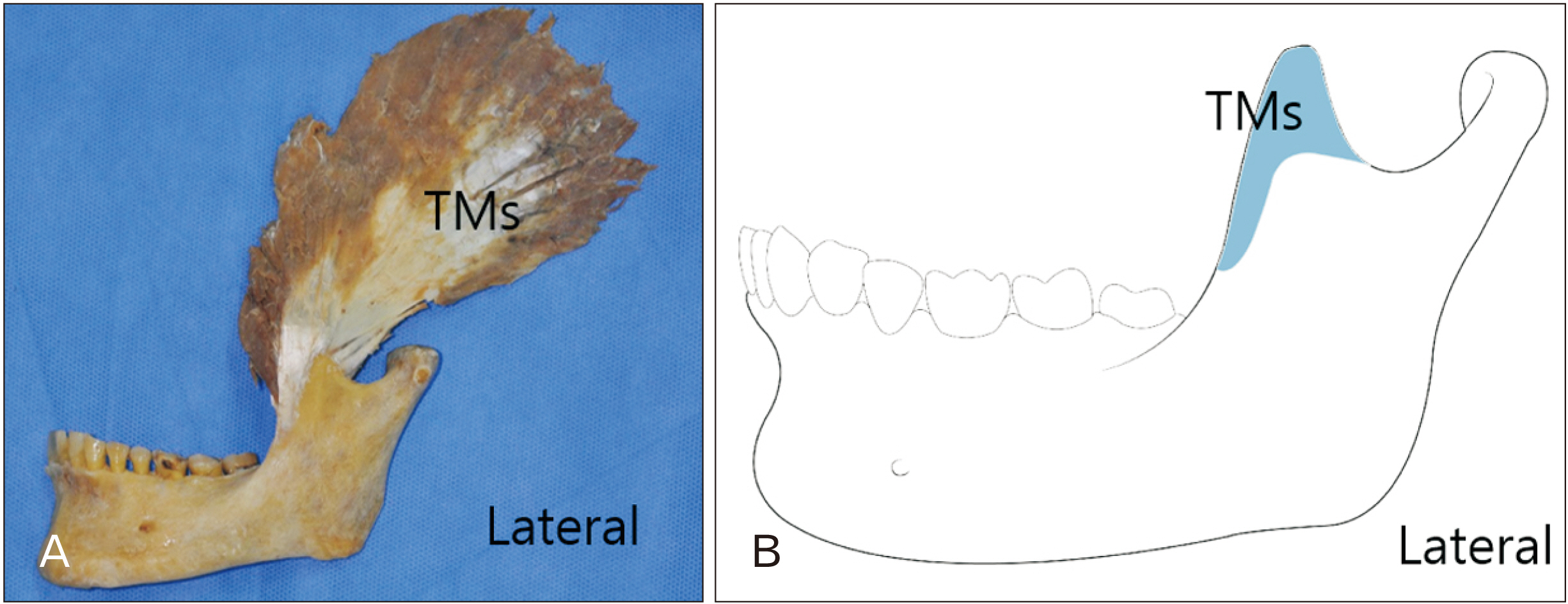
Fig. 3 Photograph (A) and schematic (B) showing the dimensions of the TMs attached onto the coronoid process on the lateral side of the mandible. TMs, superficial part of the temporalis muscle.

Fig. 4 Photographs (A–C) and schematic (D) showing the dimensions of the TMs and TMd attached onto the coronoid process on the medial side of the mandible. The dotted arrows indicated that the muscle fibers of two separate parts of the temporalis muscle were intermingled in the posterior side. TMs, superficial part of the temporalis muscle; TMd, deep part of the temporalis muscle.
Reference
-
References
1. Geers C, Nyssen-Behets C, Cosnard G, Lengelé B. 2005; The deep belly of the temporalis muscle: an anatomical, histological and MRI study. Surg Radiol Anat. 27:184–91. DOI: 10.1007/s00276-004-0306-3. PMID: 15821860.
Article2. Schön Ybarra MA, Bauer B. 2001; Medial portion of M. Temporalis and its potential involvement in facial pain. Clin Anat. 14:25–30. DOI: 10.1002/1098-2353(200101)14:1<25::AID-CA1004>3.0.CO;2-Q. PMID: 11135394.3. Sedlmayr JC, Kirsch CF, Wisco JJ. 2009; The human temporalis muscle: superficial, deep, and zygomatic parts comprise one structural unit. Clin Anat. 22:655–64. DOI: 10.1002/ca.20837. PMID: 19637294.
Article4. Lee JY, Kim JN, Kim SH, Choi HG, Hu KS, Kim HJ, Song WC, Koh KS. 2012; Anatomical verification and designation of the superficial layer of the temporalis muscle. Clin Anat. 25:176–81. DOI: 10.1002/ca.21212. PMID: 21739477.
Article5. Drake RL, Vogl W, Mitchell AWM, Gray H. 2010. Gray's anatomy for students. 2nd ed. Churchill Livingstone;Philadelphia:6. Schuenke M, Schulte E, Schumacher U. 2010. Thieme atlas of anatomy. Thieme Stuttgart;New York:7. Benninger B, Lee BI. 2012; Clinical importance of morphology and nomenclature of distal attachment of temporalis tendon. J Oral Maxillofac Surg. 70:557–61. DOI: 10.1016/j.joms.2011.02.047. PMID: 21549487.
Article8. Harn SD, Shackelford LS. 1982; Further evaluation of the superficial and deep tendons of the human temporalis muscle. Anat Rec. 202:537–48. DOI: 10.1002/ar.1092020413. PMID: 7072995.
Article9. Bressler HB, Friedman T, Friedman L. 2017; Ultrasound-guided injection of the temporalis tendon: a novel technique. J Ultrasound Med. 36:2125–31. DOI: 10.1002/jum.14232. PMID: 28504311.
Article10. Owusu Boahene KD. 2016; Temporalis muscle tendon unit transfer for smile restoration after facial paralysis. Facial Plast Surg Clin North Am. 24:37–45. DOI: 10.1016/j.fsc.2015.09.004. PMID: 26611700.
Article11. Palomari ET, Picosse LR, Tobo MP, Isayama RN, da Cunha MR. 2013; Sphenomandibular muscle or deep bundle of temporal muscle? Int J Morphol. 31:1158–61. DOI: 10.4067/S0717-95022013000400002.
Article12. Parker NP, Eisler LS, Dresner HS, Walsh WE. 2012; Orthodromic temporalis tendon transfer: anatomical considerations. Arch Facial Plast Surg. 14:39–44. DOI: 10.1001/archfaci.2011.1277. PMID: 22250268.13. Kim MK. 2017. Head and neck anatomy. 6th ed. Dental & Medical Publishing;Seoul:14. Gaudy JF, Zouaoui A, Bravetti P, Charrier JL, Laison F. 2001; Functional anatomy of the human temporal muscle. Surg Radiol Anat. 23:389–98. DOI: 10.1007/s00276-001-0389-z. PMID: 11963621.
Article15. Won SY, Kim DH, Yang HM, Park JT, Kwak HH, Hu KS, Kim HJ. 2011; Clinical and anatomical approach using Sihler's staining technique (whole mount nerve stain). Anat Cell Biol. 44:1–7. DOI: 10.5115/acb.2011.44.1.1. PMID: 21519543. PMCID: PMC3080003.
Article16. Blanksma NG, van Eijden TM. 1995; Electromyographic heterogeneity in the human temporalis and masseter muscles during static biting, open/close excursions, and chewing. J Dent Res. 74:1318–27. DOI: 10.1177/00220345950740061201. PMID: 7629340.17. Mérida-Velasco JR, Rodríguez-Vázquez JF, De La Cuadra C, Mérida-Velasco JA, Jiménez-Collado J. 2001; The course of the buccal nerve: relationships with the temporalis muscle during the prenatal period. J Anat. 198(Pt 4):423–9. DOI: 10.1017/S0021878201007567. PMID: 11327204. PMCID: PMC1468228.18. Logan BM, Hutchings RT, Reynolds PA, McMinn RMH. 2004. McMinn's Colour atlas of head & neck anatomy. Mosby;London:19. Akita K, Sakaguchi-Kuma T, Fukino K, Ono T. 2019; Masticatory muscles and branches of mandibular nerve: positional relationships between various muscle bundles and their innervating branches. Anat Rec (Hoboken). 302:609–19. DOI: 10.1002/ar.23943. PMID: 30312011.
Article20. Eisler P. von Bardeleben K, editor. 1912. Die Muskeln des stammes. Handbuch der anatomie des menschen. Gustav Fischer;Jena: p. 197–220. German.21. Yoshikawa T, Suzuki T. 1962; The lamination of the human masseter- the new identification of M. temporalis superficialis, M. maxillomandibularis and M. zygomaticomandibularis in the human anatomy. Acta Anat Nippon. 37:260–7.22. Zenker W. 1954; Function of the medial portion of the M. temporalis. Osterr Z Stomatol. 51:550–4. German. PMID: 14356656.23. Tomo S. 1990; Morphological classification of the mastication muscle based on their innervation. Ochanomizu Med J Tokyo. 38:57–71. Japanese.24. Shankland WE 2nd, Negulesco JA, O'Brian B. 1996; The "pre-anterior belly" of the temporalis muscle: a preliminary study of a newly described muscle. Cranio. 14:106–12. DOI: 10.1080/08869634.1996.11745956. PMID: 8949865.
Article25. Dunn GF, Hack GD, Robinson WL, Koritzer RT. 1996; Anatomical observation of a craniomandibular muscle originating from the skull base: the sphenomandibularis. Cranio. 14:97–103. DOI: 10.1080/08869634.1996.11745955. PMID: 8949864.
Article26. Kim HJ, Park BS, Cho YH, Yu SK. 2017; Course of buccal nerve on the anterior border of mandibular ramus related to temporalis tendon. Oral Biol Res. 41:236–9. DOI: 10.21851/obr.41.04.201712.236.
Article27. Ranganathan K, Terjimanian M, Lisiecki J, Rinkinen J, Mukkamala A, Brownley C, Buchman SR, Wang SC, Levi B. 2014; Temporalis muscle morphomics: the psoas of the craniofacial skeleton. J Surg Res. 186:246–52. DOI: 10.1016/j.jss.2013.07.059. PMID: 24079810.
Article28. Shimokawa T, Akita K, Soma K, Sato T. 1999; An anatomical study of the muscles innervated by the masseteric nerve. Okajimas Folia Anat Jpn. 75:271–80. DOI: 10.2535/ofaj1936.75.6_271. PMID: 10217945.
Article29. Khoury JN, Mihailidis S, Ghabriel M, Townsend G. 2011; Applied anatomy of the pterygomandibular space: improving the success of inferior alveolar nerve blocks. Aust Dent J. 56:112–21. DOI: 10.1111/j.1834-7819.2011.01312.x. PMID: 21623801.
Article
- Full Text Links
-
- Actions
-
Cited
- CITED
-
- Close
- Share
-
- Similar articles
-
- Orthodromic Transfer of the Temporalis Muscle in Incomplete Facial Nerve Palsy
- Reconstruction of the coronoid process using a graft from the olecranon of the same side for chronic posterior dislocation of the elbow
- Functional Analysis of the Masticatory System of the Dog with Relation to the Human
- Anatomical Location of the Tendinous Intersections of the Rectus Abdominis Muscle in Korean Women
- Trismus Due to Bilateral Coronoid Hyperplasia
